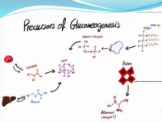
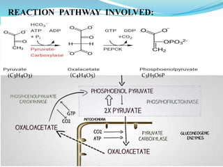
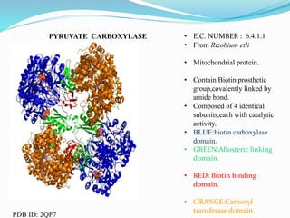
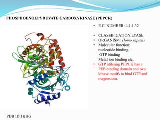
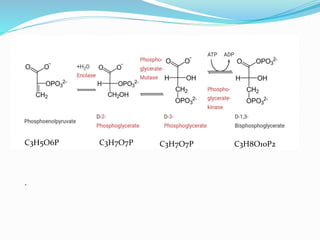
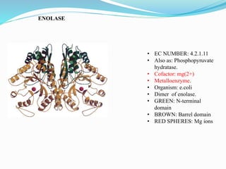
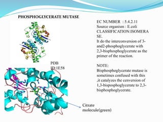
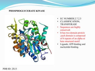
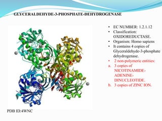
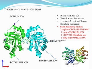
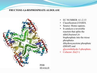
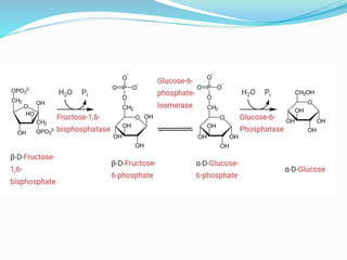
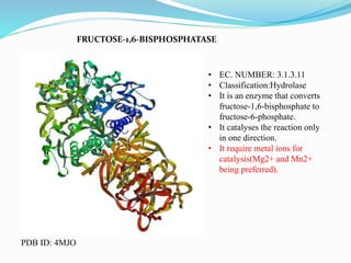
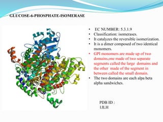
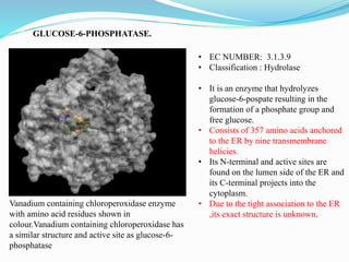
A PRESENTATION ON UNIQUENESS OF ENZYME
INVOLVED IN THE METABOLIC PATHWAY OF -
GLUCONEOGENESIS.
SUBMITTED TO: Dr. TARA KASHAV
SUBMITTED BY:
POOJA KUMARI SHASHI.
M.Sc. LIFE SCIENCE.
BATCH: 2017-2019
ROLL: 10
GLUCONEOGENESIS: An intro
 GLUCO: GLUCOSE ; NEO: NEW ; GENESIS: FORMATION.
 Defined as biosynthesis of glucose from non-carbohydrate precursors.
 The major non-carbohydrate precursors are lactate, amino acids ,glycerol and the
carbon skeleton of most amino acids.
 Non –carbohydrate precursors of glucose are first converted into pyruvate or as
oxaloacetate and DHAP.
 A ubiquitous process , present in plants ,animals ,fungi , bacteria and other
microorganisms.
 One of the main mechanism humans and many other animals use to keep blood
glucose level from dropping too low(hypoglycemia).
 Occurs mainy in liver and to some extent in the cortex of kidney.
 This process occurs during periods of fasting,starvation,low carbohydrate diet or
intense exercise and is highly endergonic.
REACTION PATHWAY INVOLVED:
(C3H4O3) (C4H4O5) C3H5O6P
.
PYRUVATE CARBOXYLASE • E.C. NUMBER : 6.4.1.1
• From Rizobium etli
• Mitochondrial protein.
• Contain Biotin prosthetic
group,covalently linked by
amide bond.
• Composed of 4 identical
subunits,each with catalytic
activity.
• BLUE:biotin carboxylase
domain.
• GREEN:Allosteric linking
domain.
• RED: Biotin binding
domain.
• ORANGE:Carboxyl
transferase domain.
PDB ID: 2QF7
PHOSPHOENOLPYRUVATE CARBOXYKINASE (PEPCK)
• E.C. NUMBER: 4.1.1.32
• CLASSIFICATION:LYASE
• ORGANISM: Homo sapiens
• Molecular function:
nucleotide binding,
GTP binding
Metal ion binding etc.
• GTP utilising PEPCK has a
PEP-binding domain and two
kinase motifs to bind GTP and
magnesium.
PDB ID:1KHG
C3H5O6P C3H7O7P C3H7O7P C3H8O10P2
.
ENOLASE
• EC NUMBER: 4.2.1.11
• Also as: Phosphopyruvate
hydratase.
• Cofactor: mg(2+)
• Metalloenzyme.
• Organism: e.coli
• Dimer of enolase.
• GREEN: N-terminal
domain
• BROWN: Barrel domain
• RED SPHERES: Mg ions
PHOSPHOGLYCERATE MUTASE
EC NUMBER : 5.4.2.11
Source organism : E.coli
CLASSIFICATION:ISOMERA
SE.
It do the interconversion of 3-
and2-phosphoglycerate with
2,3-bisphosphoglycerate as the
primer of the reaction.
NOTE:
Bisphosphoglycerate mutase is
sometimes confused with this
,it catalyzes the cenversion of
1,3-bisposphoglycerate to 2,3-
bisphosphoglycerate.
Citrate
molecule(green)
PDB
ID:1E58
PHOSPHOGLYCERATE KINASE
• EC NUMBER:2.7.2.3
• CLASSIFICATION;
TRANSFERASE
• Sequences are highly
conserved.
• It has two domain protein
;each domain is composed
of 6 repeats of an alpha or
beta structural motif.
• Ligands: ATP-binding and
nucleotide-binding.
PDB ID: 2X13
GLYCERALDEHYDE-3-PHOSPHATE-DEHYDROGENASE
• EC NUMBER: 1.2.1.12
• Classification:
OXIDOREDUCTASE.
• Organism: Homo sapiens
• It contains 4 copies of
Glyceraldehyde-3-phosphate
dehydrogenase.
• 2 non-polymeric entities:
a. 3 copies of
NICOTINAMIDE-
ADENINE-
DINUCLEOTIDE.
b. 3 copies of ZINC ION.
PDB ID:4WNC
TRIOSE-PHOSPHATE-ISOMERASE
• EC NUMBER: 5.3.1.1
• Classification : isomerases.
• It contains 2 copies of Triose-
phosphate isomerase.
• 4 non-polymeric entities:
2 copies of POTASSIUM ION.
1 copy of SODIUM ION
1 COPY OF phosphate ion.
1 copy of BROMIDE ION.
POTASSIUM ION
SODIUM ION
PHOSPHATE ION
BROMIDE ION
PDB ID:1NEY
FRUCTOSE-1,6-BISPHOSPHATE-ALDOLASE
• EC NUMBER: 4.1.2.13
• Classification:LYASES.
• Source: Homo sapiens.
• It catalyze a reversible
reaction that splits the
aldol,fructose1,6-
bisphosphate into the triose
phosphates
dihydroxyacetone phosphate
(DHAP) and
glyceraldehyde-3-phosphate.
• Cofactor: Zn(2+)
PDB
ID:4ALD
FRUCTOSE-1,6-BISPHOSPHATASE
• EC. NUMBER: 3.1.3.11
• Classification:Hydrolase
• It is an enzyme that converts
fructose-1,6-bisphosphate to
fructose-6-phosphate.
• It catalyses the reaction only
in one direction.
• It require metal ions for
catalysis(Mg2+ and Mn2+
being preferred).
PDB ID: 4MJO
GLUCOSE-6-PHOSPHATE-ISOMERASE
• EC NUMBER: 5.3.1.9
• Classification: isomerases.
• It catalyzes the reversible isomerization.
• It is a dimer composed of two identical
monomers.
• GPI monomers are made up of two
domains,one made of two separate
segments called the large domains and
the other made of the segment in
between called the small domain.
• The two domains are each alpa beta
alpha sandwiches.
PDB ID :
1JLH
GLUCOSE-6-PHOSPHATASE.
• EC NUMBER: 3.1.3.9
• Classification : Hydrolase
• It is an enzyme that hydrolyzes
glucose-6-pospate resulting in the
formation of a phosphate group and
free glucose.
• Consists of 357 amino acids anchored
to the ER by nine transmembrane
helicies.
• Its N-terminal and active sites are
found on the lumen side of the ER and
its C-terminal projects into the
cytoplasm.
• Due to the tight association to the ER
,its exact structure is unknown.
Vanadium containing chloroperoxidase enzyme
with amino acid residues shown in
colour.Vanadium containing chloroperoxidase has
a similar structure and active site as glucose-6-
phosphatase
CONCLUSION
ENZYMES CLASSIFICATION
• PYRUVATE CARBOXYLASE LIGASE
• PHOSPHOENOLPYRUVATE
CARBOXYKINASE.
• ENOLASE.
• FRUCTOSE-1,6-BISPHOSPHATE
ALDOLASE.
LYASES
• PHOSPHOGLYCERATE MUTASE
• TRIOSE-PHOSPHATE ISOMERASE.
• GLUCOSE-6-PHOSPHATE
ISOMERASE.
ISOMERASE
• PHOSPHOGLYCERATE KINASE TRANSFERASES
• GLYCERALDEHYDE-3-
PHOSPHATE DEHYDROGENASE.
OXIDOREDUCTA
SE
• FRUCTOSE-1,6-BISPHOSPHATASE
• GLUCOSE-6-PHOSPHATASE.
HYDROLASE
• As one can see
from this table
that the
gluconeogenesis
metabolic
pathway includes
all the 6 classes
of enzymes.
• Shape of an enzyme is very important and different type of enzymes have
different shapes and functions because the order and type of amino acids in
their structure is different.
The rate of gluconeogenesis is ultimately controlled by the action of a key
enzyme,fructose-1,6-bisphosphatase
Gluconeogenesis ppt

Gluconeogenesis ppt

  • 1.
    A PRESENTATION ONUNIQUENESS OF ENZYME INVOLVED IN THE METABOLIC PATHWAY OF - GLUCONEOGENESIS. SUBMITTED TO: Dr. TARA KASHAV SUBMITTED BY: POOJA KUMARI SHASHI. M.Sc. LIFE SCIENCE. BATCH: 2017-2019 ROLL: 10
  • 2.
    GLUCONEOGENESIS: An intro GLUCO: GLUCOSE ; NEO: NEW ; GENESIS: FORMATION.  Defined as biosynthesis of glucose from non-carbohydrate precursors.  The major non-carbohydrate precursors are lactate, amino acids ,glycerol and the carbon skeleton of most amino acids.  Non –carbohydrate precursors of glucose are first converted into pyruvate or as oxaloacetate and DHAP.  A ubiquitous process , present in plants ,animals ,fungi , bacteria and other microorganisms.  One of the main mechanism humans and many other animals use to keep blood glucose level from dropping too low(hypoglycemia).  Occurs mainy in liver and to some extent in the cortex of kidney.  This process occurs during periods of fasting,starvation,low carbohydrate diet or intense exercise and is highly endergonic.
  • 4.
  • 5.
    PYRUVATE CARBOXYLASE •E.C. NUMBER : 6.4.1.1 • From Rizobium etli • Mitochondrial protein. • Contain Biotin prosthetic group,covalently linked by amide bond. • Composed of 4 identical subunits,each with catalytic activity. • BLUE:biotin carboxylase domain. • GREEN:Allosteric linking domain. • RED: Biotin binding domain. • ORANGE:Carboxyl transferase domain. PDB ID: 2QF7
  • 6.
    PHOSPHOENOLPYRUVATE CARBOXYKINASE (PEPCK) •E.C. NUMBER: 4.1.1.32 • CLASSIFICATION:LYASE • ORGANISM: Homo sapiens • Molecular function: nucleotide binding, GTP binding Metal ion binding etc. • GTP utilising PEPCK has a PEP-binding domain and two kinase motifs to bind GTP and magnesium. PDB ID:1KHG
  • 7.
  • 8.
    ENOLASE • EC NUMBER:4.2.1.11 • Also as: Phosphopyruvate hydratase. • Cofactor: mg(2+) • Metalloenzyme. • Organism: e.coli • Dimer of enolase. • GREEN: N-terminal domain • BROWN: Barrel domain • RED SPHERES: Mg ions
  • 9.
    PHOSPHOGLYCERATE MUTASE EC NUMBER: 5.4.2.11 Source organism : E.coli CLASSIFICATION:ISOMERA SE. It do the interconversion of 3- and2-phosphoglycerate with 2,3-bisphosphoglycerate as the primer of the reaction. NOTE: Bisphosphoglycerate mutase is sometimes confused with this ,it catalyzes the cenversion of 1,3-bisposphoglycerate to 2,3- bisphosphoglycerate. Citrate molecule(green) PDB ID:1E58
  • 10.
    PHOSPHOGLYCERATE KINASE • ECNUMBER:2.7.2.3 • CLASSIFICATION; TRANSFERASE • Sequences are highly conserved. • It has two domain protein ;each domain is composed of 6 repeats of an alpha or beta structural motif. • Ligands: ATP-binding and nucleotide-binding. PDB ID: 2X13
  • 12.
    GLYCERALDEHYDE-3-PHOSPHATE-DEHYDROGENASE • EC NUMBER:1.2.1.12 • Classification: OXIDOREDUCTASE. • Organism: Homo sapiens • It contains 4 copies of Glyceraldehyde-3-phosphate dehydrogenase. • 2 non-polymeric entities: a. 3 copies of NICOTINAMIDE- ADENINE- DINUCLEOTIDE. b. 3 copies of ZINC ION. PDB ID:4WNC
  • 13.
    TRIOSE-PHOSPHATE-ISOMERASE • EC NUMBER:5.3.1.1 • Classification : isomerases. • It contains 2 copies of Triose- phosphate isomerase. • 4 non-polymeric entities: 2 copies of POTASSIUM ION. 1 copy of SODIUM ION 1 COPY OF phosphate ion. 1 copy of BROMIDE ION. POTASSIUM ION SODIUM ION PHOSPHATE ION BROMIDE ION PDB ID:1NEY
  • 14.
    FRUCTOSE-1,6-BISPHOSPHATE-ALDOLASE • EC NUMBER:4.1.2.13 • Classification:LYASES. • Source: Homo sapiens. • It catalyze a reversible reaction that splits the aldol,fructose1,6- bisphosphate into the triose phosphates dihydroxyacetone phosphate (DHAP) and glyceraldehyde-3-phosphate. • Cofactor: Zn(2+) PDB ID:4ALD
  • 16.
    FRUCTOSE-1,6-BISPHOSPHATASE • EC. NUMBER:3.1.3.11 • Classification:Hydrolase • It is an enzyme that converts fructose-1,6-bisphosphate to fructose-6-phosphate. • It catalyses the reaction only in one direction. • It require metal ions for catalysis(Mg2+ and Mn2+ being preferred). PDB ID: 4MJO
  • 17.
    GLUCOSE-6-PHOSPHATE-ISOMERASE • EC NUMBER:5.3.1.9 • Classification: isomerases. • It catalyzes the reversible isomerization. • It is a dimer composed of two identical monomers. • GPI monomers are made up of two domains,one made of two separate segments called the large domains and the other made of the segment in between called the small domain. • The two domains are each alpa beta alpha sandwiches. PDB ID : 1JLH
  • 18.
    GLUCOSE-6-PHOSPHATASE. • EC NUMBER:3.1.3.9 • Classification : Hydrolase • It is an enzyme that hydrolyzes glucose-6-pospate resulting in the formation of a phosphate group and free glucose. • Consists of 357 amino acids anchored to the ER by nine transmembrane helicies. • Its N-terminal and active sites are found on the lumen side of the ER and its C-terminal projects into the cytoplasm. • Due to the tight association to the ER ,its exact structure is unknown. Vanadium containing chloroperoxidase enzyme with amino acid residues shown in colour.Vanadium containing chloroperoxidase has a similar structure and active site as glucose-6- phosphatase
  • 19.
    CONCLUSION ENZYMES CLASSIFICATION • PYRUVATECARBOXYLASE LIGASE • PHOSPHOENOLPYRUVATE CARBOXYKINASE. • ENOLASE. • FRUCTOSE-1,6-BISPHOSPHATE ALDOLASE. LYASES • PHOSPHOGLYCERATE MUTASE • TRIOSE-PHOSPHATE ISOMERASE. • GLUCOSE-6-PHOSPHATE ISOMERASE. ISOMERASE • PHOSPHOGLYCERATE KINASE TRANSFERASES • GLYCERALDEHYDE-3- PHOSPHATE DEHYDROGENASE. OXIDOREDUCTA SE • FRUCTOSE-1,6-BISPHOSPHATASE • GLUCOSE-6-PHOSPHATASE. HYDROLASE • As one can see from this table that the gluconeogenesis metabolic pathway includes all the 6 classes of enzymes.
  • 20.
    • Shape ofan enzyme is very important and different type of enzymes have different shapes and functions because the order and type of amino acids in their structure is different. The rate of gluconeogenesis is ultimately controlled by the action of a key enzyme,fructose-1,6-bisphosphatase