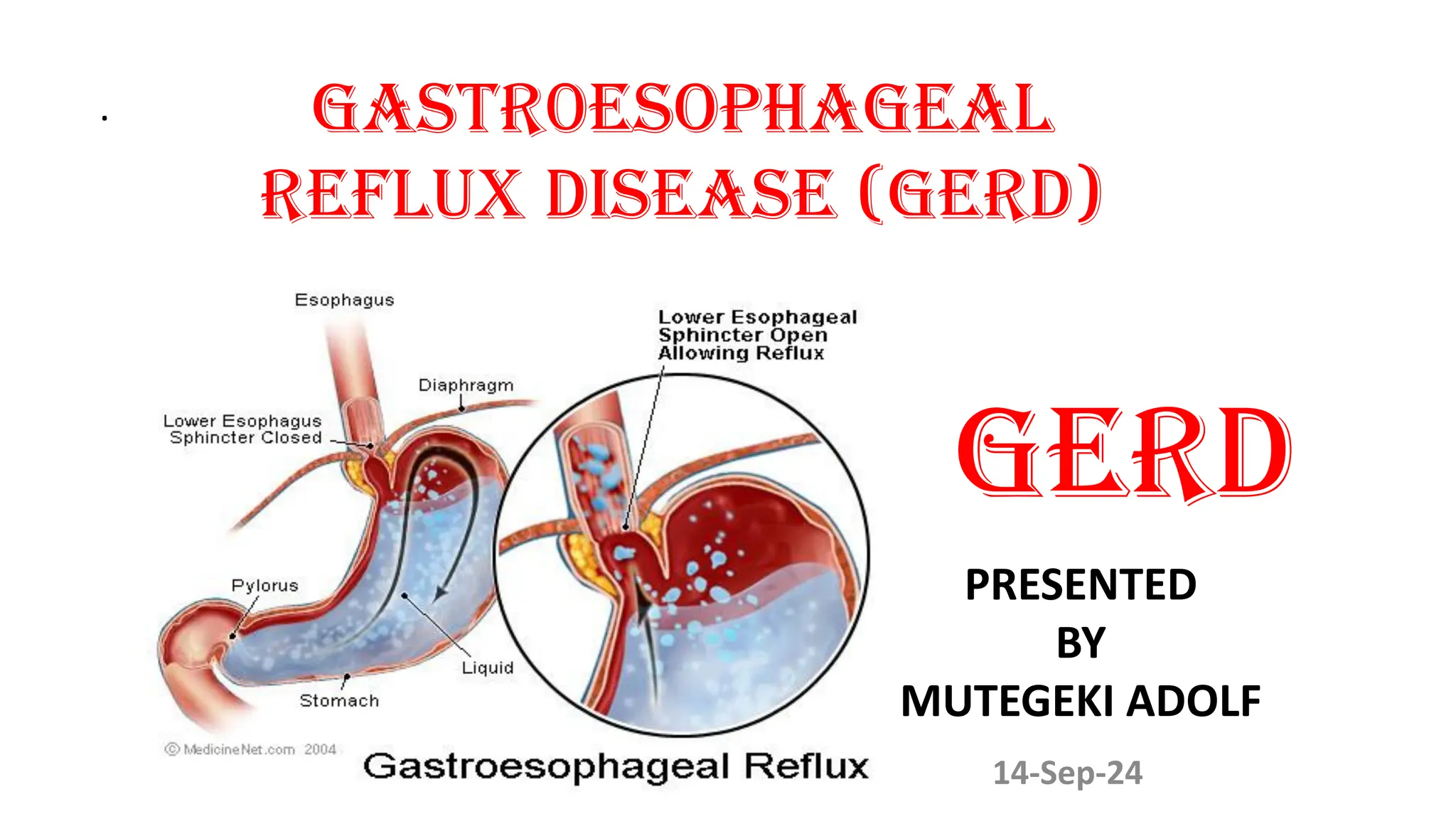
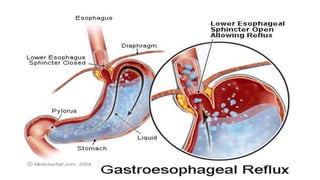
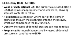
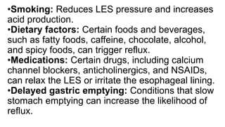
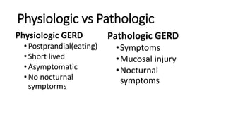
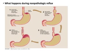
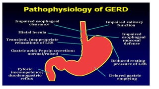
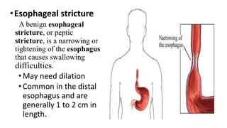
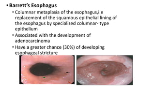
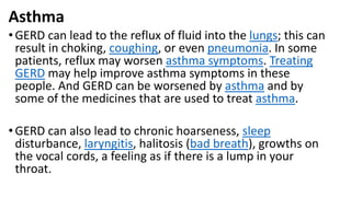
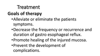
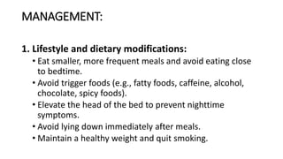
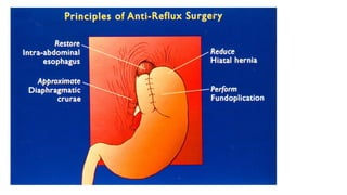
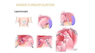
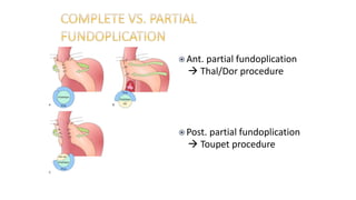
. GASTR0ESOPHAGEAL
REFLUX DISEASE (GERD)
GERD
14-Sep-24
PRESENTED
BY
MUTEGEKI ADOLF
Objectives
• Definition of GERD
• Epidemiology of GERD
• Pathophysiology of GERD
• Clinical Manisfestations
• Diagnostic Evaluation
• Treatment
• Complications
Definition
• Gastroesophageal Reflux Disease (GERD) is a chronic
condition where stomach contents, including acid and
bile, flow back (reflux) into the esophagus, leading to
symptoms like heartburn and regurgitation.
• GERD occurs when the lower esophageal sphincter
(LES) is weak or relaxes inappropriately, allowing the
stomach contents to flow back into the esophagus.
ETIOLOGY/ RISK FACTORS
•Weak or dysfunctional LES: The primary cause of GERD is a
LES that relaxes inappropriately or is weakened, allowing
stomach contents to reflux.
•Hiatal hernia: A condition where part of the stomach
pushes up through the diaphragm into the chest cavity,
which can compromise the LES function.
•Obesity: Increased abdominal pressure can lead to reflux.
•Pregnancy: Hormonal changes and increased abdominal
pressure can contribute to GERD
•Smoking: Reduces LES pressure and increases
acid production.
•Dietary factors: Certain foods and beverages,
such as fatty foods, caffeine, chocolate, alcohol,
and spicy foods, can trigger reflux.
•Medications: Certain drugs, including calcium
channel blockers, anticholinergics, and NSAIDs,
can relax the LES or irritate the esophageal lining.
•Delayed gastric emptying: Conditions that slow
stomach emptying can increase the likelihood of
reflux.
Physiologic vs Pathologic
Physiologic GERD
•Postprandial(eating)
•Short lived
•Asymptomatic
•No nocturnal
symptorms
Pathologic GERD
•Symptoms
•Mucosal injury
•Nocturnal
symptoms
• What happens during nonpathologic reflux
Epidemiology
• GERD occurs in all ages but, most common in those older than 40
years of age.
• About 10-20% of people in western countries suffer from GERD
symptoms on a weekly basis
• About 7% have symptoms daily.
• Except for NERD and pregnancy , no much difference in incidence
between men and women.
• But for Barrett’s esophagus, prevalence is more in males particularly
white adult males.
PATHOPHYSIOLOGY
1) DECREASED LOWER ESOPHAGEAL SPHINCTER PRESSURE
• Primary barrier to gastro esophageal reflux is the lower
esophageal sphincter
• LES normally works in conjunction with the diaphragm
• If barrier disrupted, acid goes from stomach to esophagus
May be due to
• Spontaneous transient LES relaxations
• Transient increase in intra abdominal pressure
• An atonic LES
Factors affecting les tone
•Drugs that reduce LES tone include calcium channel
antagonists (e.g., nifedipine, verapamil, diltiazem),
nitrates, anticholinergic agents(e.g.,tricyclic
antidepressants , antihistamines), and oral
contraceptives and estrogen.
•Foods that reduce LES tone include chocolate, fatty
foods , onions, peppermint, and garlic
•Smoking(nicotine) reduces LES tone.
2)DISRUPTION OFANATOMICAL BARRIERS
• Associated with hiatal hernia
• The size of hiatal hernia is proportional to the frequency of LES relaxations
• Hypotensive LES pressures and large hiatal hernia- more chance of GERD
following abrupt increase in intra abdominal pressure
3) ESOPHAGEAL CLEARANCE
• The GI acid produced spent too much time in contact with the esophageal
mucosa
• Normally swallowing contributes to esophageal clearance by increasing
salivary flow
• Saliva decreases with increasing age, so more often seen with elderly.
4)MUCOSAL RESISTANCE
• The mucus secreated by the mucus secreting glands involves in the
protection of esophagus
• The bicarbonates moving from the blood to the lumen can neutralize acidic
refluxate in the esophagus. On repeated exposure to the refluxate or due to
some defect in normal mucosal defenses hydrogen ions diffuse into the
mucosa, leading to cellular acidification and necrosis leading to esophagitis.
5)DELAYED GASTRIC EMPTYING
• An increase in gastric volume may increase both the frequency of reflux and
the amount of gastric fluid available to be refluxed
• Physiologic Postprandial Gastro esophageal reflux occurs
PATHOGENESIS
• Amount of esophageal damage seen
dependent on:
–Composition of refluxed material
–Volume of refluxed material
–Length of contact time
–Natural sensitivity of esophageal mucosa
–Rate of gastric emptying
COMPLICATIONS
• Esophagitis
• Esophageal strictures and ulcers
• Hemorrhage
• Perforation
• Aspiration
• Development of Barrett’s esophagus (cahage
in the lining of esophagus to be more like that
of small intestines)
• Precipitation of an asthma attack
•Erosive esophagitis
Erosive esophagitis is a condition in which areas of the
esophageal lining are inflamed and ulcerated. The most
common cause of erosive esophagitis is chronic acid reflux.
•Responsible for 40-60% of GERD symptoms
•Severity of symptoms often fail to match severity of
erosive esophagitis.
•Esophageal stricture
A benign esophageal
stricture, or peptic
stricture, is a narrowing or
tightening of the esophagus
that causes swallowing
difficulties.
•May need dilation
•Common in the distal
esophagus and are
generally 1 to 2 cm in
length.
•Barrett’s Esophagus
• Columnar metaplasia of the esophagus,i.e
replacement of the squamous epithelial lining of
the esophagus by specialized columnar- type
epithelium
• Associated with the development of
adenocarcinoma
• Have a greater chance (30%) of developing
esophageal stricture
Asthma
•GERD can lead to the reflux of fluid into the lungs; this can
result in choking, coughing, or even pneumonia. In some
patients, reflux may worsen asthma symptoms. Treating
GERD may help improve asthma symptoms in these
people. And GERD can be worsened by asthma and by
some of the medicines that are used to treat asthma.
•GERD can also lead to chronic hoarseness, sleep
disturbance, laryngitis, halitosis (bad breath), growths on
the vocal cords, a feeling as if there is a lump in your
throat.
CLINICAL PRESENTATIONS
• Heartburn: A burning sensation in the chest, usually after eating,
which might worsen when lying down or bending over.
• Regurgitation: A sour or bitter-tasting acid backing up into the
throat or mouth.
• Dysphagia: Difficulty swallowing or feeling of a lump in the throat.
• Chest pain: May mimic cardiac pain but is related to reflux.
• Chronic cough, laryngitis, or hoarseness: Due to reflux affecting
the throat and larynx.
• Nausea and vomiting: In some cases, GERD can cause persistent
nausea.
CLASSES OF SYMPTOMS
•TYPICAL SYMPTOMS
May be aggravated by activities that worsen
gastroesophageal reflux such as recumbent
position, bending over, or eating a meal high
in fat.
• Heartburn— retrosternal burning discomfort
(behind the breastbone)
• Regurgitation (spitting up of food)—effortless
return of gastric contents into the pharynx
without nausea, retching, or abdominal
contractions
• Water brash (hyper salivation)
• Belching
• ALARM SIGNS/SYMPTOMS
These symptoms may be indicative of complications of
GERD such as Barrett’s esophagus, esophageal strictures,
or esophageal cancer
• Dysphagia (difficulty swallowing )
• Early satiety (feeling full)
• GI bleeding
• Odynophagia (painful swallowing)
• Vomiting
• Unexplained Weight loss
• Iron deficiency anemia
• Choking
• Continual pain
INVESTIGATIONS
• Clinical history and physical examination: Often sufficient for diagnosis based
on symptoms.
• Upper endoscopy (EGD): To visualize the esophagus, look for inflammation,
erosions, or Barrett's esophagus, and rule out other conditions.
• 24-hour pH monitoring: Measures acid levels in the esophagus over 24 hours
to confirm abnormal acid exposure.
• Esophageal manometry: Assesses the function of the LES and esophageal
motility.
• Barium swallow (esophagram): An X-ray study to visualize reflux and assess
for structural abnormalities.
• Bravo pH monitoring: A wireless capsule attached to the esophagus that
measures acid exposure over a 48-hour period.
 An esophagram or barium swallow is an X-ray
imaging test used to visualize the structures of the
esophagus. The patient swallows liquid barium while
X-ray images are obtained. The barium fills and then
coats the lining of the esophagus so that it can diagnose
anatomical abnormalities such as tumors.
 Useful first diagnostic test for patients with dysphagia
 Stricture (location, length)
 Mass (location, length)
 Hiatal hernia (size, type)
Limitations
 Detailed mucosal exam for erosive
esophagitis, Barrett’s esophagus
 Indications
 Alarm symptoms
 Empiric therapy failure
 Preoperative evaluation
 Detection of Barrett’s
esophagus
Ambulatory 24 hr. pH Monitoring
Normal
GERD
Treatment
Goals of therapy
•Alleviate or eliminate the patients
symptoms.
•Decrease the frequency or recurrence and
duration of gastro esophageal reflux.
•Promote healing of the injured mucosa.
•Prevent the development of
complications.
MANAGEMENT:
1. Lifestyle and dietary modifications:
• Eat smaller, more frequent meals and avoid eating close
to bedtime.
• Avoid trigger foods (e.g., fatty foods, caffeine, alcohol,
chocolate, spicy foods).
• Elevate the head of the bed to prevent nighttime
symptoms.
• Avoid lying down immediately after meals.
• Maintain a healthy weight and quit smoking.
2. Medications:
•Antacids: Provide quick, short-term relief by neutralizing
stomach acid.
•H2 receptor blockers (e.g., ranitidine, famotidine):
Reduce acid production.
•Proton pump inhibitors (PPIs) (e.g., omeprazole,
esomeprazole): More effective in reducing stomach acid
production and healing the esophagus.
•Prokinetics (e.g., metoclopramide): Help strengthen the
LES and speed up gastric emptying, though used less
frequently due to side effects.
Treatment
•Antacids
• Over the counter acid
suppressants and
antacids appropriate
initial therapy
• Approx 1/3 of patients
with heartburn-related
symptoms use at least
twice weekly
• More effective than
placebo in relieving GERD
symptoms
3. Surgical interventions:
•Fundoplication (e.g., Nissen fundoplication): A
surgical procedure where the top of the stomach is
wrapped around the LES to strengthen it and
prevent reflux.
•LINX device: A ring of magnetic beads is placed
around the LES to augment its function without
inhibiting swallowing or belching.
Laparoscopic
 Ant. partial fundoplication
 Thal/Dor procedure
 Post. partial fundoplication
 Toupet procedure
TREATMENT
•Postsurgery
•10% have solid food dysphagia
•2-3% have permanent symptoms
•7-10% have gas, bloating, diarrhea, nausea, early
satiety
•Within 3-5 years 52% of patients back on antireflux
medications
PREVENTION:
• Maintain a healthy weight to reduce abdominal pressure.
• Avoid foods and drinks that trigger reflux symptoms.
•Eat meals at least 2-3 hours before lying down or going
to bed.
• Elevate the head of the bed or use wedge pillows to
prevent nighttime symptoms.
• Avoid smoking and alcohol consumption.
•Use medications like NSAIDs cautiously and under
supervision if prone to GERD symptoms.
GERD. GASTRO ESOPHAGEAL REFLUX DISEASE. GIT

GERD. GASTRO ESOPHAGEAL REFLUX DISEASE. GIT

  • 1.
    . GASTR0ESOPHAGEAL REFLUX DISEASE(GERD) GERD 14-Sep-24 PRESENTED BY MUTEGEKI ADOLF
  • 2.
    Objectives • Definition ofGERD • Epidemiology of GERD • Pathophysiology of GERD • Clinical Manisfestations • Diagnostic Evaluation • Treatment • Complications
  • 3.
    Definition • Gastroesophageal RefluxDisease (GERD) is a chronic condition where stomach contents, including acid and bile, flow back (reflux) into the esophagus, leading to symptoms like heartburn and regurgitation. • GERD occurs when the lower esophageal sphincter (LES) is weak or relaxes inappropriately, allowing the stomach contents to flow back into the esophagus.
  • 5.
    ETIOLOGY/ RISK FACTORS •Weakor dysfunctional LES: The primary cause of GERD is a LES that relaxes inappropriately or is weakened, allowing stomach contents to reflux. •Hiatal hernia: A condition where part of the stomach pushes up through the diaphragm into the chest cavity, which can compromise the LES function. •Obesity: Increased abdominal pressure can lead to reflux. •Pregnancy: Hormonal changes and increased abdominal pressure can contribute to GERD
  • 6.
    •Smoking: Reduces LESpressure and increases acid production. •Dietary factors: Certain foods and beverages, such as fatty foods, caffeine, chocolate, alcohol, and spicy foods, can trigger reflux. •Medications: Certain drugs, including calcium channel blockers, anticholinergics, and NSAIDs, can relax the LES or irritate the esophageal lining. •Delayed gastric emptying: Conditions that slow stomach emptying can increase the likelihood of reflux.
  • 7.
    Physiologic vs Pathologic PhysiologicGERD •Postprandial(eating) •Short lived •Asymptomatic •No nocturnal symptorms Pathologic GERD •Symptoms •Mucosal injury •Nocturnal symptoms
  • 8.
    • What happensduring nonpathologic reflux
  • 9.
    Epidemiology • GERD occursin all ages but, most common in those older than 40 years of age. • About 10-20% of people in western countries suffer from GERD symptoms on a weekly basis • About 7% have symptoms daily. • Except for NERD and pregnancy , no much difference in incidence between men and women. • But for Barrett’s esophagus, prevalence is more in males particularly white adult males.
  • 10.
    PATHOPHYSIOLOGY 1) DECREASED LOWERESOPHAGEAL SPHINCTER PRESSURE • Primary barrier to gastro esophageal reflux is the lower esophageal sphincter • LES normally works in conjunction with the diaphragm • If barrier disrupted, acid goes from stomach to esophagus May be due to • Spontaneous transient LES relaxations • Transient increase in intra abdominal pressure • An atonic LES
  • 11.
    Factors affecting lestone •Drugs that reduce LES tone include calcium channel antagonists (e.g., nifedipine, verapamil, diltiazem), nitrates, anticholinergic agents(e.g.,tricyclic antidepressants , antihistamines), and oral contraceptives and estrogen. •Foods that reduce LES tone include chocolate, fatty foods , onions, peppermint, and garlic •Smoking(nicotine) reduces LES tone.
  • 12.
    2)DISRUPTION OFANATOMICAL BARRIERS •Associated with hiatal hernia • The size of hiatal hernia is proportional to the frequency of LES relaxations • Hypotensive LES pressures and large hiatal hernia- more chance of GERD following abrupt increase in intra abdominal pressure 3) ESOPHAGEAL CLEARANCE • The GI acid produced spent too much time in contact with the esophageal mucosa • Normally swallowing contributes to esophageal clearance by increasing salivary flow • Saliva decreases with increasing age, so more often seen with elderly.
  • 13.
    4)MUCOSAL RESISTANCE • Themucus secreated by the mucus secreting glands involves in the protection of esophagus • The bicarbonates moving from the blood to the lumen can neutralize acidic refluxate in the esophagus. On repeated exposure to the refluxate or due to some defect in normal mucosal defenses hydrogen ions diffuse into the mucosa, leading to cellular acidification and necrosis leading to esophagitis. 5)DELAYED GASTRIC EMPTYING • An increase in gastric volume may increase both the frequency of reflux and the amount of gastric fluid available to be refluxed • Physiologic Postprandial Gastro esophageal reflux occurs
  • 15.
    PATHOGENESIS • Amount ofesophageal damage seen dependent on: –Composition of refluxed material –Volume of refluxed material –Length of contact time –Natural sensitivity of esophageal mucosa –Rate of gastric emptying
  • 16.
    COMPLICATIONS • Esophagitis • Esophagealstrictures and ulcers • Hemorrhage • Perforation • Aspiration • Development of Barrett’s esophagus (cahage in the lining of esophagus to be more like that of small intestines) • Precipitation of an asthma attack
  • 17.
    •Erosive esophagitis Erosive esophagitisis a condition in which areas of the esophageal lining are inflamed and ulcerated. The most common cause of erosive esophagitis is chronic acid reflux. •Responsible for 40-60% of GERD symptoms •Severity of symptoms often fail to match severity of erosive esophagitis.
  • 18.
    •Esophageal stricture A benignesophageal stricture, or peptic stricture, is a narrowing or tightening of the esophagus that causes swallowing difficulties. •May need dilation •Common in the distal esophagus and are generally 1 to 2 cm in length.
  • 19.
    •Barrett’s Esophagus • Columnarmetaplasia of the esophagus,i.e replacement of the squamous epithelial lining of the esophagus by specialized columnar- type epithelium • Associated with the development of adenocarcinoma • Have a greater chance (30%) of developing esophageal stricture
  • 20.
    Asthma •GERD can leadto the reflux of fluid into the lungs; this can result in choking, coughing, or even pneumonia. In some patients, reflux may worsen asthma symptoms. Treating GERD may help improve asthma symptoms in these people. And GERD can be worsened by asthma and by some of the medicines that are used to treat asthma. •GERD can also lead to chronic hoarseness, sleep disturbance, laryngitis, halitosis (bad breath), growths on the vocal cords, a feeling as if there is a lump in your throat.
  • 21.
    CLINICAL PRESENTATIONS • Heartburn:A burning sensation in the chest, usually after eating, which might worsen when lying down or bending over. • Regurgitation: A sour or bitter-tasting acid backing up into the throat or mouth. • Dysphagia: Difficulty swallowing or feeling of a lump in the throat. • Chest pain: May mimic cardiac pain but is related to reflux. • Chronic cough, laryngitis, or hoarseness: Due to reflux affecting the throat and larynx. • Nausea and vomiting: In some cases, GERD can cause persistent nausea.
  • 22.
    CLASSES OF SYMPTOMS •TYPICALSYMPTOMS May be aggravated by activities that worsen gastroesophageal reflux such as recumbent position, bending over, or eating a meal high in fat. • Heartburn— retrosternal burning discomfort (behind the breastbone) • Regurgitation (spitting up of food)—effortless return of gastric contents into the pharynx without nausea, retching, or abdominal contractions • Water brash (hyper salivation) • Belching
  • 23.
    • ALARM SIGNS/SYMPTOMS Thesesymptoms may be indicative of complications of GERD such as Barrett’s esophagus, esophageal strictures, or esophageal cancer • Dysphagia (difficulty swallowing ) • Early satiety (feeling full) • GI bleeding • Odynophagia (painful swallowing) • Vomiting • Unexplained Weight loss • Iron deficiency anemia • Choking • Continual pain
  • 24.
    INVESTIGATIONS • Clinical historyand physical examination: Often sufficient for diagnosis based on symptoms. • Upper endoscopy (EGD): To visualize the esophagus, look for inflammation, erosions, or Barrett's esophagus, and rule out other conditions. • 24-hour pH monitoring: Measures acid levels in the esophagus over 24 hours to confirm abnormal acid exposure. • Esophageal manometry: Assesses the function of the LES and esophageal motility. • Barium swallow (esophagram): An X-ray study to visualize reflux and assess for structural abnormalities. • Bravo pH monitoring: A wireless capsule attached to the esophagus that measures acid exposure over a 48-hour period.
  • 25.
     An esophagramor barium swallow is an X-ray imaging test used to visualize the structures of the esophagus. The patient swallows liquid barium while X-ray images are obtained. The barium fills and then coats the lining of the esophagus so that it can diagnose anatomical abnormalities such as tumors.  Useful first diagnostic test for patients with dysphagia  Stricture (location, length)  Mass (location, length)  Hiatal hernia (size, type) Limitations  Detailed mucosal exam for erosive esophagitis, Barrett’s esophagus
  • 26.
     Indications  Alarmsymptoms  Empiric therapy failure  Preoperative evaluation  Detection of Barrett’s esophagus
  • 27.
    Ambulatory 24 hr.pH Monitoring Normal GERD
  • 28.
    Treatment Goals of therapy •Alleviateor eliminate the patients symptoms. •Decrease the frequency or recurrence and duration of gastro esophageal reflux. •Promote healing of the injured mucosa. •Prevent the development of complications.
  • 29.
    MANAGEMENT: 1. Lifestyle anddietary modifications: • Eat smaller, more frequent meals and avoid eating close to bedtime. • Avoid trigger foods (e.g., fatty foods, caffeine, alcohol, chocolate, spicy foods). • Elevate the head of the bed to prevent nighttime symptoms. • Avoid lying down immediately after meals. • Maintain a healthy weight and quit smoking.
  • 30.
    2. Medications: •Antacids: Providequick, short-term relief by neutralizing stomach acid. •H2 receptor blockers (e.g., ranitidine, famotidine): Reduce acid production. •Proton pump inhibitors (PPIs) (e.g., omeprazole, esomeprazole): More effective in reducing stomach acid production and healing the esophagus. •Prokinetics (e.g., metoclopramide): Help strengthen the LES and speed up gastric emptying, though used less frequently due to side effects.
  • 31.
    Treatment •Antacids • Over thecounter acid suppressants and antacids appropriate initial therapy • Approx 1/3 of patients with heartburn-related symptoms use at least twice weekly • More effective than placebo in relieving GERD symptoms
  • 32.
    3. Surgical interventions: •Fundoplication(e.g., Nissen fundoplication): A surgical procedure where the top of the stomach is wrapped around the LES to strengthen it and prevent reflux. •LINX device: A ring of magnetic beads is placed around the LES to augment its function without inhibiting swallowing or belching.
  • 34.
  • 35.
     Ant. partialfundoplication  Thal/Dor procedure  Post. partial fundoplication  Toupet procedure
  • 36.
    TREATMENT •Postsurgery •10% have solidfood dysphagia •2-3% have permanent symptoms •7-10% have gas, bloating, diarrhea, nausea, early satiety •Within 3-5 years 52% of patients back on antireflux medications
  • 38.
    PREVENTION: • Maintain ahealthy weight to reduce abdominal pressure. • Avoid foods and drinks that trigger reflux symptoms. •Eat meals at least 2-3 hours before lying down or going to bed. • Elevate the head of the bed or use wedge pillows to prevent nighttime symptoms. • Avoid smoking and alcohol consumption. •Use medications like NSAIDs cautiously and under supervision if prone to GERD symptoms.