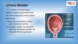
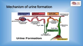
BY,
MR. BASAVARAJ
NURSINGTUTOR,
NAVODAYACON RAICHUR.
LEARNING OBJECTIVES
• Review of anatomy and physiology
• Composition and characteristics of urine.
• Factors influencing Urination
• Alteration in Urinary Elimination
• Types and Collection of urine specimen, Observation, urine testing
• Facilitating urine elimination: (assessment, types, equipment's,
procedures and special considerations)
• Providing urinal/bed pan, Condom drainage.
• Catheterization, Care of urinary drainage, and perineal care.
INTRODUCTION
• Urinary elimination is defined as expulsion of waste products from
the body through the urinary system.
• It is essential to maintain homeostasis of the body.
• It helps in removal of metabolic waste products from the body.
Review of anatomy and physiology
Organs of urine elimination
• Kidney:
• Ureters
• Urinary Bladder
• Urethra
Kidney
• Kidney: there are two kidneys present retroperitoneally in each side
• It consists of two parts i.e Medulla and cortex.
• Nephron is the Structural and functional unit of urinary system.
• Blood supply is by Renal artery and venous blood
is drained to renal vein.
• Renal pelvis is the point where renal artery enters
the kidney, and renal vein and ureters leave
the kidney.
Functions of kidney
1.Excratory function:
• Excretion of metabolites.
• drugs and
• Toxins from the body.
2. Homeostatic function:
• Maintenance of water balance.
• Maintenance of electrolyte balance.
• Maintenance of acid-base balance.
3. Endocrine (Hormonal) function.
• Renin secretion by JG cells.
• Secretion of Erythropoietin Hormone.
• Secretion of prostaglandins.
4. The kidney converts Vitamin D3 active 1,25-dihydroxycholecalciferol.
Ureters:
• There are two ureters descends from each kidney.
• They are made-up of Smooth muscles and inner lining is by transitional
epithelium.
• These ureters carries the urine from kidney to urinary Bladder.
• Each ureters are around 10 to 12 (20 to 30cm) inch longer.
• Upper half of the ureter located in Abdominal cavity and lower half is present in
Pelvic cavity.
Clinical significance
• Ureteral stones
• Reflux of Urine
• Congenital malformation
• Ca ureters
Urinary Bladder
• It is a hallow muscular organ
present in pelvic cavity which store
the urine produced by kidney before
eliminating.
• Superiorly connected to ureters
and inferiorly to Urethra.
• Ca bladder, cystitis, incontinence,
retention and Spastic bladder are
the main clinical significance.
Mechanism of urine formation
Nephron structure
Composition of urine
• 95% of volume of normal urine is due to water.
• Organic components
Urea (end product of protein metabolism)
Uric acid (end product of purines)
Creatinine
Amino acids
Metabolites of Hormones
• In-organics
Cations: Na2+, K+, Ca2+ etc
Anions: Cl-, SO4
2-, HCO3
- etc
Characteristic of Urine
Physical characters
• Color : pale yellow to deep Amber
• Odor : odorless
• Volume : 1 to 2 liters per 24 hours
• Specific gravity : 1.003-1.032 (1.010)
Other characters
• pH : 4.5 to 8.0 (6.8)
• Blood cells : nil
• Protein : nil
• Glucose : nil
• Ketone bodies : nil
Factors influencing urination
• Lifestyle
• Fluid and food intake
• Environment
• Psychological factor
• Medications
• Muscle tone and activity
• Pathological condition
• Surgical and diagnostic procedures
Alteration in urine elimination
• Polyuria: urine volume in excess of 3L/day.
• Oliguria : urine volume less than 500 ml/day.
• Anuria : urine volume less than 100 ml/day.
• Nocturia : frequent night time urination.
• Dysuria :difficulty in urination/ burning maturation.
• Enuresis :bed wetting.
• Urinary incontinence and :involuntary dribbling of urine.
• Urinary retention : inability to void the urine/ empty the bladder
completely.
Cont……..
• Haematuria : blood in the urine.
• Proteinuria : presence of protein in the urine.
• Glycosuria : presence of glucose in urine.
Urine specimen collection
• Proper collection of specimen is important to maximize the
outcome of laboratory test for the diagnosis of infectious
diseases.
• A variety of laboratory test can be performed to make a
presumptive or definitive diagnosis so that therapy can begin.
Types of urine specimen collection
• Random specimen collection.
• First morning specimen.
• Clean catch or midstream urine.
• Urine sample collection from catheter.
• Supra pubic aspiration.
Diagnostic tests
• Routine urine analysis.
• Blood test (BUN and Creatinine Clearance).
• Cystoscopy.
• Intravenous pyelogram IVP.
• Urine culture and sensitivity.
• CT Scan.
Facilitating urine elimination
• Providing urinal/bed pan,
• Condom drainage.
• Catheterization,
• Care of urinary drainage, and perineal care.
Catheterization
“Urinary catheterization is the insertion of a hollow tube through the
urethra into the bladder for removing urine”.
• It is a aseptic procedure for which sterile equipment's are required
Size of the catheter used
8-10 f are used for children's.
12-14 are used for female adults.
14,16, and 18 are used for male adults.
Purposes of urinary catheter
• To relieve from urinary retention.
• To obtain a sterile urine specimen.
• To measure residual urine.
• To empty the bladder before, during and after the surgery.
• To measure the urine output accurately.
Types of catheterization
• Intermittent catheter: an intermittent
catheter is used to drain the bladder for
short period or at once. It will have only
single lumen.
• Indwelling/retention catheter:
This type of catheter placed in to bladder
and secured there for a period of time.
• Supra pubic catheterization:
In this catheter is used to bladder by making
a small incision above the pubic area.
Procedure of catheterization
• Preparation .
• Insertion of catheter.
• After care and removal of urinary catheter.
Preparation
Preparation of patient:
• Prepare the patient mentally by explaining the procedure to gain cooperation.
• Prepare the part (urethral opening).
• Provide privacy and position the patient.
Prepare the articles:
• Catheter
• Bladder wash set
• 10cc/20cc syringe
• Sterile water
• Cotton balls with betadine
• Lubricant
• Sterile gloves
• Urine bag
• Micropore
Procedure of inserting catheter
• Explain the procedure to the patient.
• Provide privacy and adequate lightening and collect all articles.
• Position the male patient in supine position and female patient in
dorsal recommend position.
• Wash the hands.
• Drape the perineal area.
• Open the sterile catheter kit, using sterile technique.
• Put on the sterile gloves.
• Lubricate the catheter with sterile lubricant.
• Retract the foreskin of the penis in male and open the labial folds in
female.
Continued……..
• Clean the urethra in a circular manner from inside to outer.
• Hold the penis in 900 angle, insert the catheter and allow urinary
sphincter to relax.
• Lower the catheter and continue to advance the catheter.
Note: never force the catheter to advance and discontinue the
procedure if there is resistance.
• When the catheter reaches bladder urine starts to flow, gently insert
until 1-2 inches beyond where urine is noted.
• Inflate the balloon, using correct amount of sterile liquid.
Continued………
• Gently pull the catheter until inflation balloon is sung against bladder
neck, and connect the catheter to drainage system.
• Fix the tube with micropore and keep bag below the bladder level.
After care of procedure and articles
• Discard the waste, Remove gloves and replace the articles.
• Wash hands, and document the procedure.
Catheter care
• Fix the catheter to thigh or abdominal wall of the patient.
• Always keep urine bag below the bladder level.
• Everyday morning catheter care should be given with aseptic
techniques.
• Maintain close drainage system.
• Irrigate bladder with antimicrobials.
• Routinely examine for any signs of infection.
• Don’t collect urine sample from urine bag or catheter directly.
• Provide Perineal care.
• Don’t allow the faecal matter to contaminate the catheter.
Removal of Urinary catheter
• Once patient got relieved from the condition physician can plan to
remove the catheter.
• Take a Sterile 10/20cc syringe.
• Deflate the catheter.
• Pull the catheter gently until catheter come out.
• Discard the catheter, wash hands and document the procedure.
Urinary elimination

Urinary elimination

  • 1.
  • 2.
    LEARNING OBJECTIVES • Reviewof anatomy and physiology • Composition and characteristics of urine. • Factors influencing Urination • Alteration in Urinary Elimination • Types and Collection of urine specimen, Observation, urine testing • Facilitating urine elimination: (assessment, types, equipment's, procedures and special considerations) • Providing urinal/bed pan, Condom drainage. • Catheterization, Care of urinary drainage, and perineal care.
  • 3.
    INTRODUCTION • Urinary eliminationis defined as expulsion of waste products from the body through the urinary system. • It is essential to maintain homeostasis of the body. • It helps in removal of metabolic waste products from the body.
  • 4.
    Review of anatomyand physiology Organs of urine elimination • Kidney: • Ureters • Urinary Bladder • Urethra
  • 5.
    Kidney • Kidney: thereare two kidneys present retroperitoneally in each side • It consists of two parts i.e Medulla and cortex. • Nephron is the Structural and functional unit of urinary system. • Blood supply is by Renal artery and venous blood is drained to renal vein. • Renal pelvis is the point where renal artery enters the kidney, and renal vein and ureters leave the kidney.
  • 6.
    Functions of kidney 1.Excratoryfunction: • Excretion of metabolites. • drugs and • Toxins from the body. 2. Homeostatic function: • Maintenance of water balance. • Maintenance of electrolyte balance. • Maintenance of acid-base balance. 3. Endocrine (Hormonal) function. • Renin secretion by JG cells. • Secretion of Erythropoietin Hormone. • Secretion of prostaglandins. 4. The kidney converts Vitamin D3 active 1,25-dihydroxycholecalciferol.
  • 7.
    Ureters: • There aretwo ureters descends from each kidney. • They are made-up of Smooth muscles and inner lining is by transitional epithelium. • These ureters carries the urine from kidney to urinary Bladder. • Each ureters are around 10 to 12 (20 to 30cm) inch longer. • Upper half of the ureter located in Abdominal cavity and lower half is present in Pelvic cavity. Clinical significance • Ureteral stones • Reflux of Urine • Congenital malformation • Ca ureters
  • 8.
    Urinary Bladder • Itis a hallow muscular organ present in pelvic cavity which store the urine produced by kidney before eliminating. • Superiorly connected to ureters and inferiorly to Urethra. • Ca bladder, cystitis, incontinence, retention and Spastic bladder are the main clinical significance.
  • 9.
  • 10.
  • 11.
    Composition of urine •95% of volume of normal urine is due to water. • Organic components Urea (end product of protein metabolism) Uric acid (end product of purines) Creatinine Amino acids Metabolites of Hormones • In-organics Cations: Na2+, K+, Ca2+ etc Anions: Cl-, SO4 2-, HCO3 - etc
  • 12.
    Characteristic of Urine Physicalcharacters • Color : pale yellow to deep Amber • Odor : odorless • Volume : 1 to 2 liters per 24 hours • Specific gravity : 1.003-1.032 (1.010) Other characters • pH : 4.5 to 8.0 (6.8) • Blood cells : nil • Protein : nil • Glucose : nil • Ketone bodies : nil
  • 13.
    Factors influencing urination •Lifestyle • Fluid and food intake • Environment • Psychological factor • Medications • Muscle tone and activity • Pathological condition • Surgical and diagnostic procedures
  • 14.
    Alteration in urineelimination • Polyuria: urine volume in excess of 3L/day. • Oliguria : urine volume less than 500 ml/day. • Anuria : urine volume less than 100 ml/day. • Nocturia : frequent night time urination. • Dysuria :difficulty in urination/ burning maturation. • Enuresis :bed wetting. • Urinary incontinence and :involuntary dribbling of urine. • Urinary retention : inability to void the urine/ empty the bladder completely.
  • 15.
    Cont…….. • Haematuria :blood in the urine. • Proteinuria : presence of protein in the urine. • Glycosuria : presence of glucose in urine.
  • 16.
    Urine specimen collection •Proper collection of specimen is important to maximize the outcome of laboratory test for the diagnosis of infectious diseases. • A variety of laboratory test can be performed to make a presumptive or definitive diagnosis so that therapy can begin.
  • 17.
    Types of urinespecimen collection • Random specimen collection. • First morning specimen. • Clean catch or midstream urine. • Urine sample collection from catheter. • Supra pubic aspiration.
  • 18.
    Diagnostic tests • Routineurine analysis. • Blood test (BUN and Creatinine Clearance). • Cystoscopy. • Intravenous pyelogram IVP. • Urine culture and sensitivity. • CT Scan.
  • 19.
    Facilitating urine elimination •Providing urinal/bed pan, • Condom drainage. • Catheterization, • Care of urinary drainage, and perineal care.
  • 20.
    Catheterization “Urinary catheterization isthe insertion of a hollow tube through the urethra into the bladder for removing urine”. • It is a aseptic procedure for which sterile equipment's are required Size of the catheter used 8-10 f are used for children's. 12-14 are used for female adults. 14,16, and 18 are used for male adults.
  • 21.
    Purposes of urinarycatheter • To relieve from urinary retention. • To obtain a sterile urine specimen. • To measure residual urine. • To empty the bladder before, during and after the surgery. • To measure the urine output accurately.
  • 22.
    Types of catheterization •Intermittent catheter: an intermittent catheter is used to drain the bladder for short period or at once. It will have only single lumen. • Indwelling/retention catheter: This type of catheter placed in to bladder and secured there for a period of time. • Supra pubic catheterization: In this catheter is used to bladder by making a small incision above the pubic area.
  • 23.
    Procedure of catheterization •Preparation . • Insertion of catheter. • After care and removal of urinary catheter.
  • 24.
    Preparation Preparation of patient: •Prepare the patient mentally by explaining the procedure to gain cooperation. • Prepare the part (urethral opening). • Provide privacy and position the patient. Prepare the articles: • Catheter • Bladder wash set • 10cc/20cc syringe • Sterile water • Cotton balls with betadine • Lubricant • Sterile gloves • Urine bag • Micropore
  • 25.
    Procedure of insertingcatheter • Explain the procedure to the patient. • Provide privacy and adequate lightening and collect all articles. • Position the male patient in supine position and female patient in dorsal recommend position. • Wash the hands. • Drape the perineal area. • Open the sterile catheter kit, using sterile technique. • Put on the sterile gloves. • Lubricate the catheter with sterile lubricant. • Retract the foreskin of the penis in male and open the labial folds in female.
  • 26.
    Continued…….. • Clean theurethra in a circular manner from inside to outer. • Hold the penis in 900 angle, insert the catheter and allow urinary sphincter to relax. • Lower the catheter and continue to advance the catheter. Note: never force the catheter to advance and discontinue the procedure if there is resistance. • When the catheter reaches bladder urine starts to flow, gently insert until 1-2 inches beyond where urine is noted. • Inflate the balloon, using correct amount of sterile liquid.
  • 27.
    Continued……… • Gently pullthe catheter until inflation balloon is sung against bladder neck, and connect the catheter to drainage system. • Fix the tube with micropore and keep bag below the bladder level. After care of procedure and articles • Discard the waste, Remove gloves and replace the articles. • Wash hands, and document the procedure.
  • 28.
    Catheter care • Fixthe catheter to thigh or abdominal wall of the patient. • Always keep urine bag below the bladder level. • Everyday morning catheter care should be given with aseptic techniques. • Maintain close drainage system. • Irrigate bladder with antimicrobials. • Routinely examine for any signs of infection. • Don’t collect urine sample from urine bag or catheter directly. • Provide Perineal care. • Don’t allow the faecal matter to contaminate the catheter.
  • 29.
    Removal of Urinarycatheter • Once patient got relieved from the condition physician can plan to remove the catheter. • Take a Sterile 10/20cc syringe. • Deflate the catheter. • Pull the catheter gently until catheter come out. • Discard the catheter, wash hands and document the procedure.