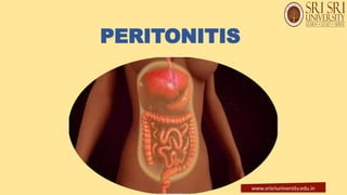
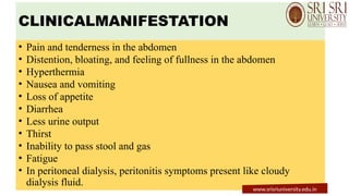
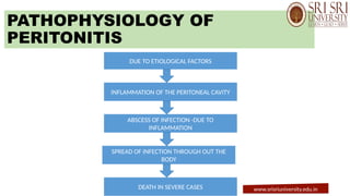
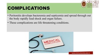
www.srisriuniversity.edu.in
GASTRO-INTESTINAL
PERFORATION
PRESENTED BY:
Ms.RAJASHREE PRIYADARSINI
Msc. TUTOR
SRI SRI COLLEGE OF NURSING,
SRI SRI UNIVERSITY.
www.srisriuniversity.edu.in
INTRODUCTION
• Gastrointestinal perforation is known as ruptured bowel,
intestinal perforation and perforation of the intestines.
• It occurs from the upper esophagus to the anorectal junction
at anywhere in the structural anatomical location.
• It is caused by peptic ulcers and sigmoid diverticulum.
• Without proper surgical management lead septic shock,
multi organ dysfunction and death.
www.srisriuniversity.edu.in
• Esophagus: Perforation can occur in the upper part of the esophagus.
• Stomach: Common sites include the gastric antrum and duodenal bulb.
• Small Intestine: Perforations can happen in the jejunum and ileum.
• Large Intestine: The ascending colon, transverse colon, descending
colon, sigmoid colon, and rectum are potential sites.
• Appendix: The appendix itself can perforate.
SITES OF GI PERFORATION
www.srisriuniversity.edu.in
DEFINITION
• Intestinal perforation is a serious
condition in which a hole in the
duodenum, jejunum and illiac allows
the intestinal contents to leak out the
abdominal cavity.and causes
Peritonities.
www.srisriuniversity.edu.in
ETIOLOGY
• Inflammatory bowel diseases (Crohn's disease and ulcerative colitis)
• Congenital abnormality of the small intestine - Meckel's diverticulum
inflammation
• Gastrointestinal tract cancer
• Volvulus-bowel obstruction that happens when the large bowel becomes twisted
• Diverticulitis-inflammatory condition that affects the large bowel
• Peptic ulcers in the stomach and small intestine
• Ischemia colitis inflammation of the large bowel due to an inadequate supply of
blood
• Foreign body ingestion.
• Iatrogenic injuries
• Neoplasm
www.srisriuniversity.edu.in
• Abdomen injury by knife, and gunshot
• Abdominal surgery
• NSAID drugs and steroids cause stomach ulcers
• Smoking and excessive use of alcohol increase risk of gastric perforation
www.srisriuniversity.edu.in
PATHOPHYSIOLOGY OF
PERFORATION.
Obstruction or Injury
↓
↑ Intraluminal Pressure or Direct Mucosal Damage
↓
Venous Congestion → Ischemia
↓
Mucosal Breakdown → Necrosis
↓
Full-Thickness GI Wall Breach
↓
Leakage of Contents into Peritoneal Cavity
↓
Peritonitis → Sepsis → Septic Shock (if untreated)
www.srisriuniversity.edu.in
CLINICAL MANIFESTATIONS.
• Rapid pain develops and sharp in nature and associated with malaise,
vomiting, and lethargy.
• Rigid abdomen
• Develop peritonitis always required urgent surgery.
• Esophageal rupture in thoracic region perforation causes radiating pain from
chest, neck and the back, pain worsening during inspiration. Pain associated
with vomiting and respiratory symptoms. Percussion may reveal signs of a
pleural effusion, with the potential for palpable crepitus.
• Abdominal Distension and Tenderness
• Loss of Bowel Sounds
• Shortness of Breath: This can occur as the inflammation and fluid buildup in
the abdomen put pressure on the lungs.
• Dizziness and Fatigue: These are general signs of a serious medical condition.
www.srisriuniversity.edu.in
DIAGNOSTIC EVALUATION
• History collection related to the etiological factors.
• Physiological examination-abdominal rigidity, rebound tenderness, board
like abdomen, dull and irritable appearance.
• Assess the patient's health status appearance, sign of injury, abrasion,
ecchymosis, medical history, any comorbid illness like diabetes,
hypertension, GIT problems.
• Observe patient's breathing patterns, abdominal movements with breathing,
abdominal distention, discoloration
• Abdominal tenderness on percussion evidence for peritoneal inflammation.
• Bowel sounds are usually absent in peritonitis.
• Assess any other conditions like acute appendicitis, ruptured tubo-ovarian
abscess, and perforated diverticulitis.
www.srisriuniversity.edu.in
• Raised white blood cells and C-reactive protein dependent
on duration and severity of microbial contamination and
amylase is often mildly elevated in perforation.
• Routine urinalysis helps to differentiate to exclude both renal
and tubo-ovarian pathology.
• Chest X-ray-show free air under the diaphragm,
pneumomediastinum, widened mediastinum if the perforation is
thoracic in origin.
www.srisriuniversity.edu.in
• CT scan helps to confirming free air presence and location of the
perforation.
• Abdominal X-ray-show signs of perforation.
www.srisriuniversity.edu.in
TREATMENT:
• Surgery: To close the hole
• antibiotics
• supportive care : Iv fluid, nutritional support.
www.srisriuniversity.edu.in
MANAGEMENT
• Keep patient Nil per oral.
• NG tube insertion.
• Start intravenous line and administrate broad spectrum antibiotics (metronidazole, gentamicin,
cefotetan, cefoperazone) to patient with signs of septicemia.
• Initiate crystalloid patient with dehydration or septicemia.
• Surgical management-required surgical emergency surgery.
www.srisriuniversity.edu.in
• Conventional laparotomy open the abdominal cavity and remove the
contaminated necrotic material, contaminated fluid and lavage the abdominal
cavity by using antibiotic, and repair the perforation.
• Emergency surgery required when patient not respond to resuscitation,
following stabilization and maintenance of adequate urine output.
• Laparoscopic laparotomy or mini laparotomy, a camera present in the
laparoscopy to visualize inside the abdomen. Small incisions with this minimally
invasive surgery.
• Colostomy is creates a small hole in the abdomen called a stoma, and attaches a
piece of the small intestine to the stoma, then connects a colostomy bag, to the
outside of the stoma. The colostomy bag is a sealed, discreet pouch that collects
stool.
www.srisriuniversity.edu.in
• Peritonitis
• sepsis
• hemorrhage.
COMPLICATIONS:
www.srisriuniversity.edu.in
PERITONITIS
www.srisriuniversity.edu.in
INTRODUCTION
Peritoneum is an inner abdominal membrane and it covers all the
abdominal organs.
Peritonitis is inflammation of the peritoneum caused by bacterial and
fungal infection.
It is occurring due to rupture and perforation of the abdominal organs
and its complications.
www.srisriuniversity.edu.in
DEFINITION
Inflammation of the peritoneum, the serosal
membrane lining the abdominal cavity and covering
the abdominal organs, which can be caused by
infection or other irritants.
www.srisriuniversity.edu.in
ETIOLOGY
• Medical procedures, such as peritoneal dialysis,
• gastrointestinal surgery, paracentesis, colonoscopy and endoscopy.
• Inflamed appendix ruptured, stomach ulcer and perforated colon.
• Inflammation of pancreas.
• Diverticulitis, inflammation of the diverticula. It is pouches like
projection in the intestinal wall. The intestinal waste content leaking
into abdominal cavity due to inflammation and ruptures of diverticula.
• Trauma by allowing bacteria and chemicals from other parts of the
body to enter the peritoneum.
• Peritonitis.
www.srisriuniversity.edu.in
RISK FACTORS
• Peritoneal dialysis
• Inflammatory and perforation disease like cirrhosis, appendicitis, Crohn's disease,
stomach ulcers, diverticulitis and pancreatitis.
• Past history of peritonitis
Types of Peritonitis:
• Spontaneous bacterial peritonitis: Inflammation of peritoneum caused by
bacteria. It can occur due to renal disorders and liver disease, such as cirrhosis.
• Secondary peritonitis: It happens due to rupture of inside abdominal organ and
other complications of other disease like peritoneal dialysis and abdominal injury.
www.srisriuniversity.edu.in
CLINICALMANIFESTATION
• Pain and tenderness in the abdomen
• Distention, bloating, and feeling of fullness in the abdomen
• Hyperthermia
• Nausea and vomiting
• Loss of appetite
• Diarrhea
• Less urine output
• Thirst
• Inability to pass stool and gas
• Fatigue
• In peritoneal dialysis, peritonitis symptoms present like cloudy
dialysis fluid.
www.srisriuniversity.edu.in
PATHOPHYSIOLOGY OF
PERITONITIS
DUE TO ETIOLOGICAL FACTORS
INFLAMMATION OF THE PERITONEAL CAVITY
ABSCESS OF INFECTION -DUE TO
INFLAMMATION
SPREAD OF INFECTION THROUGH OUT THE
BODY
DEATH IN SEVERE CASES www.srisriuniversity.edu.in
COMPLICATIONS
• Peritonitis develops bacteremia and septicemia and spread through out
the body rapidly lead shock and organ failure.
• These complications are life threatening conditions.
www.srisriuniversity.edu.in
DIAGNOSTIC EVALUATIONS
history collection
physical examination
blood test- to check for a high white blood cell count and blood
culture to find the organism bacteria in blood causes peritonities.
X-ray of chest and abdominal cavity.
peritoneal fluid analysis.
www.srisriuniversity.edu.in
MANAGEMENT
antibiotics therapy
pain medications
IV fluids
Supplimental oxygen
blood transfusion
surgical treatment
www.srisriuniversity.edu.in
PREVENTIVE MEASURES
Follow proper hand washing
 follow asceptic technique before any invasive procedure.
wash hand before and after the procedure
clean the skin around the catheter with an antiseptic everyday
store the supplies in a clean place
wear a surgical mask during the dialysis fluid exchanges .
www.srisriuniversity.edu.in

GASTROINTESTINAL PERFORATION & PERITONITIS.pptx

  • 1.
  • 2.
    GASTRO-INTESTINAL PERFORATION PRESENTED BY: Ms.RAJASHREE PRIYADARSINI Msc.TUTOR SRI SRI COLLEGE OF NURSING, SRI SRI UNIVERSITY. www.srisriuniversity.edu.in
  • 3.
    INTRODUCTION • Gastrointestinal perforationis known as ruptured bowel, intestinal perforation and perforation of the intestines. • It occurs from the upper esophagus to the anorectal junction at anywhere in the structural anatomical location. • It is caused by peptic ulcers and sigmoid diverticulum. • Without proper surgical management lead septic shock, multi organ dysfunction and death. www.srisriuniversity.edu.in
  • 4.
    • Esophagus: Perforationcan occur in the upper part of the esophagus. • Stomach: Common sites include the gastric antrum and duodenal bulb. • Small Intestine: Perforations can happen in the jejunum and ileum. • Large Intestine: The ascending colon, transverse colon, descending colon, sigmoid colon, and rectum are potential sites. • Appendix: The appendix itself can perforate. SITES OF GI PERFORATION www.srisriuniversity.edu.in
  • 5.
    DEFINITION • Intestinal perforationis a serious condition in which a hole in the duodenum, jejunum and illiac allows the intestinal contents to leak out the abdominal cavity.and causes Peritonities. www.srisriuniversity.edu.in
  • 6.
    ETIOLOGY • Inflammatory boweldiseases (Crohn's disease and ulcerative colitis) • Congenital abnormality of the small intestine - Meckel's diverticulum inflammation • Gastrointestinal tract cancer • Volvulus-bowel obstruction that happens when the large bowel becomes twisted • Diverticulitis-inflammatory condition that affects the large bowel • Peptic ulcers in the stomach and small intestine • Ischemia colitis inflammation of the large bowel due to an inadequate supply of blood • Foreign body ingestion. • Iatrogenic injuries • Neoplasm www.srisriuniversity.edu.in
  • 7.
    • Abdomen injuryby knife, and gunshot • Abdominal surgery • NSAID drugs and steroids cause stomach ulcers • Smoking and excessive use of alcohol increase risk of gastric perforation www.srisriuniversity.edu.in
  • 8.
    PATHOPHYSIOLOGY OF PERFORATION. Obstruction orInjury ↓ ↑ Intraluminal Pressure or Direct Mucosal Damage ↓ Venous Congestion → Ischemia ↓ Mucosal Breakdown → Necrosis ↓ Full-Thickness GI Wall Breach ↓ Leakage of Contents into Peritoneal Cavity ↓ Peritonitis → Sepsis → Septic Shock (if untreated) www.srisriuniversity.edu.in
  • 9.
    CLINICAL MANIFESTATIONS. • Rapidpain develops and sharp in nature and associated with malaise, vomiting, and lethargy. • Rigid abdomen • Develop peritonitis always required urgent surgery. • Esophageal rupture in thoracic region perforation causes radiating pain from chest, neck and the back, pain worsening during inspiration. Pain associated with vomiting and respiratory symptoms. Percussion may reveal signs of a pleural effusion, with the potential for palpable crepitus. • Abdominal Distension and Tenderness • Loss of Bowel Sounds • Shortness of Breath: This can occur as the inflammation and fluid buildup in the abdomen put pressure on the lungs. • Dizziness and Fatigue: These are general signs of a serious medical condition. www.srisriuniversity.edu.in
  • 10.
    DIAGNOSTIC EVALUATION • Historycollection related to the etiological factors. • Physiological examination-abdominal rigidity, rebound tenderness, board like abdomen, dull and irritable appearance. • Assess the patient's health status appearance, sign of injury, abrasion, ecchymosis, medical history, any comorbid illness like diabetes, hypertension, GIT problems. • Observe patient's breathing patterns, abdominal movements with breathing, abdominal distention, discoloration • Abdominal tenderness on percussion evidence for peritoneal inflammation. • Bowel sounds are usually absent in peritonitis. • Assess any other conditions like acute appendicitis, ruptured tubo-ovarian abscess, and perforated diverticulitis. www.srisriuniversity.edu.in
  • 11.
    • Raised whiteblood cells and C-reactive protein dependent on duration and severity of microbial contamination and amylase is often mildly elevated in perforation. • Routine urinalysis helps to differentiate to exclude both renal and tubo-ovarian pathology. • Chest X-ray-show free air under the diaphragm, pneumomediastinum, widened mediastinum if the perforation is thoracic in origin. www.srisriuniversity.edu.in
  • 12.
    • CT scanhelps to confirming free air presence and location of the perforation. • Abdominal X-ray-show signs of perforation. www.srisriuniversity.edu.in
  • 13.
    TREATMENT: • Surgery: Toclose the hole • antibiotics • supportive care : Iv fluid, nutritional support. www.srisriuniversity.edu.in
  • 14.
    MANAGEMENT • Keep patientNil per oral. • NG tube insertion. • Start intravenous line and administrate broad spectrum antibiotics (metronidazole, gentamicin, cefotetan, cefoperazone) to patient with signs of septicemia. • Initiate crystalloid patient with dehydration or septicemia. • Surgical management-required surgical emergency surgery. www.srisriuniversity.edu.in
  • 15.
    • Conventional laparotomyopen the abdominal cavity and remove the contaminated necrotic material, contaminated fluid and lavage the abdominal cavity by using antibiotic, and repair the perforation. • Emergency surgery required when patient not respond to resuscitation, following stabilization and maintenance of adequate urine output. • Laparoscopic laparotomy or mini laparotomy, a camera present in the laparoscopy to visualize inside the abdomen. Small incisions with this minimally invasive surgery. • Colostomy is creates a small hole in the abdomen called a stoma, and attaches a piece of the small intestine to the stoma, then connects a colostomy bag, to the outside of the stoma. The colostomy bag is a sealed, discreet pouch that collects stool. www.srisriuniversity.edu.in
  • 16.
    • Peritonitis • sepsis •hemorrhage. COMPLICATIONS: www.srisriuniversity.edu.in
  • 17.
  • 18.
    INTRODUCTION Peritoneum is aninner abdominal membrane and it covers all the abdominal organs. Peritonitis is inflammation of the peritoneum caused by bacterial and fungal infection. It is occurring due to rupture and perforation of the abdominal organs and its complications. www.srisriuniversity.edu.in
  • 19.
    DEFINITION Inflammation of theperitoneum, the serosal membrane lining the abdominal cavity and covering the abdominal organs, which can be caused by infection or other irritants. www.srisriuniversity.edu.in
  • 20.
    ETIOLOGY • Medical procedures,such as peritoneal dialysis, • gastrointestinal surgery, paracentesis, colonoscopy and endoscopy. • Inflamed appendix ruptured, stomach ulcer and perforated colon. • Inflammation of pancreas. • Diverticulitis, inflammation of the diverticula. It is pouches like projection in the intestinal wall. The intestinal waste content leaking into abdominal cavity due to inflammation and ruptures of diverticula. • Trauma by allowing bacteria and chemicals from other parts of the body to enter the peritoneum. • Peritonitis. www.srisriuniversity.edu.in
  • 21.
    RISK FACTORS • Peritonealdialysis • Inflammatory and perforation disease like cirrhosis, appendicitis, Crohn's disease, stomach ulcers, diverticulitis and pancreatitis. • Past history of peritonitis Types of Peritonitis: • Spontaneous bacterial peritonitis: Inflammation of peritoneum caused by bacteria. It can occur due to renal disorders and liver disease, such as cirrhosis. • Secondary peritonitis: It happens due to rupture of inside abdominal organ and other complications of other disease like peritoneal dialysis and abdominal injury. www.srisriuniversity.edu.in
  • 22.
    CLINICALMANIFESTATION • Pain andtenderness in the abdomen • Distention, bloating, and feeling of fullness in the abdomen • Hyperthermia • Nausea and vomiting • Loss of appetite • Diarrhea • Less urine output • Thirst • Inability to pass stool and gas • Fatigue • In peritoneal dialysis, peritonitis symptoms present like cloudy dialysis fluid. www.srisriuniversity.edu.in
  • 23.
    PATHOPHYSIOLOGY OF PERITONITIS DUE TOETIOLOGICAL FACTORS INFLAMMATION OF THE PERITONEAL CAVITY ABSCESS OF INFECTION -DUE TO INFLAMMATION SPREAD OF INFECTION THROUGH OUT THE BODY DEATH IN SEVERE CASES www.srisriuniversity.edu.in
  • 24.
    COMPLICATIONS • Peritonitis developsbacteremia and septicemia and spread through out the body rapidly lead shock and organ failure. • These complications are life threatening conditions. www.srisriuniversity.edu.in
  • 25.
    DIAGNOSTIC EVALUATIONS history collection physicalexamination blood test- to check for a high white blood cell count and blood culture to find the organism bacteria in blood causes peritonities. X-ray of chest and abdominal cavity. peritoneal fluid analysis. www.srisriuniversity.edu.in
  • 26.
    MANAGEMENT antibiotics therapy pain medications IVfluids Supplimental oxygen blood transfusion surgical treatment www.srisriuniversity.edu.in
  • 27.
    PREVENTIVE MEASURES Follow properhand washing  follow asceptic technique before any invasive procedure. wash hand before and after the procedure clean the skin around the catheter with an antiseptic everyday store the supplies in a clean place wear a surgical mask during the dialysis fluid exchanges . www.srisriuniversity.edu.in
  • 28.