Download to read offline


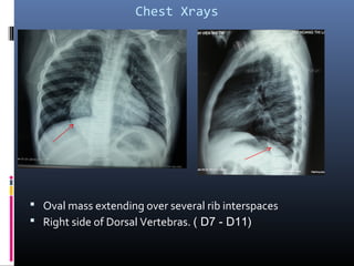
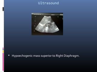
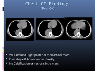
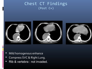
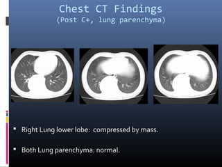

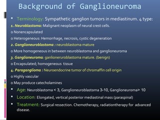

A 5-year-old female presented with coughing and vomiting blood. Imaging showed an oval mass in her right posterior mediastinum compressing her right lung and superior vena cava. CT and ultrasound imaging characterized the mass as well-defined, homogeneous and hypoechoic without calcification or necrosis. She was diagnosed with ganglioneuroma, a benign sympathetic ganglion tumor most common in older children.


