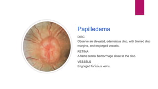
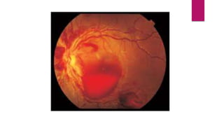
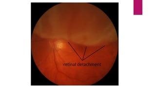
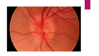
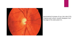
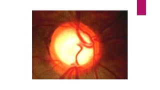
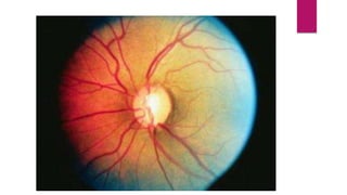
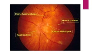
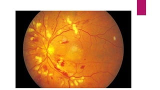
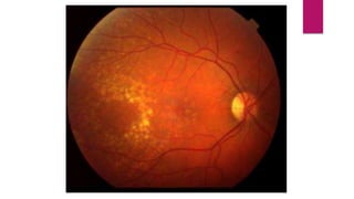
Fundoscopy images
SANA RAFIQUE
Normal fundus
Normal Fundus
DISC
Clear; the central area of the cup is pale.
RETINA
A normal red/orange color; the macula is dark.
VESSELS
Arteries appear bright red, vein slightly purple. Arterial to
venous ratio is about 2 to 3.
papilledema
Swelling of the optic disc and anterior bulging of the
physiologic cup. Color is pink and hyperemic with disc
margins blurred. Disc vessels are more visible, numerous,
and curve over disc borders. It is a sign not a Dx = can be
seen in more then 1 eye diseases.
Papilledema
DISC
Observe an elevated, edematous disc, with blurred disc
margins, and engorged vessels.
RETINA
A flame retinal hemorrhage close to the disc.
VESSELS
Engorged tortuous veins.
Retinal haemorrhage
Superficial:Small, linear, flame-shaped red streaks in
fundi shaped by superficial nerve fibers that radiate
from optic disc. HTN related.
Deep:Small, rounded, slightly irregular spots. Occur
in deeper layer of retina. Diabetes related.
Retinal detachment
Retinal Detachment
DISC
The disc appears normal.
RETINA
Observation of the retina shows a gray elevation in the
temporal area with folds in the detached section.
VESSELS
The vessels are totuous and elevated over the detached
retina
Optic neuritis
Optic Neuritis
DISC
The disc is elevated, with blurred margins.
RETINA
The retina is normal.
VESSELS
The arteries are attentuated; the veins are normal.
Glaucoma
Glaucomatous Cupping of
the Disc
DISC
Observe a large cup, with disc vessels displaced
peripherally; a pale white color; and pigmenting
surrounding the disc.
RETINA
The retina appears normal.
VESSELS
The vessels are normal.
Asymmetrical increase of cup: disc ratio (0.8).
Cupping seen where vessels disappear over
the edge of the attenuated rim.
Central Retinal vein occlusion
Central Retinal Vein Occlusion
DISC
The disc is virtually obscured by edema and hemorrhages.
RETINA
There are extensive blot retinal hemorrhages in all quadrants
to the periphery.
VESSELS
Observe dilated totuous veins. The vessels are partially
obscured by retinal hemorrhages.
Central Retinal artery occlusion
•Cherry red spot: The lack of arterial perfusion to the inner
layers of the retina results in edema of the retinal nerve
fiber layer and ganglion cell layer, which makes it appear
opaque. The inner retina is multi-layered throughout the
posterior pole except at the fovea; thus, the retinal edema
surrounds the fovea. In the fovea, the underlying choroid
with intact RPE is visible and appears as a "cherry red
spot" when compared to the cloudy, edematous retina
adjacent to it.
•Vascular attenuation and/or "box-carring" of flow in
arteries and veins.
•May see an embolus in vessel on optic nerve.
Hypertensive Retinopathy
Diabetic Retinopathy
Macular Degeneration
Fundoscopy images-1.pptx

Fundoscopy images-1.pptx

  • 1.
  • 2.
    Normal fundus Normal Fundus DISC Clear;the central area of the cup is pale. RETINA A normal red/orange color; the macula is dark. VESSELS Arteries appear bright red, vein slightly purple. Arterial to venous ratio is about 2 to 3.
  • 3.
    papilledema Swelling of theoptic disc and anterior bulging of the physiologic cup. Color is pink and hyperemic with disc margins blurred. Disc vessels are more visible, numerous, and curve over disc borders. It is a sign not a Dx = can be seen in more then 1 eye diseases.
  • 4.
    Papilledema DISC Observe an elevated,edematous disc, with blurred disc margins, and engorged vessels. RETINA A flame retinal hemorrhage close to the disc. VESSELS Engorged tortuous veins.
  • 7.
    Retinal haemorrhage Superficial:Small, linear,flame-shaped red streaks in fundi shaped by superficial nerve fibers that radiate from optic disc. HTN related. Deep:Small, rounded, slightly irregular spots. Occur in deeper layer of retina. Diabetes related.
  • 9.
    Retinal detachment Retinal Detachment DISC Thedisc appears normal. RETINA Observation of the retina shows a gray elevation in the temporal area with folds in the detached section. VESSELS The vessels are totuous and elevated over the detached retina
  • 13.
    Optic neuritis Optic Neuritis DISC Thedisc is elevated, with blurred margins. RETINA The retina is normal. VESSELS The arteries are attentuated; the veins are normal.
  • 17.
    Glaucoma Glaucomatous Cupping of theDisc DISC Observe a large cup, with disc vessels displaced peripherally; a pale white color; and pigmenting surrounding the disc. RETINA The retina appears normal. VESSELS The vessels are normal.
  • 18.
    Asymmetrical increase ofcup: disc ratio (0.8). Cupping seen where vessels disappear over the edge of the attenuated rim.
  • 22.
    Central Retinal veinocclusion Central Retinal Vein Occlusion DISC The disc is virtually obscured by edema and hemorrhages. RETINA There are extensive blot retinal hemorrhages in all quadrants to the periphery. VESSELS Observe dilated totuous veins. The vessels are partially obscured by retinal hemorrhages.
  • 26.
    Central Retinal arteryocclusion •Cherry red spot: The lack of arterial perfusion to the inner layers of the retina results in edema of the retinal nerve fiber layer and ganglion cell layer, which makes it appear opaque. The inner retina is multi-layered throughout the posterior pole except at the fovea; thus, the retinal edema surrounds the fovea. In the fovea, the underlying choroid with intact RPE is visible and appears as a "cherry red spot" when compared to the cloudy, edematous retina adjacent to it. •Vascular attenuation and/or "box-carring" of flow in arteries and veins. •May see an embolus in vessel on optic nerve.
  • 30.
  • 35.
  • 41.