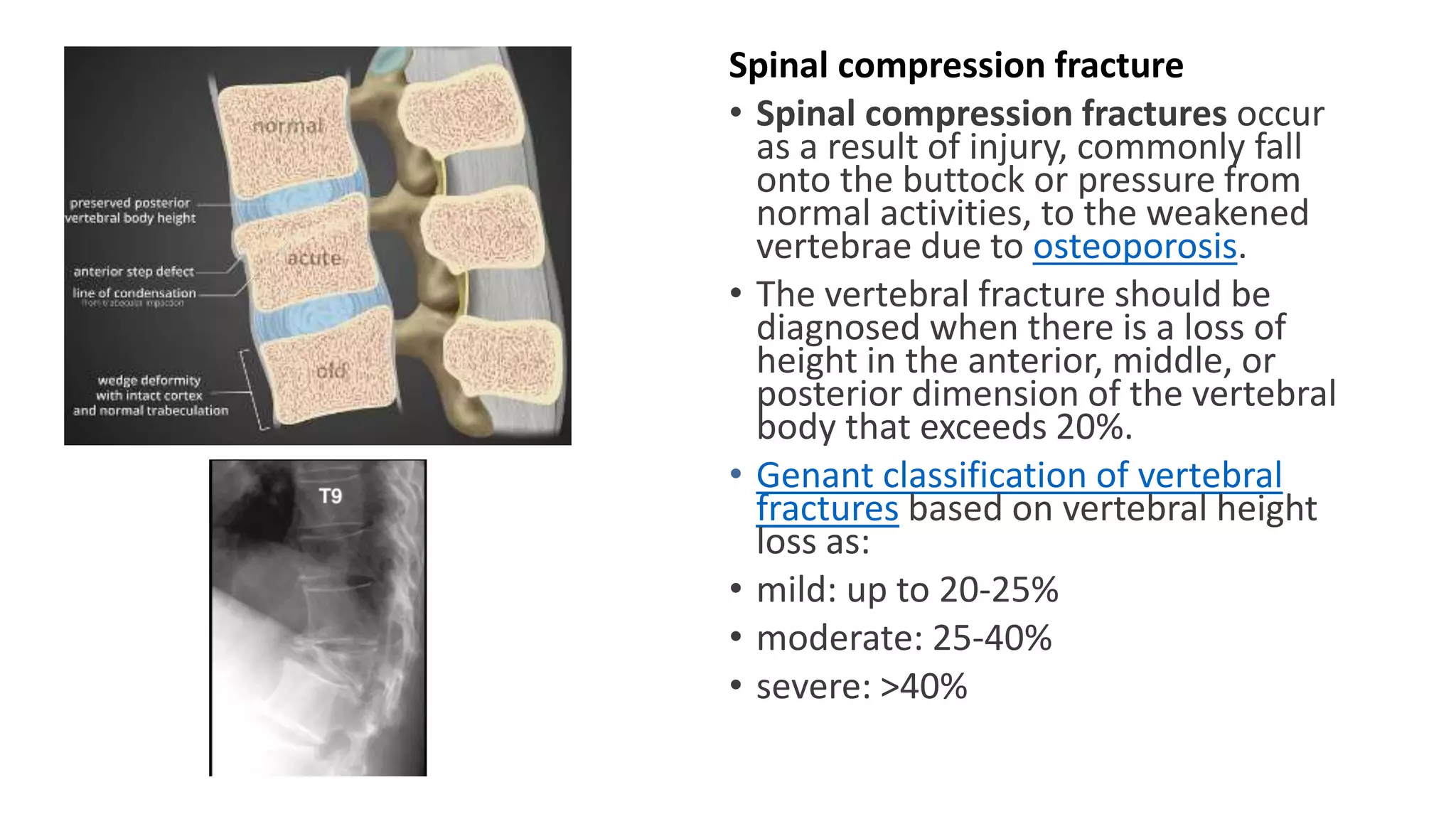
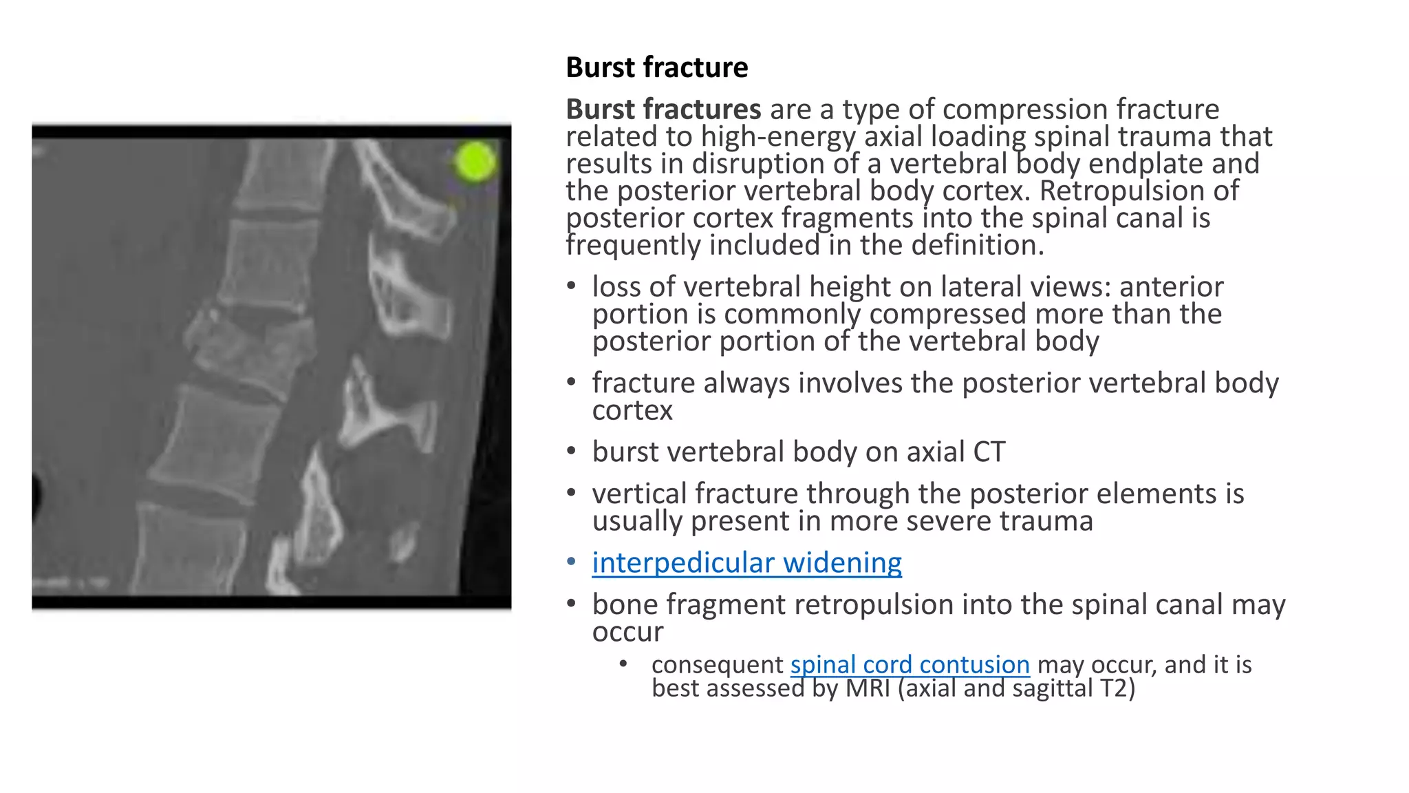
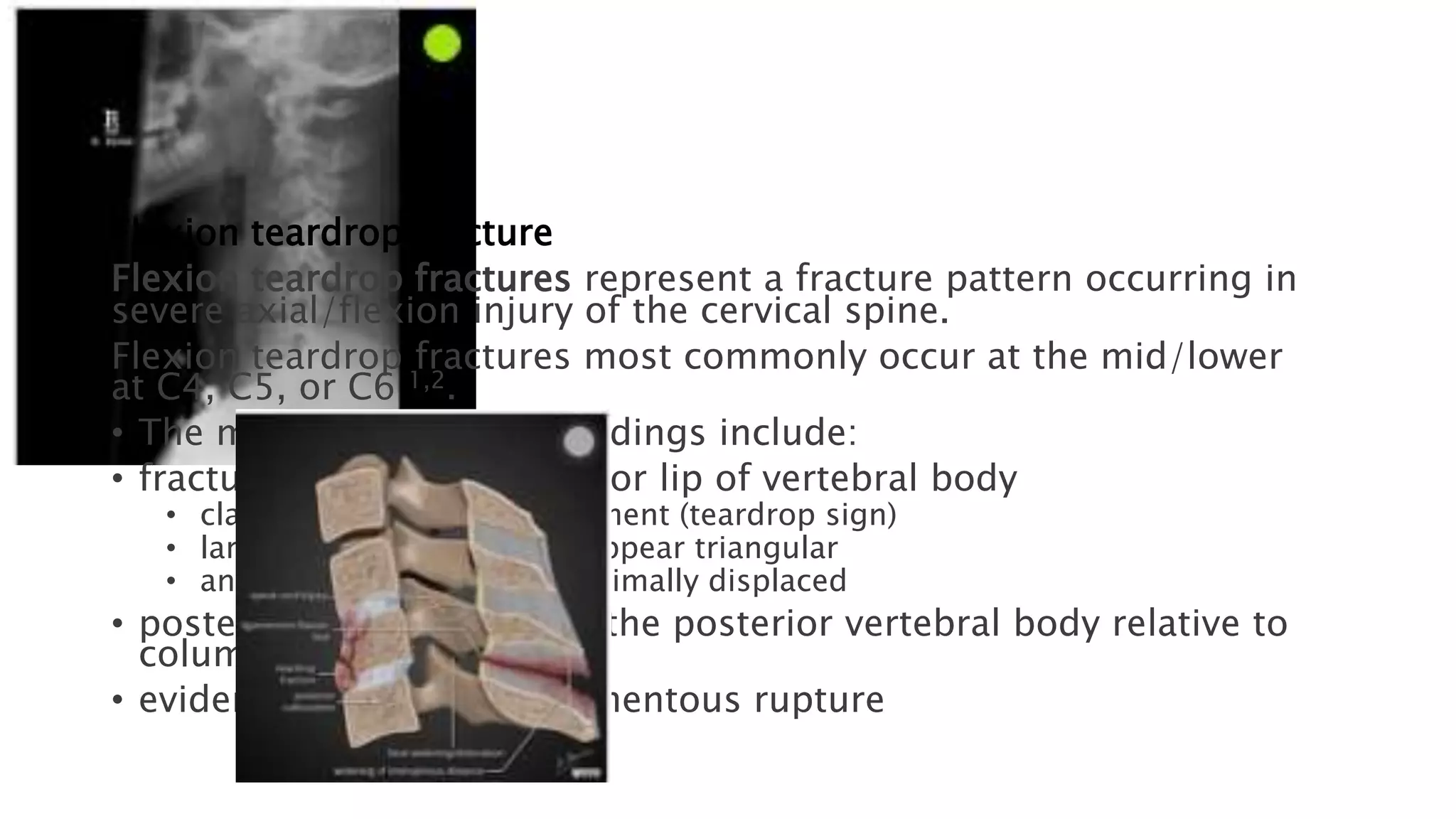
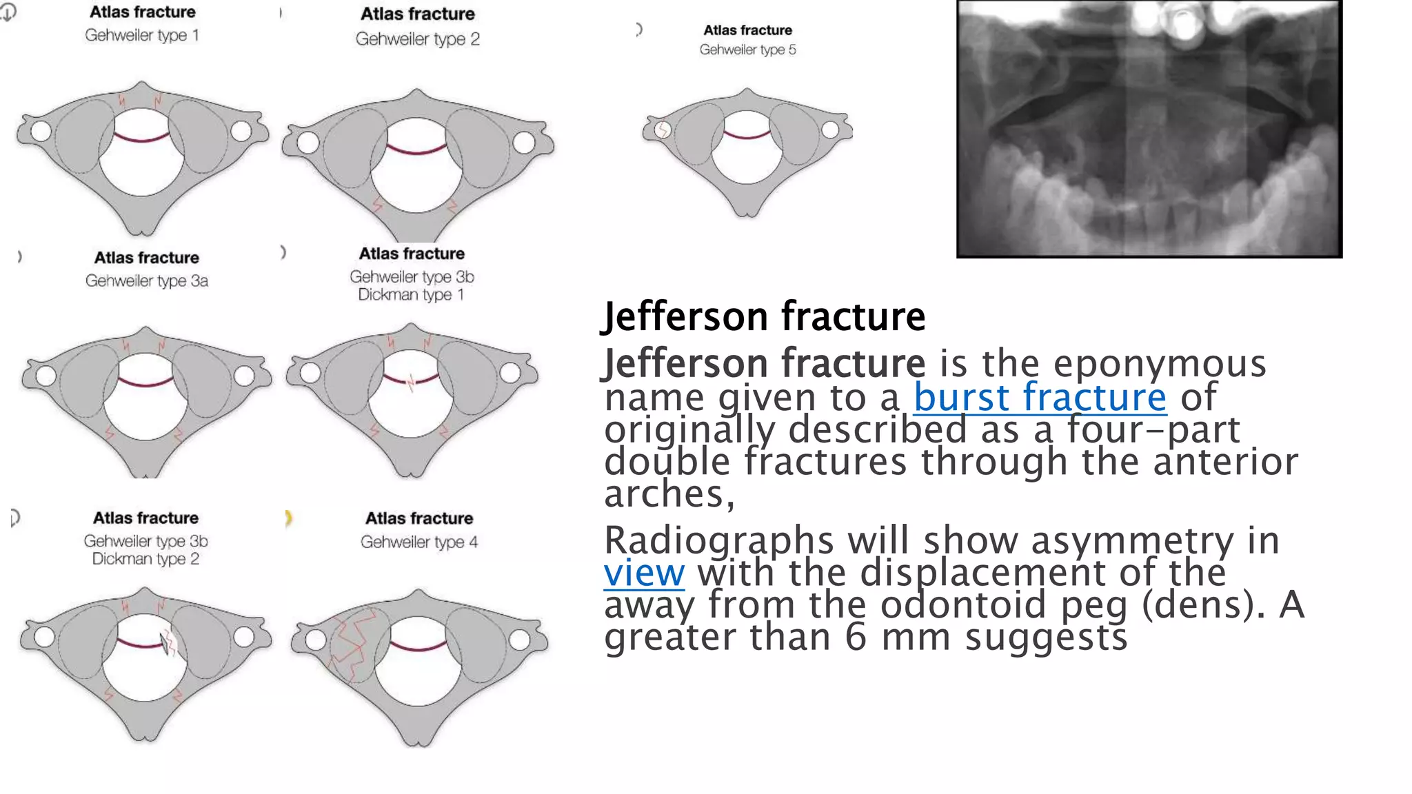
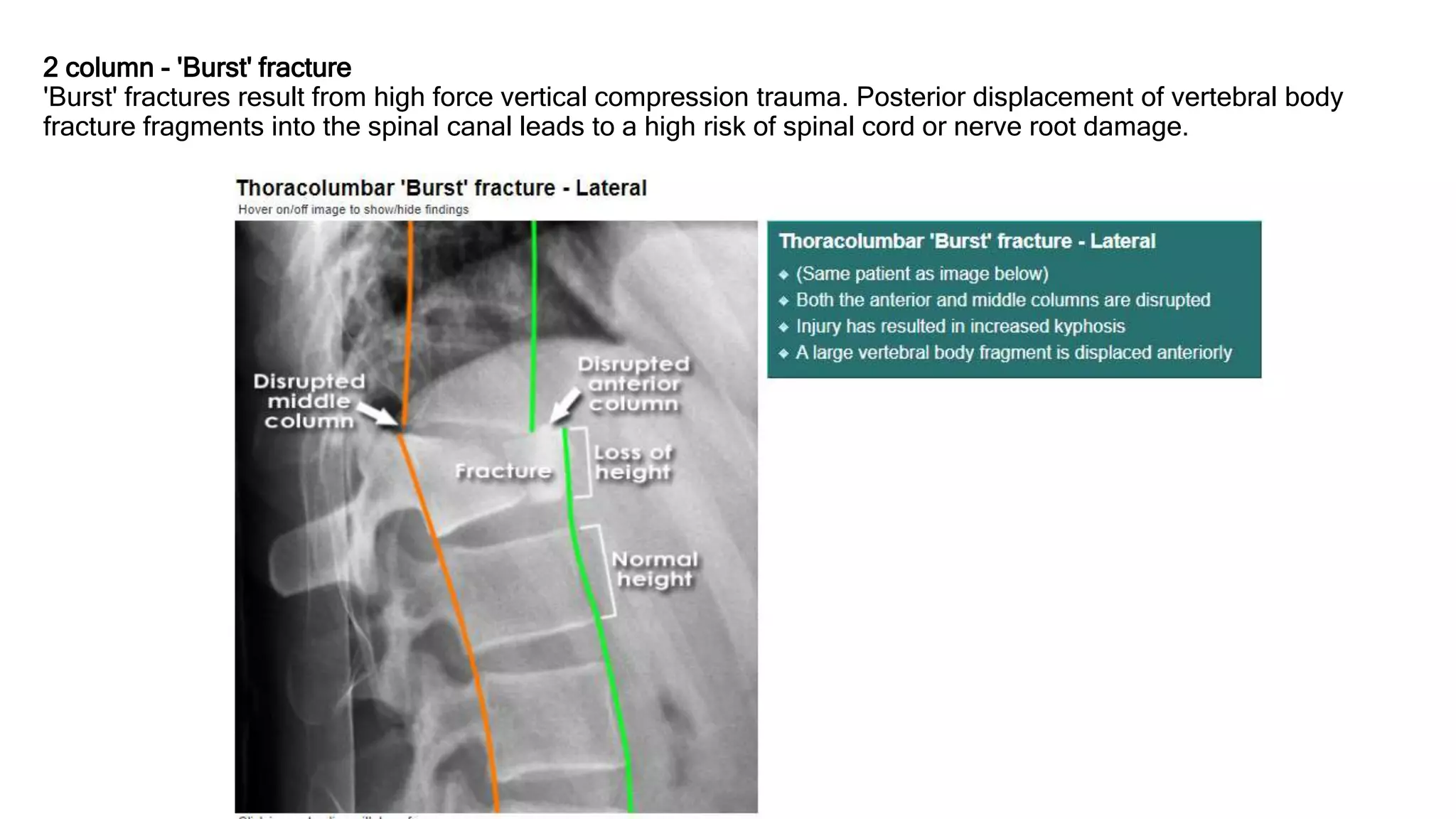
SlideShare a Scribd company logo
Spinal Fractures
Morphology
• Chalk stick fracture
• Spinal compression fracture
• burst fracture
• wedge fracture
• vertebra plana (mnemonic)
Fractures by location
• cervical spine fracture
• dens fracture
• extension teardrop fracture
• ​flexion teardrop fracture
• hangman fracture
• Jefferson fracture
• clay-shoveler's fracture
• thoracolumbar spine fracture
• Chance fracture
• transverse process fracture
• spondylolysis
• limbus fractures
• sacral fracture
• sacral insufficiency fractures
Chalk stick, also known as carrot stick
fractures, are fractures of the fused spine,
classically seen in ankylosing spondylitis
Chalk stick fractures are most commonly
encountered in ankylosing spondylitis but
may also been seen in the fused spines in
patients with 2:
• diffuse idiopathic skeletal hyperostosis
• ossification of the ligamentum flavum
• ossification of the posterior longitudinal
ligament
• surgical spinal fusion
Spinal compression fracture
• Spinal compression fractures occur
as a result of injury, commonly fall
onto the buttock or pressure from
normal activities, to the weakened
vertebrae due to osteoporosis.
• The vertebral fracture should be
diagnosed when there is a loss of
height in the anterior, middle, or
posterior dimension of the vertebral
body that exceeds 20%.
• Genant classification of vertebral
fractures based on vertebral height
loss as:
• mild: up to 20-25%
• moderate: 25-40%
• severe: >40%
Burst fracture
Burst fractures are a type of compression fracture
related to high-energy axial loading spinal trauma that
results in disruption of a vertebral body endplate and
the posterior vertebral body cortex. Retropulsion of
posterior cortex fragments into the spinal canal is
frequently included in the definition.
• loss of vertebral height on lateral views: anterior
portion is commonly compressed more than the
posterior portion of the vertebral body
• fracture always involves the posterior vertebral body
cortex
• burst vertebral body on axial CT
• vertical fracture through the posterior elements is
usually present in more severe trauma
• interpedicular widening
• bone fragment retropulsion into the spinal canal may
occur
• consequent spinal cord contusion may occur, and it is
best assessed by MRI (axial and sagittal T2)
Wedge fracture
• Spinal wedge (compression)
fractures are hyperflexion injuries
to the vertebral body resulting
loading.
• Radiographs, CT, and MRI may
disruption with impaction of one
without the involvement of the
wall 6. This results in the
"wedged" appearance
Vertebra plana
• Vertebra plana (plural:
vertebrae planae), also known
the pancake, silver
edge vertebra, is the term given
when a vertebral body has lost
entire height anteriorly and
representing a very advanced
fracture
Fractures by location
• cervical spine fracture
• dens fracture
• extension teardrop fracture
• flexion teardrop fracture
• hangman fracture
• Jefferson fracture
• clay-shoveler's fracture
• thoracolumbar spine fracture
• Chance fracture
• transverse process fracture
• spondylolysis
• limbus fractures
• sacral fracture
• sacral insufficiency fractures
Fractures by location
• cervical spine fracture
• dens fracture
• extension teardrop fracture
• flexion teardrop fracture
• hangman fracture
• Jefferson fracture
• clay-shoveler's fracture
Odontoid fracture
Odontoid process fracture, also known as a peg or dens fracture, occurs where there is a
fracture through the odontoid process of C2.
Classification Anderson and D'Alonzo
•type I
•rare
•fracture of the upper part of the odontoid peg (generally oblique)
•above the level of the transverse band of the cruciform ligament
•usually considered stable
•type II
•most common
•transverse course fracture at the base of the odontoid
•below the level of the transverse band of the cruciform ligament
•unstable
•high risk of non-union
•type IIa
•​type II fractures with comminution at the odontoid base
•Hadley 2 described this type of fracture which has a significantly increased risk of nonunion when
classical type II fractures
•represents 5-10% of type II fractures
•type III
•through the odontoid and into the lateral masses of C2
•relatively stable if not excessively displaced
•best prognosis for healing because of the larger surface area of the fracture
Extension teardrop fracture
Extension teardrop fracture typically occurs due to forced
extension of the neck with resulting avulsion of the
vertebral body
anterior-inferior corner fracture 3
• avulsion fracture from the attachment of the anterior
inferior corner of the vertebral body, usually a thin fracture
• the fragment is triangular in a shape reminiscent of a teardrop
• vertical height of fragment is equal to or greater than width
• anterior disc space widening
Flexion teardrop fracture
Flexion teardrop fractures represent a fracture pattern occurring in
severe axial/flexion injury of the cervical spine.
Flexion teardrop fractures most commonly occur at the mid/lower
at C4, C5, or C6 1,2.
• The most characteristic findings include:
• fracture of the anteroinferior lip of vertebral body
• classically a triangular fragment (teardrop sign)
• larger fragments may not appear triangular
• anterior fragment often minimally displaced
• posterior displacement of the posterior vertebral body relative to
column
• evidence of posterior ligamentous rupture
Hangman fracture
Hangman fracture, also known as traumatic spondylolisthesis of
the axis, is a fracture which involves the pars
interarticularis of C2 on both sides, and is a result of
Radiography and CT demonstrate the findings:
• typical: bilateral C2 pars interarticularis fractures
• atypical variant: one or both sides of C2 has a coronal plane
fracture through the posterior vertebral body instead of the
• possible alignment abnormality: anterolisthesis of C2 on C3 or
• Extension of the fracture to the transverse foramina should be
possibility of vertebral artery injury.
Jefferson fracture
Jefferson fracture is the eponymous
name given to a burst fracture of
originally described as a four-part
double fractures through the anterior
arches,
Radiographs will show asymmetry in
view with the displacement of the
away from the odontoid peg (dens). A
greater than 6 mm suggests
Jefferson fracture
• type 1: fractures of the anterior arch
• type 2: fractures of the posterior arch and are
usually bilateral
• type 3: fractures involving the anterior and
posterior arch (Jefferson burst fracture)
• type 3a: intact transverse atlantal ligament
• type 3b: disrupted transverse atlantal ligament
complex
• Dickman type 1: ligamentous disruption
• Dickman type 2: bony avulsion with an intact transverse
atlantal ligament
• type 4: fractures of the lateral mass
• type 5: isolated fractures of the C1 transverse
process
Clay-shoveler fracture
Clay-shoveler fractures are fractures of the spinous
process of a lower cervical vertebra.
The fracture is seen on lateral radiographs as an oblique
spinous process, usually of C7. There is usually significant
Fractures by location
• thoracolumbar
fracture
• Chance fracture
• transverse process
fracture
• spondylolysis
• limbus fractures
Chance fracture
Chance fractures also referred to as seatbelt fractures, are flexion-
distraction type injuries of the spine that extend to involve all three
unstable injuries and have a high association with intra-abdominal
Anterior wedge fracture of the vertebral body with a horizontal
elements or distraction of facet joints and spinous processes.
• empty vertebral body sign: can be seen on an AP radiograph and
separation of the posterior elements displacing the spinous
fracture fragments of the vertebral body on the AP projection
• transverse fractures across the transverse processes, laminae, and
• widening of the interpedicular distance: often suggests a burst
• widening of the facet joints and increased intercostal spacing
• widening of the interspinous spaces
Transverse process fracture
Transverse process fractures are common sequelae of
trauma, although they are considered minor and
Transverse process fracture most commonly occurs in the
spine and are commonly multiple 2. The fracture line can
the transverse foramen, and in the cervical spine, there is a
complicating vertebral artery dissection.
Spondylolysis
Spondylolysis is a defect in the pars interarticularis of the
neural arch, the portion of the neural arch that connects
articular facets. It is commonly known as pars
simply as pars defect.
• Scotty dog sign: on oblique radiographs, a break in the
can have the appearance of a collar around the dog's neck
• inverted Napoleon hat sign
Posterior ring apophyseal fracture
Posterior ring apophyseal fracture or separation, also called limbus fracture, occur in the
immature skeleton, most commonly in the lumbar spine. They represent bony fractures of
of attachment of the Sharpey fibers of the intervertebral disc.
They can be classified as follows:
• type I: avulsions of the posterior cortical vertebral rim
• type II: central cortical and cancellous bone fractures
• type III: lateralized chip fractures
• type IV: span the entire length and breadth of the posterior vertebral margin between the
endplates
CT
• CT is excellent for bony detail and is therefore usually the first line imaging modality.
• osseous fragment displaced posteriorly to endplate with rectangular or arc-shaped
• posterior endplate defect
• posterior disc herniation
• Sacral fracture
• sacral
fractures
Sacral insufficiency fracture
Sacral insufficiency fractures are a subtype of stress fractures, which
are the result of normal stresses on abnormal bone, most frequently
of osteoporosis. They fall under the broader group of pelvic
Plain radiograph
• They may be normal, or a sclerotic line may be noted in the
CT
• May show a fracture line along with sclerosis that is parallel to the
even CT imaging is less sensitive as compared to MRI and nuclear
MRI
• Can depict bone marrow edema as early as 18 days after the
Le Fort fracture classification
Le Fort fractures are fractures of the midface, which collectively
involve separation of all or a portion of the midface from
• Le Fort type I
• horizontal maxillary fracture, separating the teeth from the upper
• fracture line passes through the alveolar ridge, lateral nose and
the maxillary sinus
• also known as a Guerin fracture
• Le Fort type II
• pyramidal fracture, with the teeth at the pyramid base, and
• fracture arch passes through the posterior alveolar ridge, lateral
sinuses, inferior orbital rim and nasal bones
• uppermost fracture line can pass through the nasofrontal junction
the maxilla 3
• Le Fort type III
• craniofacial disjunction
• transverse fracture line passes through nasofrontal
wall, and zygomatic arch/zygomaticofrontal suture
• because of the involvement of the zygomatic arch, there is a risk
impingement
• unsurprisingly type III fractures have the highest rate of CSF leak
1 Column - Anterior compression injury
Anterior compression injury is a common fracture pattern which results from traumatic hyper-flexion with compression.
Although considered 'stable' the greater the loss of height anteriorly the greater the risk of middle column involvement.
X-ray may underestimate the extent of injury and so if there has been high risk injury or other suspicion of instability then
CT should be considered.
2 column - 'Burst' fracture
'Burst' fractures result from high force vertical compression trauma. Posterior displacement of vertebral body
fracture fragments into the spinal canal leads to a high risk of spinal cord or nerve root damage.
Spinal Fractures.pptx

More Related Content

What's hot

Hallux valgus.pptx
Hallux valgus.pptxHallux valgus.pptx
Hallux valgus.pptx
Siwaporn Khureerung
 
Spinal cord injury (sci) Rehab
Spinal cord injury (sci) RehabSpinal cord injury (sci) Rehab
Spinal cord injury (sci) Rehab
Quan Fu Gan
 
MCL,LCL & ALL injuries of the knee
MCL,LCL & ALL injuries of the knee MCL,LCL & ALL injuries of the knee
MCL,LCL & ALL injuries of the knee
Mohamed Abulsoud
 
Spinal cord injury
Spinal cord injury Spinal cord injury
Brachial plexus injuries by krr
Brachial plexus injuries by krrBrachial plexus injuries by krr
Brachial plexus injuries by krr
ramachandra reddy
 
Brachial Plexus Injury - An Introduction to the Physiotherapists
Brachial Plexus Injury - An Introduction to the PhysiotherapistsBrachial Plexus Injury - An Introduction to the Physiotherapists
Brachial Plexus Injury - An Introduction to the Physiotherapists
JebarajFletcher
 
Thoraco lumbar fractures
Thoraco lumbar fracturesThoraco lumbar fractures
Thoraco lumbar fractures
MOHAMED HASSANEIN
 
Rotator cuff injuries
Rotator cuff injuriesRotator cuff injuries
Rotator cuff injuries
pratigya deuja
 
Hip biomechanics
Hip biomechanicsHip biomechanics
Hip biomechanics
Sudheer Kumar
 
Peripheral nerve injury
Peripheral nerve injuryPeripheral nerve injury
Peripheral nerve injuryPrateek Singh
 
Classification of spinal fracture
Classification of spinal fractureClassification of spinal fracture
Classification of spinal fracture
BipulBorthakur
 
Fracture neck of femur
Fracture neck of femurFracture neck of femur
Fracture neck of femurRenuga Sri
 
Fracture of neck of femur
Fracture of neck of femurFracture of neck of femur
Cervical Fractures(clay shovelers,hangmans,odontoid)
Cervical Fractures(clay shovelers,hangmans,odontoid)Cervical Fractures(clay shovelers,hangmans,odontoid)
Cervical Fractures(clay shovelers,hangmans,odontoid)
rineeshk19
 
Posterior Cruciate Ligament Injury
Posterior Cruciate Ligament InjuryPosterior Cruciate Ligament Injury
Posterior Cruciate Ligament Injury
Arslan Luqman
 
Foot Deformities
Foot DeformitiesFoot Deformities
Foot Deformities
suravipramanik
 
Radial head fracture
Radial head fractureRadial head fracture
Radial head fracture
Md Ashiqur Rahman
 
Patellar fractures & Physiotherapy
Patellar fractures & PhysiotherapyPatellar fractures & Physiotherapy
Patellar fractures & Physiotherapy
Dibyendunarayan Bid
 
Spondylolisthesis
Spondylolisthesis Spondylolisthesis
Spondylolisthesis
Mahak Jain
 
Clay shoveler's fracture
Clay shoveler's fracture Clay shoveler's fracture
Clay shoveler's fracture
monirul islam
 

What's hot (20)

Hallux valgus.pptx
Hallux valgus.pptxHallux valgus.pptx
Hallux valgus.pptx
 
Spinal cord injury (sci) Rehab
Spinal cord injury (sci) RehabSpinal cord injury (sci) Rehab
Spinal cord injury (sci) Rehab
 
MCL,LCL & ALL injuries of the knee
MCL,LCL & ALL injuries of the knee MCL,LCL & ALL injuries of the knee
MCL,LCL & ALL injuries of the knee
 
Spinal cord injury
Spinal cord injury Spinal cord injury
Spinal cord injury
 
Brachial plexus injuries by krr
Brachial plexus injuries by krrBrachial plexus injuries by krr
Brachial plexus injuries by krr
 
Brachial Plexus Injury - An Introduction to the Physiotherapists
Brachial Plexus Injury - An Introduction to the PhysiotherapistsBrachial Plexus Injury - An Introduction to the Physiotherapists
Brachial Plexus Injury - An Introduction to the Physiotherapists
 
Thoraco lumbar fractures
Thoraco lumbar fracturesThoraco lumbar fractures
Thoraco lumbar fractures
 
Rotator cuff injuries
Rotator cuff injuriesRotator cuff injuries
Rotator cuff injuries
 
Hip biomechanics
Hip biomechanicsHip biomechanics
Hip biomechanics
 
Peripheral nerve injury
Peripheral nerve injuryPeripheral nerve injury
Peripheral nerve injury
 
Classification of spinal fracture
Classification of spinal fractureClassification of spinal fracture
Classification of spinal fracture
 
Fracture neck of femur
Fracture neck of femurFracture neck of femur
Fracture neck of femur
 
Fracture of neck of femur
Fracture of neck of femurFracture of neck of femur
Fracture of neck of femur
 
Cervical Fractures(clay shovelers,hangmans,odontoid)
Cervical Fractures(clay shovelers,hangmans,odontoid)Cervical Fractures(clay shovelers,hangmans,odontoid)
Cervical Fractures(clay shovelers,hangmans,odontoid)
 
Posterior Cruciate Ligament Injury
Posterior Cruciate Ligament InjuryPosterior Cruciate Ligament Injury
Posterior Cruciate Ligament Injury
 
Foot Deformities
Foot DeformitiesFoot Deformities
Foot Deformities
 
Radial head fracture
Radial head fractureRadial head fracture
Radial head fracture
 
Patellar fractures & Physiotherapy
Patellar fractures & PhysiotherapyPatellar fractures & Physiotherapy
Patellar fractures & Physiotherapy
 
Spondylolisthesis
Spondylolisthesis Spondylolisthesis
Spondylolisthesis
 
Clay shoveler's fracture
Clay shoveler's fracture Clay shoveler's fracture
Clay shoveler's fracture
 

Similar to Spinal Fractures.pptx

neck x ray.pptx
neck x ray.pptxneck x ray.pptx
neck x ray.pptx
FatimaAmirlou
 
upper limb trauma.pptx
upper limb trauma.pptxupper limb trauma.pptx
upper limb trauma.pptx
rohanjohnjacob
 
clinical anatomy (upper limb)
clinical anatomy (upper limb)clinical anatomy (upper limb)
clinical anatomy (upper limb)
Dr Neeraj Tiwari
 
Fractures and dislocation upper limb .pptx
Fractures and dislocation upper limb .pptxFractures and dislocation upper limb .pptx
Fractures and dislocation upper limb .pptx
Dr vaibhavi patel
 
fRACTURES.pptx
fRACTURES.pptxfRACTURES.pptx
fRACTURES.pptx
Sandra710258
 
FRACTURE (Sites)
FRACTURE (Sites)FRACTURE (Sites)
FRACTURE (Sites)
Julie Jane
 
Cervical trauma
Cervical traumaCervical trauma
Cervical trauma
Ali Jiwani
 
Fractures & dislocations general principles
Fractures & dislocations general principlesFractures & dislocations general principles
Fractures & dislocations general principles
Dr KAMBLE
 
shoulder injury.pptx
shoulder injury.pptxshoulder injury.pptx
shoulder injury.pptx
shaleen rakheja
 
Radiological presentation fractures.pptx
Radiological presentation fractures.pptxRadiological presentation fractures.pptx
Radiological presentation fractures.pptx
AhsanUllahKhan27
 
Radiology of the Elbow Joint. Dr. Sumit Sharma
Radiology of the Elbow Joint. Dr. Sumit SharmaRadiology of the Elbow Joint. Dr. Sumit Sharma
Radiology of the Elbow Joint. Dr. Sumit Sharma
Sumit Sharma
 
AJM Sheet: Talar fracture
AJM Sheet: Talar fractureAJM Sheet: Talar fracture
AJM Sheet: Talar fracture
Podiatry Town
 
16-Clinical Anatomy of The Upper Limb - Dr Akalanka Jayasinghe.pdf
16-Clinical Anatomy of The Upper Limb - Dr Akalanka Jayasinghe.pdf16-Clinical Anatomy of The Upper Limb - Dr Akalanka Jayasinghe.pdf
16-Clinical Anatomy of The Upper Limb - Dr Akalanka Jayasinghe.pdf
DilankaMadhushan1
 
fracture and dislocation ppt . Almas khan. khorfakkhan hospital dubai
fracture and dislocation ppt . Almas khan. khorfakkhan hospital dubaifracture and dislocation ppt . Almas khan. khorfakkhan hospital dubai
fracture and dislocation ppt . Almas khan. khorfakkhan hospital dubai
almasmkm
 
Vertebral column associated pathology and radiographic appearance
Vertebral column associated pathology and radiographic appearanceVertebral column associated pathology and radiographic appearance
Vertebral column associated pathology and radiographic appearance
Swapnil Shetty
 
femoral neck fracture .pptx
femoral neck fracture .pptxfemoral neck fracture .pptx
femoral neck fracture .pptx
BiehasAlwakaf1
 
Neck of Femur, IT and Subtrochanteric fracture- Dr Sundar Ortho.pptx
Neck of Femur, IT and Subtrochanteric fracture- Dr Sundar Ortho.pptxNeck of Femur, IT and Subtrochanteric fracture- Dr Sundar Ortho.pptx
Neck of Femur, IT and Subtrochanteric fracture- Dr Sundar Ortho.pptx
Dr. Sundar Karki
 
Femoral fracture
Femoral fractureFemoral fracture
Femoral fracture
aya tya
 
fractures around Elbow.pdf
fractures around Elbow.pdffractures around Elbow.pdf
fractures around Elbow.pdf
FuadNaji1
 
Radiological Imaging of Musculoskeletal Trauma.pdf
Radiological Imaging of Musculoskeletal Trauma.pdfRadiological Imaging of Musculoskeletal Trauma.pdf
Radiological Imaging of Musculoskeletal Trauma.pdf
KhairilAnwar764824
 

Similar to Spinal Fractures.pptx (20)

neck x ray.pptx
neck x ray.pptxneck x ray.pptx
neck x ray.pptx
 
upper limb trauma.pptx
upper limb trauma.pptxupper limb trauma.pptx
upper limb trauma.pptx
 
clinical anatomy (upper limb)
clinical anatomy (upper limb)clinical anatomy (upper limb)
clinical anatomy (upper limb)
 
Fractures and dislocation upper limb .pptx
Fractures and dislocation upper limb .pptxFractures and dislocation upper limb .pptx
Fractures and dislocation upper limb .pptx
 
fRACTURES.pptx
fRACTURES.pptxfRACTURES.pptx
fRACTURES.pptx
 
FRACTURE (Sites)
FRACTURE (Sites)FRACTURE (Sites)
FRACTURE (Sites)
 
Cervical trauma
Cervical traumaCervical trauma
Cervical trauma
 
Fractures & dislocations general principles
Fractures & dislocations general principlesFractures & dislocations general principles
Fractures & dislocations general principles
 
shoulder injury.pptx
shoulder injury.pptxshoulder injury.pptx
shoulder injury.pptx
 
Radiological presentation fractures.pptx
Radiological presentation fractures.pptxRadiological presentation fractures.pptx
Radiological presentation fractures.pptx
 
Radiology of the Elbow Joint. Dr. Sumit Sharma
Radiology of the Elbow Joint. Dr. Sumit SharmaRadiology of the Elbow Joint. Dr. Sumit Sharma
Radiology of the Elbow Joint. Dr. Sumit Sharma
 
AJM Sheet: Talar fracture
AJM Sheet: Talar fractureAJM Sheet: Talar fracture
AJM Sheet: Talar fracture
 
16-Clinical Anatomy of The Upper Limb - Dr Akalanka Jayasinghe.pdf
16-Clinical Anatomy of The Upper Limb - Dr Akalanka Jayasinghe.pdf16-Clinical Anatomy of The Upper Limb - Dr Akalanka Jayasinghe.pdf
16-Clinical Anatomy of The Upper Limb - Dr Akalanka Jayasinghe.pdf
 
fracture and dislocation ppt . Almas khan. khorfakkhan hospital dubai
fracture and dislocation ppt . Almas khan. khorfakkhan hospital dubaifracture and dislocation ppt . Almas khan. khorfakkhan hospital dubai
fracture and dislocation ppt . Almas khan. khorfakkhan hospital dubai
 
Vertebral column associated pathology and radiographic appearance
Vertebral column associated pathology and radiographic appearanceVertebral column associated pathology and radiographic appearance
Vertebral column associated pathology and radiographic appearance
 
femoral neck fracture .pptx
femoral neck fracture .pptxfemoral neck fracture .pptx
femoral neck fracture .pptx
 
Neck of Femur, IT and Subtrochanteric fracture- Dr Sundar Ortho.pptx
Neck of Femur, IT and Subtrochanteric fracture- Dr Sundar Ortho.pptxNeck of Femur, IT and Subtrochanteric fracture- Dr Sundar Ortho.pptx
Neck of Femur, IT and Subtrochanteric fracture- Dr Sundar Ortho.pptx
 
Femoral fracture
Femoral fractureFemoral fracture
Femoral fracture
 
fractures around Elbow.pdf
fractures around Elbow.pdffractures around Elbow.pdf
fractures around Elbow.pdf
 
Radiological Imaging of Musculoskeletal Trauma.pdf
Radiological Imaging of Musculoskeletal Trauma.pdfRadiological Imaging of Musculoskeletal Trauma.pdf
Radiological Imaging of Musculoskeletal Trauma.pdf
 

Recently uploaded

The Importance of COVID-19 PCR Tests for Travel in 2024.pptx
The Importance of COVID-19 PCR Tests for Travel in 2024.pptxThe Importance of COVID-19 PCR Tests for Travel in 2024.pptx
The Importance of COVID-19 PCR Tests for Travel in 2024.pptx
Global Travel Clinics
 
Deep Leg Vein Thrombosis (DVT): Meaning, Causes, Symptoms, Treatment, and Mor...
Deep Leg Vein Thrombosis (DVT): Meaning, Causes, Symptoms, Treatment, and Mor...Deep Leg Vein Thrombosis (DVT): Meaning, Causes, Symptoms, Treatment, and Mor...
Deep Leg Vein Thrombosis (DVT): Meaning, Causes, Symptoms, Treatment, and Mor...
The Lifesciences Magazine
 
LGBTQ+ Adults: Unique Opportunities and Inclusive Approaches to Care
LGBTQ+ Adults: Unique Opportunities and Inclusive Approaches to CareLGBTQ+ Adults: Unique Opportunities and Inclusive Approaches to Care
LGBTQ+ Adults: Unique Opportunities and Inclusive Approaches to Care
VITASAuthor
 
DRAFT Ventilator Rapid Reference version 2.4.pdf
DRAFT Ventilator Rapid Reference  version  2.4.pdfDRAFT Ventilator Rapid Reference  version  2.4.pdf
DRAFT Ventilator Rapid Reference version 2.4.pdf
Robert Cole
 
Child Welfare Clinic and Well baby clinicin Sri Lanka.ppsx
Child Welfare Clinic and Well baby clinicin Sri Lanka.ppsxChild Welfare Clinic and Well baby clinicin Sri Lanka.ppsx
Child Welfare Clinic and Well baby clinicin Sri Lanka.ppsx
Sankalpa Gunathilaka
 
Artificial Intelligence to Optimize Cardiovascular Therapy
Artificial Intelligence to Optimize Cardiovascular TherapyArtificial Intelligence to Optimize Cardiovascular Therapy
Artificial Intelligence to Optimize Cardiovascular Therapy
Iris Thiele Isip-Tan
 
Letter to MREC - application to conduct study
Letter to MREC - application to conduct studyLetter to MREC - application to conduct study
Letter to MREC - application to conduct study
Azreen Aj
 
Under Pressure : Kenneth Kruk's Strategy
Under Pressure : Kenneth Kruk's StrategyUnder Pressure : Kenneth Kruk's Strategy
Under Pressure : Kenneth Kruk's Strategy
Kenneth Kruk
 
Global launch of the Healthy Ageing and Prevention Index 2nd wave – alongside...
Global launch of the Healthy Ageing and Prevention Index 2nd wave – alongside...Global launch of the Healthy Ageing and Prevention Index 2nd wave – alongside...
Global launch of the Healthy Ageing and Prevention Index 2nd wave – alongside...
ILC- UK
 
Suraj Goswami Journey From Guru Kashi University
Suraj Goswami Journey From Guru Kashi UniversitySuraj Goswami Journey From Guru Kashi University
Suraj Goswami Journey From Guru Kashi University
Suraj Goswami
 
Mastoid cavity problem and obilteration presentation by Dr Salison Salim Pani...
Mastoid cavity problem and obilteration presentation by Dr Salison Salim Pani...Mastoid cavity problem and obilteration presentation by Dr Salison Salim Pani...
Mastoid cavity problem and obilteration presentation by Dr Salison Salim Pani...
salisonsalim1
 
The Impact of Meeting: How It Can Change Your Life
The Impact of Meeting: How It Can Change Your LifeThe Impact of Meeting: How It Can Change Your Life
The Impact of Meeting: How It Can Change Your Life
ranishasharma67
 
CANSA support - Caring for Cancer Patients' Caregivers
CANSA support - Caring for Cancer Patients' CaregiversCANSA support - Caring for Cancer Patients' Caregivers
CANSA support - Caring for Cancer Patients' Caregivers
CANSA The Cancer Association of South Africa
 
HEAT WAVE presented by priya bhojwani..pptx
HEAT WAVE presented by priya bhojwani..pptxHEAT WAVE presented by priya bhojwani..pptx
HEAT WAVE presented by priya bhojwani..pptx
priyabhojwani1200
 
ventilator, child on ventilator, newborn
ventilator, child on ventilator, newbornventilator, child on ventilator, newborn
ventilator, child on ventilator, newborn
Pooja Rani
 
Contact ME {89011**83002} Haridwar ℂall Girls By Full Service Call Girl In Ha...
Contact ME {89011**83002} Haridwar ℂall Girls By Full Service Call Girl In Ha...Contact ME {89011**83002} Haridwar ℂall Girls By Full Service Call Girl In Ha...
Contact ME {89011**83002} Haridwar ℂall Girls By Full Service Call Girl In Ha...
ranishasharma67
 
Rate Controlled Drug Delivery Systems.pdf
Rate Controlled Drug Delivery Systems.pdfRate Controlled Drug Delivery Systems.pdf
Rate Controlled Drug Delivery Systems.pdf
Rajarambapu College of Pharmacy Kasegaon Dist Sangli
 
ICH Guidelines for Pharmacovigilance.pdf
ICH Guidelines for Pharmacovigilance.pdfICH Guidelines for Pharmacovigilance.pdf
ICH Guidelines for Pharmacovigilance.pdf
NEHA GUPTA
 
Surgery-Mini-OSCE-All-Past-Years-Questions-Modified.
Surgery-Mini-OSCE-All-Past-Years-Questions-Modified.Surgery-Mini-OSCE-All-Past-Years-Questions-Modified.
Surgery-Mini-OSCE-All-Past-Years-Questions-Modified.
preciousstephanie75
 
CHAPTER 1 SEMESTER V PREVENTIVE-PEDIATRICS.pdf
CHAPTER 1 SEMESTER V PREVENTIVE-PEDIATRICS.pdfCHAPTER 1 SEMESTER V PREVENTIVE-PEDIATRICS.pdf
CHAPTER 1 SEMESTER V PREVENTIVE-PEDIATRICS.pdf
Sachin Sharma
 

Recently uploaded (20)

The Importance of COVID-19 PCR Tests for Travel in 2024.pptx
The Importance of COVID-19 PCR Tests for Travel in 2024.pptxThe Importance of COVID-19 PCR Tests for Travel in 2024.pptx
The Importance of COVID-19 PCR Tests for Travel in 2024.pptx
 
Deep Leg Vein Thrombosis (DVT): Meaning, Causes, Symptoms, Treatment, and Mor...
Deep Leg Vein Thrombosis (DVT): Meaning, Causes, Symptoms, Treatment, and Mor...Deep Leg Vein Thrombosis (DVT): Meaning, Causes, Symptoms, Treatment, and Mor...
Deep Leg Vein Thrombosis (DVT): Meaning, Causes, Symptoms, Treatment, and Mor...
 
LGBTQ+ Adults: Unique Opportunities and Inclusive Approaches to Care
LGBTQ+ Adults: Unique Opportunities and Inclusive Approaches to CareLGBTQ+ Adults: Unique Opportunities and Inclusive Approaches to Care
LGBTQ+ Adults: Unique Opportunities and Inclusive Approaches to Care
 
DRAFT Ventilator Rapid Reference version 2.4.pdf
DRAFT Ventilator Rapid Reference  version  2.4.pdfDRAFT Ventilator Rapid Reference  version  2.4.pdf
DRAFT Ventilator Rapid Reference version 2.4.pdf
 
Child Welfare Clinic and Well baby clinicin Sri Lanka.ppsx
Child Welfare Clinic and Well baby clinicin Sri Lanka.ppsxChild Welfare Clinic and Well baby clinicin Sri Lanka.ppsx
Child Welfare Clinic and Well baby clinicin Sri Lanka.ppsx
 
Artificial Intelligence to Optimize Cardiovascular Therapy
Artificial Intelligence to Optimize Cardiovascular TherapyArtificial Intelligence to Optimize Cardiovascular Therapy
Artificial Intelligence to Optimize Cardiovascular Therapy
 
Letter to MREC - application to conduct study
Letter to MREC - application to conduct studyLetter to MREC - application to conduct study
Letter to MREC - application to conduct study
 
Under Pressure : Kenneth Kruk's Strategy
Under Pressure : Kenneth Kruk's StrategyUnder Pressure : Kenneth Kruk's Strategy
Under Pressure : Kenneth Kruk's Strategy
 
Global launch of the Healthy Ageing and Prevention Index 2nd wave – alongside...
Global launch of the Healthy Ageing and Prevention Index 2nd wave – alongside...Global launch of the Healthy Ageing and Prevention Index 2nd wave – alongside...
Global launch of the Healthy Ageing and Prevention Index 2nd wave – alongside...
 
Suraj Goswami Journey From Guru Kashi University
Suraj Goswami Journey From Guru Kashi UniversitySuraj Goswami Journey From Guru Kashi University
Suraj Goswami Journey From Guru Kashi University
 
Mastoid cavity problem and obilteration presentation by Dr Salison Salim Pani...
Mastoid cavity problem and obilteration presentation by Dr Salison Salim Pani...Mastoid cavity problem and obilteration presentation by Dr Salison Salim Pani...
Mastoid cavity problem and obilteration presentation by Dr Salison Salim Pani...
 
The Impact of Meeting: How It Can Change Your Life
The Impact of Meeting: How It Can Change Your LifeThe Impact of Meeting: How It Can Change Your Life
The Impact of Meeting: How It Can Change Your Life
 
CANSA support - Caring for Cancer Patients' Caregivers
CANSA support - Caring for Cancer Patients' CaregiversCANSA support - Caring for Cancer Patients' Caregivers
CANSA support - Caring for Cancer Patients' Caregivers
 
HEAT WAVE presented by priya bhojwani..pptx
HEAT WAVE presented by priya bhojwani..pptxHEAT WAVE presented by priya bhojwani..pptx
HEAT WAVE presented by priya bhojwani..pptx
 
ventilator, child on ventilator, newborn
ventilator, child on ventilator, newbornventilator, child on ventilator, newborn
ventilator, child on ventilator, newborn
 
Contact ME {89011**83002} Haridwar ℂall Girls By Full Service Call Girl In Ha...
Contact ME {89011**83002} Haridwar ℂall Girls By Full Service Call Girl In Ha...Contact ME {89011**83002} Haridwar ℂall Girls By Full Service Call Girl In Ha...
Contact ME {89011**83002} Haridwar ℂall Girls By Full Service Call Girl In Ha...
 
Rate Controlled Drug Delivery Systems.pdf
Rate Controlled Drug Delivery Systems.pdfRate Controlled Drug Delivery Systems.pdf
Rate Controlled Drug Delivery Systems.pdf
 
ICH Guidelines for Pharmacovigilance.pdf
ICH Guidelines for Pharmacovigilance.pdfICH Guidelines for Pharmacovigilance.pdf
ICH Guidelines for Pharmacovigilance.pdf
 
Surgery-Mini-OSCE-All-Past-Years-Questions-Modified.
Surgery-Mini-OSCE-All-Past-Years-Questions-Modified.Surgery-Mini-OSCE-All-Past-Years-Questions-Modified.
Surgery-Mini-OSCE-All-Past-Years-Questions-Modified.
 
CHAPTER 1 SEMESTER V PREVENTIVE-PEDIATRICS.pdf
CHAPTER 1 SEMESTER V PREVENTIVE-PEDIATRICS.pdfCHAPTER 1 SEMESTER V PREVENTIVE-PEDIATRICS.pdf
CHAPTER 1 SEMESTER V PREVENTIVE-PEDIATRICS.pdf
 

Spinal Fractures.pptx

  • 2. Morphology • Chalk stick fracture • Spinal compression fracture • burst fracture • wedge fracture • vertebra plana (mnemonic) Fractures by location • cervical spine fracture • dens fracture • extension teardrop fracture • ​flexion teardrop fracture • hangman fracture • Jefferson fracture • clay-shoveler's fracture • thoracolumbar spine fracture • Chance fracture • transverse process fracture • spondylolysis • limbus fractures • sacral fracture • sacral insufficiency fractures
  • 3. Chalk stick, also known as carrot stick fractures, are fractures of the fused spine, classically seen in ankylosing spondylitis Chalk stick fractures are most commonly encountered in ankylosing spondylitis but may also been seen in the fused spines in patients with 2: • diffuse idiopathic skeletal hyperostosis • ossification of the ligamentum flavum • ossification of the posterior longitudinal ligament • surgical spinal fusion
  • 4. Spinal compression fracture • Spinal compression fractures occur as a result of injury, commonly fall onto the buttock or pressure from normal activities, to the weakened vertebrae due to osteoporosis. • The vertebral fracture should be diagnosed when there is a loss of height in the anterior, middle, or posterior dimension of the vertebral body that exceeds 20%. • Genant classification of vertebral fractures based on vertebral height loss as: • mild: up to 20-25% • moderate: 25-40% • severe: >40%
  • 5. Burst fracture Burst fractures are a type of compression fracture related to high-energy axial loading spinal trauma that results in disruption of a vertebral body endplate and the posterior vertebral body cortex. Retropulsion of posterior cortex fragments into the spinal canal is frequently included in the definition. • loss of vertebral height on lateral views: anterior portion is commonly compressed more than the posterior portion of the vertebral body • fracture always involves the posterior vertebral body cortex • burst vertebral body on axial CT • vertical fracture through the posterior elements is usually present in more severe trauma • interpedicular widening • bone fragment retropulsion into the spinal canal may occur • consequent spinal cord contusion may occur, and it is best assessed by MRI (axial and sagittal T2)
  • 6. Wedge fracture • Spinal wedge (compression) fractures are hyperflexion injuries to the vertebral body resulting loading. • Radiographs, CT, and MRI may disruption with impaction of one without the involvement of the wall 6. This results in the "wedged" appearance
  • 7. Vertebra plana • Vertebra plana (plural: vertebrae planae), also known the pancake, silver edge vertebra, is the term given when a vertebral body has lost entire height anteriorly and representing a very advanced fracture
  • 8. Fractures by location • cervical spine fracture • dens fracture • extension teardrop fracture • flexion teardrop fracture • hangman fracture • Jefferson fracture • clay-shoveler's fracture • thoracolumbar spine fracture • Chance fracture • transverse process fracture • spondylolysis • limbus fractures • sacral fracture • sacral insufficiency fractures
  • 9. Fractures by location • cervical spine fracture • dens fracture • extension teardrop fracture • flexion teardrop fracture • hangman fracture • Jefferson fracture • clay-shoveler's fracture
  • 10. Odontoid fracture Odontoid process fracture, also known as a peg or dens fracture, occurs where there is a fracture through the odontoid process of C2. Classification Anderson and D'Alonzo •type I •rare •fracture of the upper part of the odontoid peg (generally oblique) •above the level of the transverse band of the cruciform ligament •usually considered stable •type II •most common •transverse course fracture at the base of the odontoid •below the level of the transverse band of the cruciform ligament •unstable •high risk of non-union •type IIa •​type II fractures with comminution at the odontoid base •Hadley 2 described this type of fracture which has a significantly increased risk of nonunion when classical type II fractures •represents 5-10% of type II fractures •type III •through the odontoid and into the lateral masses of C2 •relatively stable if not excessively displaced •best prognosis for healing because of the larger surface area of the fracture
  • 11. Extension teardrop fracture Extension teardrop fracture typically occurs due to forced extension of the neck with resulting avulsion of the vertebral body anterior-inferior corner fracture 3 • avulsion fracture from the attachment of the anterior inferior corner of the vertebral body, usually a thin fracture • the fragment is triangular in a shape reminiscent of a teardrop • vertical height of fragment is equal to or greater than width • anterior disc space widening
  • 12. Flexion teardrop fracture Flexion teardrop fractures represent a fracture pattern occurring in severe axial/flexion injury of the cervical spine. Flexion teardrop fractures most commonly occur at the mid/lower at C4, C5, or C6 1,2. • The most characteristic findings include: • fracture of the anteroinferior lip of vertebral body • classically a triangular fragment (teardrop sign) • larger fragments may not appear triangular • anterior fragment often minimally displaced • posterior displacement of the posterior vertebral body relative to column • evidence of posterior ligamentous rupture
  • 13. Hangman fracture Hangman fracture, also known as traumatic spondylolisthesis of the axis, is a fracture which involves the pars interarticularis of C2 on both sides, and is a result of Radiography and CT demonstrate the findings: • typical: bilateral C2 pars interarticularis fractures • atypical variant: one or both sides of C2 has a coronal plane fracture through the posterior vertebral body instead of the • possible alignment abnormality: anterolisthesis of C2 on C3 or • Extension of the fracture to the transverse foramina should be possibility of vertebral artery injury.
  • 14. Jefferson fracture Jefferson fracture is the eponymous name given to a burst fracture of originally described as a four-part double fractures through the anterior arches, Radiographs will show asymmetry in view with the displacement of the away from the odontoid peg (dens). A greater than 6 mm suggests
  • 15. Jefferson fracture • type 1: fractures of the anterior arch • type 2: fractures of the posterior arch and are usually bilateral • type 3: fractures involving the anterior and posterior arch (Jefferson burst fracture) • type 3a: intact transverse atlantal ligament • type 3b: disrupted transverse atlantal ligament complex • Dickman type 1: ligamentous disruption • Dickman type 2: bony avulsion with an intact transverse atlantal ligament • type 4: fractures of the lateral mass • type 5: isolated fractures of the C1 transverse process
  • 16. Clay-shoveler fracture Clay-shoveler fractures are fractures of the spinous process of a lower cervical vertebra. The fracture is seen on lateral radiographs as an oblique spinous process, usually of C7. There is usually significant
  • 17. Fractures by location • thoracolumbar fracture • Chance fracture • transverse process fracture • spondylolysis • limbus fractures
  • 18. Chance fracture Chance fractures also referred to as seatbelt fractures, are flexion- distraction type injuries of the spine that extend to involve all three unstable injuries and have a high association with intra-abdominal Anterior wedge fracture of the vertebral body with a horizontal elements or distraction of facet joints and spinous processes. • empty vertebral body sign: can be seen on an AP radiograph and separation of the posterior elements displacing the spinous fracture fragments of the vertebral body on the AP projection • transverse fractures across the transverse processes, laminae, and • widening of the interpedicular distance: often suggests a burst • widening of the facet joints and increased intercostal spacing • widening of the interspinous spaces
  • 19. Transverse process fracture Transverse process fractures are common sequelae of trauma, although they are considered minor and Transverse process fracture most commonly occurs in the spine and are commonly multiple 2. The fracture line can the transverse foramen, and in the cervical spine, there is a complicating vertebral artery dissection.
  • 20. Spondylolysis Spondylolysis is a defect in the pars interarticularis of the neural arch, the portion of the neural arch that connects articular facets. It is commonly known as pars simply as pars defect. • Scotty dog sign: on oblique radiographs, a break in the can have the appearance of a collar around the dog's neck • inverted Napoleon hat sign
  • 21. Posterior ring apophyseal fracture Posterior ring apophyseal fracture or separation, also called limbus fracture, occur in the immature skeleton, most commonly in the lumbar spine. They represent bony fractures of of attachment of the Sharpey fibers of the intervertebral disc. They can be classified as follows: • type I: avulsions of the posterior cortical vertebral rim • type II: central cortical and cancellous bone fractures • type III: lateralized chip fractures • type IV: span the entire length and breadth of the posterior vertebral margin between the endplates CT • CT is excellent for bony detail and is therefore usually the first line imaging modality. • osseous fragment displaced posteriorly to endplate with rectangular or arc-shaped • posterior endplate defect • posterior disc herniation
  • 22. • Sacral fracture • sacral fractures
  • 23. Sacral insufficiency fracture Sacral insufficiency fractures are a subtype of stress fractures, which are the result of normal stresses on abnormal bone, most frequently of osteoporosis. They fall under the broader group of pelvic Plain radiograph • They may be normal, or a sclerotic line may be noted in the CT • May show a fracture line along with sclerosis that is parallel to the even CT imaging is less sensitive as compared to MRI and nuclear MRI • Can depict bone marrow edema as early as 18 days after the
  • 24. Le Fort fracture classification Le Fort fractures are fractures of the midface, which collectively involve separation of all or a portion of the midface from • Le Fort type I • horizontal maxillary fracture, separating the teeth from the upper • fracture line passes through the alveolar ridge, lateral nose and the maxillary sinus • also known as a Guerin fracture • Le Fort type II • pyramidal fracture, with the teeth at the pyramid base, and • fracture arch passes through the posterior alveolar ridge, lateral sinuses, inferior orbital rim and nasal bones • uppermost fracture line can pass through the nasofrontal junction the maxilla 3 • Le Fort type III • craniofacial disjunction • transverse fracture line passes through nasofrontal wall, and zygomatic arch/zygomaticofrontal suture • because of the involvement of the zygomatic arch, there is a risk impingement • unsurprisingly type III fractures have the highest rate of CSF leak
  • 25.
  • 26.
  • 27. 1 Column - Anterior compression injury Anterior compression injury is a common fracture pattern which results from traumatic hyper-flexion with compression. Although considered 'stable' the greater the loss of height anteriorly the greater the risk of middle column involvement. X-ray may underestimate the extent of injury and so if there has been high risk injury or other suspicion of instability then CT should be considered.
  • 28.
  • 29. 2 column - 'Burst' fracture 'Burst' fractures result from high force vertical compression trauma. Posterior displacement of vertebral body fracture fragments into the spinal canal leads to a high risk of spinal cord or nerve root damage.