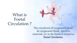
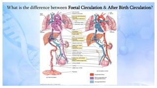
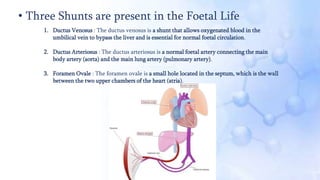
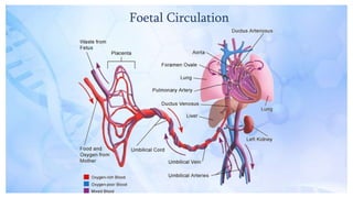
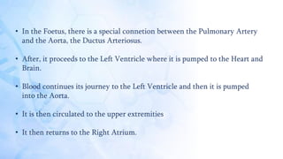
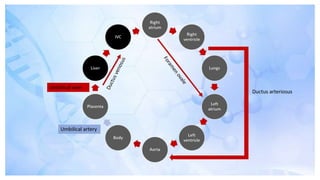
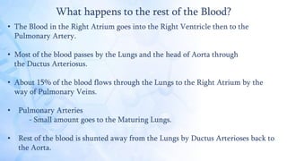
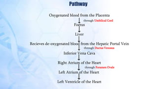
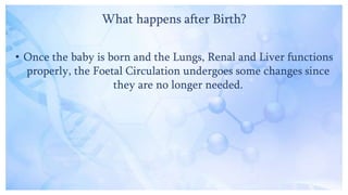
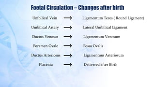
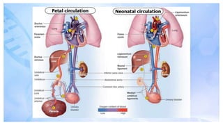
Embark on a captivating exploration of #FetalCirculation in this presentation. Delve into the intricacies of the developing cardiovascular system, understanding how the fetus receives oxygen and nutrients for optimal growth within the womb. Uncover the role of critical structures such as the ductus venosus and foramen ovale in facilitating unique circulatory patterns. Gain insights into the transition from fetal to neonatal circulation and its crucial significance for newborns. This presentation provides a comprehensive overview of the physiological marvel that sustains life before the first breath.
2. What is
Foetal
Circulation ?
The circulation of oxygenated blood,
de-oxygenated blood, nutritive
materials, etc in the foetus is termed as
Foetal Circulation.
3. What is the difference between Foetal Circulation & After Birth Circulation?
4. • Placenta
The placenta is a temporary organ that connects the baby to
the uterus during pregnancy. The placenta develops shortly
after conception and attaches to the wall of the uterus.
The baby is connected to the placenta by the umbilical cord.
Together, the placenta and umbilical cord act as the baby's
lifeline while in the womb. Functions of the placenta include:
• Provides the baby with oxygen and nutrients.
• Removes harmful waste and carbon dioxide from the baby.
• Produces hormones that help the baby grow.
• Passes immunity from you to the baby.
• Helps protect the baby.
5. Anatomy & Physiology of Foetal Circulation
• Umbilical Cords
- 2 Umbilical Arteries : The umbilical vein carries oxygenated, nutrient-rich blood from
the placenta to the foetus, and the umbilical arteries carry deoxygenated, nutrient-depleted blood
from the foetus to the placenta.
- 1 Umbilical Vein : The umbilical vein is a vein present during foetal development that
carries oxygenated blood from the placenta into the growing foetus.
6. • Three Shunts are present in the Foetal Life
1. Ductus Venosus : The ductus venosus is a shunt that allows oxygenated blood in the
umbilical vein to bypass the liver and is essential for normal foetal circulation.
2. Ductus Arteriosus : The ductus arteriosus is a normal foetal artery connecting the main
body artery (aorta) and the main lung artery (pulmonary artery).
3. Foramen Ovale : The foramen ovale is a small hole located in the septum, which is the wall
between the two upper chambers of the heart (atria).
8. How does the Blood move?
• Umbilical Vein carries oxygenated blood and nutrients from the placenta
to the foetus.
• Blood continues to travel from the Inferior Vena Cava to the Ductus
Venosus.
• Ductus Venosus allows the oxygenated blood from Placenta to bypass
through the liver and leads the blood to the Hepatic Portal Veins.
- Small amount of bloodis routed to the Sinosoids of the Liver.
- Increased blood flow leads to a large liver in Newborns.
• The blood then reaches the Right Atrium.
9. What happens to the blood after it reaches to the Right Atrium?
• Blood flows from Right Atrium to the Left Atrium through Foramen
Ovale.
• Here it again mixes with a smaller amount of deoxygenated blood
from the Pulmonary Veins.
• Some of the blood entering the Right Atrium does not pass directly to
the Left Atrium through the Foramen Ovale, but enters the Right
Ventricles and then pumped into the Pulmonary Artery.
10. • In the Foetus, there is a special connetion between the Pulmonary Artery
and the Aorta, the Ductus Arteriosus.
• After, it proceeds to the Left Ventricle where it is pumped to the Heart and
Brain.
• Blood continues its journey to the Left Ventricle and then it is pumped
into the Aorta.
• It is then circulated to the upper extremities
• It then returns to the Right Atrium.
11.
12. What happens to the rest of the Blood?
• The Blood in the Right Atrium goes into the Right Ventricle then to the
Pulmonary Artery.
• Most of the blood passes by the Lungs and the head of Aorta through
the Ductus Arteriosus.
• About 15% of the blood flows through the Lungs to the Right Atrium by the
way of Pulmonary Veins.
• Pulmonary Arteries
- Small amount goes to the Maturing Lungs.
• Rest of the blood is shunted away from the Lungs by Ductus Arterioses back to
the Aorta.
13. What happens further?
• Some of the blood moves from the Aorta through the
Internal Iliac Arteries to the Umbilical Arteries, and
re-enters the Placenta, the Maternal Cycle.
14. Pathway
Oxygenated blood from the Placenta
Foetus
Liver
Recieves de-oxygenated blood from the Hepatic Portal Vein
Inferior Vena Cava
Right Atrium of the Heart
Left Atrium of the Heart
Left Ventricle of the Heart
through Umbilical Cord
through Ductus Venosus
through Foramen Ovale
15. What happens after Birth?
• Once the baby is born and the Lungs, Renal and Liver functions
properly, the Foetal Circulation undergoes some changes since
they are no longer needed.
18. Summary
• Foetal Circulation differs from the postpartum circulation to accommodate the interaction
of the foetus and mother through the Placenta.
• There are 4 bypasses ( shunts) in the Foetus for this purpose :
1) Placental circulational – 2 Umbilical Arteries and 1 Vein.
2) Ductus Arteriosus
3) Formen Ovale
4) Dctus Venosus – Major bypass through the Liver.
• Subsequent to the newborn's first breath and clamping of the Umbilical Cord, these
bypasses within the Foetal Circulation are eliminated to adjusted life outside the Uterus.
} Bypass through Pulmonary Circulation