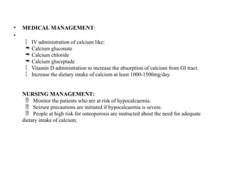
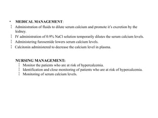
FLUID AND ELECTROLYTES IMBALANCE
BODY FLUIDS: Total body fluid amounts to about
• 60% of body weight in the adult,
• 55% in the older adult.
• 80% in the infants.
• It helps to maintain body temperature and cell shape. It helps transports nutrients,
gases and waste.
• The desired average fluid intake and loss in adult ranges 1500 to 3500 ml daily.
Intake=output.
ELECTROLYTES : Electrolytes are minerals in the body with positive (+ve) or
negative (-ve) charge. They are in the blood, urine, tissue and other body fluids.
CATIONS: (+Ve)Charge ANIONS: (-Ve) Charge
Potassium (k+) Chloride
Calcium (Ca2+) Phosphate
Magnesium (Mg+) Bicarbonate (HCo3-)
Hydrogen (H+) Sulphate
Sodium (Na+)
• Fluid and electrolytes are the basic elements of life. Fluid and electrolyte balance is
the equilibrium state between the fluids and electrolytes within the body. Hence,
when there a change in the equilibrium state is termed as fluid and electrolyte
imbalance.
• The primary fluid of the body is water. It comprises of 70% of living cells and
60% of an average adult’s total body weight. An average person needs about 2 to
2.5 litters of water per day to meet the body’s fluid requirements
BODY FULIDS COMPOSITION AND FUNCTION
•  Approximately 60% of a typical adult’s weight consists of fluid (water and
electrolytes).
•  The amount of body fluids can be influenced by age, gender and body fat.
•  In general, younger people have higher percentage of body fluids than older
people and male have proportionately more body fluids than females.
•  Body fat: obese people have less fluid than thin people because fat cell contains
little water.
COMPARTMENTS:
The body fluids are located in two compartments:
• 1. Intra cellular space (fluids inside the cell) 67%
• 2. Extra cellular space (fluids outside the cell) 33%
a) Intravascular space (fluids with in blood vessels)
b) Interstitial space (fluids around the cell)
FUNCTIONS OF BODY FLUIDS
a) Enzyme in saliva and mucus assist the passage of food into the gut and
provide mechanisms for hydrolysing food into elements that the body
can absorb.
b) ECF in blood and CSF serve as a transport mechanism to deliver
electrolytes, oxygen , nutrients and hormones to tissues.
c) Carry immune system cells to injury site for the body’s defence.
d) ECF eliminates the body’s waste products through kidneys, bowel and
perspiration.
e) Helps in the movement of electrolytes from one body compartment to
another ECF plays an important role in regulation of body temperature.
f) Acts as a lubricant.
NORMAL FLUID EXCHANGE
1. Oncotic pressure: It is the pressure exerted by chemical constituent of
plasma fluid, i.e. crystalloids and colloids
2. Hydrostatic pressure: It is the pressure exerted by the blood flowing
through the capillaries.
OSMOSIS AND OSMOLALITY
• Osmosis = is the process of water movement through a
semipermeable membrane from an area of low solute concentration
to an area of high solute concentration.
Osmolality = measures the number of dissolved particles contained
in a unit of fluid.
Normal Plasma
 Osmolality is between 275 to 295mOsm/kg.
If it goes beyond 295mOsm/kg indicates that the concentration

of particles is too great or that the water content is too little- water
deficit.
•  If it goes below 275mOsm/kg indicates too little solute for the
amount of water or too much of water for the amount of solute-
water excess.
• HYPOTONIC: the solution in which the solute is less concentrated
than in the cells.
 HYPERTONIC: The solutes are more concentrated in the fluids than
in the cell.
 ISOTONIC: The solutes in the fluids are equally concentrated as
in the cell.
• DIFFUSION:
The natural tendency of substances to move from an area of higher
concentration to one of lower concentration.
• E.g., exchange of oxygen and carbon dioxide between the pulmonary
capillaries and alveoli.
FILTRATION
• The hydrostatic pressure in capillaries tends to filter fluid out of the vascula
compartment into the interstitial fluid.
E.g., the passage of water and electrolytes from the arterial capillary bed to the

interstitial fluid.
•  The hydrostatic pressure is famished by the pumping action of the heart.
• SODIUM-POTASSIUM PUMP:
• The sodium-potassium pump is membrane protein that actively transports sodium
ions out of a cell and potassium ions into the cell against their respectiv
concentration gradients.
• The energy source for Sodium-Potassium Pump mechanism is ATP (Adenosine
triphosphate).
• It is situated in the outer plasma membrane of the cells.
• This process helps maintain the electrochemical balance essential for cell
function and it is crucial for nerve impulses and muscle contraction.
HOMEOSTATIC MECHANISM
The following organs are involved in homeostasis. They are:
Kidney
 The kidney normally filters 170 litters of plasma every day in the adult, while
excretion only1.5 L of urine
 Regulation of ECF volume and osmolality by selective retention and excretion of body
fluids.
 Regulation of electrolytes level in ECF by means of selective retention of needed
substancesand excretion of unneeded substances.
 Regulation of pH of ECF by retention of hydrogen ions.
 Excretion of metabolic waste and toxic substances.
Heart and blood vessels:
 The pumping action of the heart circulates blood through kidney under sufficient
pressure to allow for urine formation.
 Failure of this pumping action interferes with renal perfusion and thus with water
and electrolyte regulation.
Pituitary gland
 The hypothalamus malfunction ADH (Anti-Diuretic Hormone), which is stored in posterior
pituitary gland and released as needed.
 ADH is sometimes refereed as water conserving hormone since it cause the body to retain
water.
 The function of the ADH is maintaining the osmotic pressure by controlling the retention
of water by the kidney and by regulating blood volume.

Adrenal gland
 Aldosterone, an hormone secreted by adrenal gland has a profound effect on fluid balance
Increased secretion of aldosterone causes sodium retention (thus water retention) and
potassium loss.
 Decreased secretion leads to sodium and water loss and potassium retention
Parathyroid gland:
 The parathyroid hormone in the thyroid gland regulates the calcium and phosphate balance
by means of parathyroid hormone (PTH). PTH influences bone reabsorption, calcium
absorption from the intestine and calcium reabsorption from the renal tubules.
FLUID VOLUME DISTURBANCES
• Hypovolemia = Hypovolemia is refers to as lack of fluid volume. It occurs when the water
and electrolytes are lost in the same proportions as they exist in normal body fluids
• Causes:
Increase insensible water loss or perspiration

Haemorrhage

Osmotic diuretics

Vomiting, Diarrhoea

Inadequate fluid intake

Diabetic insipidus

Burns

Intestinal obstruction

Clinical Manifestations:
Restlessness, drowsy, lethargy, confusion

Thrust, dry mucus Membranes

Decreased skin turgor, reduced capillary refill

Decreased urine output

Postural hypotension, increased pulse, Decreased CVP

Increased respiratory rate

Weight loss, weakness

Seizure, coma

• 1. Balanced IV solution: Ringer’s Lactate Solution.
• 2. Isotonic (0.9%) sodium chloride is used when rapid volume replacement is
required.
3. Blood administration when the volume loss is due to blood loss.
Nursing diagnosis:
•  Deficit fluid volume related to excessive fluid loss or decreased fluid intake.
•  Decreased cardiac output related to excessive fluid loss or decreased fluid
intake.
•  Risk for deficit fluid volume
Potential complication: Hypovolemic shock

HYPERVOLEMIYA : Hypervolemia is refers to as fluid volume excess. It may result from
excessive intake of fluids,abnormal retention of fluids or fluid shift from intracellular fluid
into plasma fluid.
Causes:
 Excessive Isotonic or hypotonic IV fluids.
Heart failure

Rental failure

Primary polydipsia

SIADH

Long term use of corticosteroids

• Clinical Manifestations:
Headache, confusion, lethargy

Jugular vein distension

Bounding pulse, increased BP, increased CVP

Polyuria

Peripheral edema

Dyspnoea, Crackles

Muscle spasm

Weight gain

Seizure, coma

• Primary care:
1. Fluid restriction.
2. Discontinue the IV infusion when the fluid excess is related to excessive administration of
sodium containing fluids.
3. Restriction of sodium intake.
•
 Other measures:
1. Diuretics are prescribed, when dietary restriction of sodium alone is insufficient to reduc
edema.
2. The choice of diuretics is based on the severity
3. Of the Hypervolemic state, the degree of impairment of renal function & the potency
diuretic.
• Haemodialysis = Haemodialysis or peritoneal dialysis is advisable when the renal
function is severel impaired and so the pharmacological agents can not act efficiently.
Abdominal parenthesis:
If fluid excess leads to ascites.

Thoracentesis:
When fluid excess leads to pleural effusion.

Nursing diagnosis:
Excess fluid volume related to increased water/sodium retention.

Activity intolerance related increased water retention and fatigue/weakness.

Impaired gas exchange related to water retention leading to pulmonary edema.

Disturbed body image related to altered body appearance secondary to edema.

Potential complication: pulmonary edema, ascites.

ELECTROLYTE IMBALANCE
• SODIUM (Normal range: 135-145mEq/L)
 Hypernatremia: Hypernatremia is defined as a plasma sodium level greater than 145mEq/L. It is usually
associated with water loss and sodium gain. As a result, the sodium concentration increases and the increased
concentration pull fluid out of the cell.
CAUSES:
 Excessive sodium intake:
1. IV fluids: hypertonic NaCl, excessive Isotonic NaCl, IV Sodium Bicarbonate
2. Hypertonic tube feeding without water supplements
3. Near drowning in the salt water
 Inadequate water intake
1. Excessive water loss:
2. Increased insensible water loss
3. Osmotic diuretic therapy
4. Diarrhoea
 Disease status:
1. Diabetes Insipidus
2. Cushing syndrome
3. Uncontrolled Diabetes Mellitus
4. Primary hyperaldosteronism
• CLINICAL MANIFESTATIONS:
Decreased ECF volume:

a. Restlessness, agitation, twitching, Seizure, coma
b. Intense thirst, dry & swollen tongue, stick mucus membrane
c. Postural hypotension, increased CVP, weight loss, increased pulse
d. Weakness, lethargy
Normal or increased ECF volume
 :
a. Restlessness, agitation, twitching, Seizure, coma
b. Intense thirst, flushed skin
c. Weight gain, peripheral and pulmonary edema, increased BP, increased CVP
MEDICAL MANAGEMENT:
Gradual lowering of the Sodium level by the infusion of hypotonic electrolyte

solution
Isotonic solution

Diuretics

NURSING MANAGEMENT:
Monitor the serum sodium levels and the patient’s response to the therapy

Restrict the dietary intake of sodium

Prevent the ingestion of over-the-counter medications with high Sodium content

Note the patient’s thirst and elevated body temperature

Monitor for changes in behaviour such as Restlessness, disorientation and lethargy

• NURSING DIAGNOSIS:
Risk for acute confusion related to electrolyte imbalance.

Risk for injury related to altered sensorium and decreased level of consciousness.

Risk for electrolyte imbalance related to excessive loss of sodium/ excessive

Intake/ retention of water.
Potential complication: severe neurologic changes.

Hyponatremia (Sodium deficit): Hypernatremia is defined as a plasma sodium level
less than 135mEq/L. It is one of the most common electrolyte disorders in adults,
especially older adults. Hyponatremia is usually associated with changes in fluid
volume status.
CAUSES:
Excessive sodium loss:

1. GI loss: diarrhoea, vomiting
2. Renal loses: Diuretics, Adrenal insufficiency, Renal disease
3. Skin loses: Burns, wound drainage
Inadequate sodium intake

Excessive water Loss

1. Excessive hypotonic IV fluids
2. Primary polydipsia
Disease status

1. SIADH
2. Heart failure
3. Primary hyperaldosteronism
• CLINICAL MANIFESTATIONS:
 Decreased ECF volume
1. Irritability, confusion, Dizziness, tremors
2. Dry mucus membranes
3. Postural hypotension, decreased BP, decreased CVP
4. Cold, clamp skin
 Normal or increased ECF volume
1. Headache, apathy, confusion, muscle spasm, Seizure, coma
2. Nausea, vomiting, diarrhoea, abdominal cramps
3. Weight gain, increased BP, increased CVP
MEDICAL MANAGEMENT:
Sodium replacement therapy:

Oral intakes of sodium by eat or drink.

Administration of Small amount of IV hypertonic saline solution.

Lactate Ringer’s solution

The correction of serum sodium must not Increase greater than 12mEq/L in 24 hours

to avoid neurological damage.
• NURSING MANAGEMENT:
 Monitor serum sodium levels and the patient’s response to therapy.
Avoid adding up salt in diet.

Record the lab values and inform to the physician
 .
NURSING DIAGNOSIS:
Risk for fluid volume deficit related to excessive intake of sodium/ excessive loss

of water.
Risk for electrolyte imbalance related to excessive intake sodium/ excessive loss of

water.
Potential complication: brain damage, Seizure, coma.

Potassium(Normal range: 3.5-5.5mEq/L)
HYPOKALEMIA: Hypokalaemia is defined as a plasma potassium level less than
3.5mEq/L. It is a common electrolyte disorder especially in the older adult population. It
results from an increased loss of potassium ,from an increased shift of potassium from
ECF to ICF or rarely from deficient dietary potassium intake.
• CAUSES:
Potassium loss

i. GI loss: diarrhoea, vomiting
ii. Renal losses: diuretics, hyperaldosteronism
iii. Skin losses: diaphoresis, dialysis
Shift of potassium into cells

i. Increased insulin e.g. dextrose load
ii. Alkalosis
iii. Tissue repair
iv. Increased epinephrine e.g. stress
Lack of potassium intake

i. Starvation
ii. Low potassium in diet
CLINICAL MANIFESTATION:
Fatigue

Muscle weakness, leg cramps

Nausea, vomiting, paralytic ileus

Soft, flabby muscle

Paraesthesia, decreased reflexes

Polyuria

weak irregular pulse

hyperglycaemia

• MEDICAL MANAGEMENT:
Treated with oral and IV replacement therapy.

Administer 40 to 80 mEq/L day of potassium.

IV route is indicated if oral potassium therapy is not feasible.

Potassium rich diet for patients at risk of hypokalaemia.

NURSING MANAGEMENT:
Monitor for the presence of hypokalaemia in the patients at risk.

Encourage the patients for potassium rich diet (bananas, malen, citrus fruit, fresh

and frozen vegetables, fresh meats).
Monitor the patients who are taking digitalis which may cause potassium deficiency.

NURSING DIAGNOSIS:
Risk for electrolyte imbalance related to excessive loss of potassium.

Risk for injury related to muscle weakness and hyporeflexes.

Potential complication: dysrhythmias.

• HYPERKALEMIA: -Hyperkalaemia is defined as an elevation of the potassium level greater than
5mEq/L. It is rare electrolyte disorder but it affects people with acute and chronic renal failure.
Hyperkalemia is results from impaired renal excretion.
CAUSES:
Excessive potassium intake

i. Excessive or massive parenteral administration
ii. Potassium containing drugs. E.g. [potassium penicillin
iii. Potassium containing salt substitute
Shift of potassium out of cells

a. Acidosis
b. Tissue catabolism
Failure to eliminate potassium

a. Renal disease
b. Potassium-sparing diuretics
CLINICAL MANIFESTATIONS:
Irritability, anxiety

Abdominal cramping, diarrhoea

Weakness of lower extremities

Paraesthesia

Irregular pulse

Cardiac arrest

• MEDICAL MANAGEMENT:
Immediate ECG should be obtained to detect the changes.

Restriction of dietary potassium.

Potassium containing diuretics.

Calcium gluconate administered in case of dangerously elevated serum potassium

level.
NURSING MANAGEMENT:
Identification and close monitoring of patients who are at risk of hyperkalemia.

Observe the sign of muscle weakness and dysrhythmias.

Monitoring of serum potassium levels.

NURSING DIAGNOSIS:
Activity intolerance related to lower extremity muscle weakness.

Risk for electrolyte imbalance related to excessive retention/ cellular release of

potassium.
Risk for injury related to altered sensorium and decreased level of consciousness.

Potential complication: Dysrhythmias.

CALCIUM (Normal range: 8.5-10.5mg/dl)
 HYPOCALCEMIA: - Hypercalcemia defined as plasma calcium LESS than 8.5mEq/L.
Hypocalcemia can occur in any age group. It is a common electrolyte disorder that can have
serious physical complications.
CAUSES:
1. Decreased total calcium:
Chronic kidney disease

Elevated phosphorus

 Primary hypoparathyroidism
Vitamin D deficiency

Magnesium deficiency

Acute pancreatitis

Chronic alcoholism

Diarrhoea

2. Decreased ionized calcium; Alkalosis

CLINICAL MANIFESTATIONS:
Easy fatigability

Depression, anxiety confusion

Numbness, tingling in extremities %h region around mouth

Hyperreflexia, muscle cramps

Chvostek’s sign

Trousseau’s sign

Laryngeal spasm, tetany, Seizure

• MEDICAL MANAGEMENT:
•
 IV administration of calcium like:
Calcium gluconate

Calcium chloride

Calcium gluceptade

Vitamin D administration to increase the absorption of calcium from GI tract.

Increase the dietary intake of calcium at least 1000-1500mg/day.

NURSING MANAGEMENT:
Monitor the patients who are at risk of hypocalcaemia.

Seizure precautions are initiated if hypocalcaemia is severe.

People at high risk for osteoporosis are instructed about the need for adequate

dietary intake of calcium.
• HYPERCALCEMIA: - Hypocalcaemia is defined as a plasma calcium level less than
10.5mEq/L. It results fromVitamin D deficiency, underactive PTH glands, kidney disorders,
inadequate intake of calcium or diseases that impair calcium absorption.
CAUSES:
1. Increased total calcium:
Malignancy with bone metastasis

Prolonged immobilization

Hyperparathyroidism

Vitamin D overdose

Thiazide diuretics

Milk-alkali syndrome

Multiple myeloma

2. Increased ionized calcium:
Acidosis

CLINICAL MANIFESTATIONS:
Lethargy, weakness

Depressed reflexes

Decreased memory

Confusion, psychosis

Anorexia, Nausea, vomiting

Bone pain, fractures

Polyuria, dehydration

Stupor, coma

• MEDICAL MANAGEMENT:
 Administration of fluids to dilute serum calcium and promote it’s excretion by the
kidney.
 IV administration of 0.9% NaCl solution temporarily dilutes the serum calcium levels.
 Administering furosemide lowers serum calcium levels.
 Calcitonin administered to decrease the calcium level in plasma.
NURSING MANAGEMENT:
Monitor the patients who are at risk of hypercalcemia.

Identification and close monitoring of patients who are at risk of hypercalcemia.

Monitoring of serum calcium levels.


fluid and electrolyte.pptx for gnm 1`yrs

  • 2.
    FLUID AND ELECTROLYTESIMBALANCE BODY FLUIDS: Total body fluid amounts to about • 60% of body weight in the adult, • 55% in the older adult. • 80% in the infants. • It helps to maintain body temperature and cell shape. It helps transports nutrients, gases and waste. • The desired average fluid intake and loss in adult ranges 1500 to 3500 ml daily. Intake=output. ELECTROLYTES : Electrolytes are minerals in the body with positive (+ve) or negative (-ve) charge. They are in the blood, urine, tissue and other body fluids.
  • 3.
    CATIONS: (+Ve)Charge ANIONS:(-Ve) Charge Potassium (k+) Chloride Calcium (Ca2+) Phosphate Magnesium (Mg+) Bicarbonate (HCo3-) Hydrogen (H+) Sulphate Sodium (Na+)
  • 4.
    • Fluid andelectrolytes are the basic elements of life. Fluid and electrolyte balance is the equilibrium state between the fluids and electrolytes within the body. Hence, when there a change in the equilibrium state is termed as fluid and electrolyte imbalance. • The primary fluid of the body is water. It comprises of 70% of living cells and 60% of an average adult’s total body weight. An average person needs about 2 to 2.5 litters of water per day to meet the body’s fluid requirements BODY FULIDS COMPOSITION AND FUNCTION •  Approximately 60% of a typical adult’s weight consists of fluid (water and electrolytes). •  The amount of body fluids can be influenced by age, gender and body fat. •  In general, younger people have higher percentage of body fluids than older people and male have proportionately more body fluids than females. •  Body fat: obese people have less fluid than thin people because fat cell contains little water.
  • 5.
    COMPARTMENTS: The body fluidsare located in two compartments: • 1. Intra cellular space (fluids inside the cell) 67% • 2. Extra cellular space (fluids outside the cell) 33% a) Intravascular space (fluids with in blood vessels) b) Interstitial space (fluids around the cell)
  • 6.
    FUNCTIONS OF BODYFLUIDS a) Enzyme in saliva and mucus assist the passage of food into the gut and provide mechanisms for hydrolysing food into elements that the body can absorb. b) ECF in blood and CSF serve as a transport mechanism to deliver electrolytes, oxygen , nutrients and hormones to tissues. c) Carry immune system cells to injury site for the body’s defence. d) ECF eliminates the body’s waste products through kidneys, bowel and perspiration. e) Helps in the movement of electrolytes from one body compartment to another ECF plays an important role in regulation of body temperature. f) Acts as a lubricant.
  • 7.
    NORMAL FLUID EXCHANGE 1.Oncotic pressure: It is the pressure exerted by chemical constituent of plasma fluid, i.e. crystalloids and colloids 2. Hydrostatic pressure: It is the pressure exerted by the blood flowing through the capillaries.
  • 8.
    OSMOSIS AND OSMOLALITY •Osmosis = is the process of water movement through a semipermeable membrane from an area of low solute concentration to an area of high solute concentration. Osmolality = measures the number of dissolved particles contained in a unit of fluid. Normal Plasma  Osmolality is between 275 to 295mOsm/kg. If it goes beyond 295mOsm/kg indicates that the concentration  of particles is too great or that the water content is too little- water deficit. •  If it goes below 275mOsm/kg indicates too little solute for the amount of water or too much of water for the amount of solute- water excess.
  • 9.
    • HYPOTONIC: thesolution in which the solute is less concentrated than in the cells.  HYPERTONIC: The solutes are more concentrated in the fluids than in the cell.  ISOTONIC: The solutes in the fluids are equally concentrated as in the cell. • DIFFUSION: The natural tendency of substances to move from an area of higher concentration to one of lower concentration. • E.g., exchange of oxygen and carbon dioxide between the pulmonary capillaries and alveoli.
  • 11.
    FILTRATION • The hydrostaticpressure in capillaries tends to filter fluid out of the vascula compartment into the interstitial fluid. E.g., the passage of water and electrolytes from the arterial capillary bed to the  interstitial fluid. •  The hydrostatic pressure is famished by the pumping action of the heart. • SODIUM-POTASSIUM PUMP: • The sodium-potassium pump is membrane protein that actively transports sodium ions out of a cell and potassium ions into the cell against their respectiv concentration gradients. • The energy source for Sodium-Potassium Pump mechanism is ATP (Adenosine triphosphate). • It is situated in the outer plasma membrane of the cells. • This process helps maintain the electrochemical balance essential for cell function and it is crucial for nerve impulses and muscle contraction.
  • 12.
    HOMEOSTATIC MECHANISM The followingorgans are involved in homeostasis. They are: Kidney  The kidney normally filters 170 litters of plasma every day in the adult, while excretion only1.5 L of urine  Regulation of ECF volume and osmolality by selective retention and excretion of body fluids.  Regulation of electrolytes level in ECF by means of selective retention of needed substancesand excretion of unneeded substances.  Regulation of pH of ECF by retention of hydrogen ions.  Excretion of metabolic waste and toxic substances. Heart and blood vessels:  The pumping action of the heart circulates blood through kidney under sufficient pressure to allow for urine formation.  Failure of this pumping action interferes with renal perfusion and thus with water and electrolyte regulation.
  • 13.
    Pituitary gland  Thehypothalamus malfunction ADH (Anti-Diuretic Hormone), which is stored in posterior pituitary gland and released as needed.  ADH is sometimes refereed as water conserving hormone since it cause the body to retain water.  The function of the ADH is maintaining the osmotic pressure by controlling the retention of water by the kidney and by regulating blood volume.  Adrenal gland  Aldosterone, an hormone secreted by adrenal gland has a profound effect on fluid balance Increased secretion of aldosterone causes sodium retention (thus water retention) and potassium loss.  Decreased secretion leads to sodium and water loss and potassium retention Parathyroid gland:  The parathyroid hormone in the thyroid gland regulates the calcium and phosphate balance by means of parathyroid hormone (PTH). PTH influences bone reabsorption, calcium absorption from the intestine and calcium reabsorption from the renal tubules.
  • 14.
    FLUID VOLUME DISTURBANCES •Hypovolemia = Hypovolemia is refers to as lack of fluid volume. It occurs when the water and electrolytes are lost in the same proportions as they exist in normal body fluids • Causes: Increase insensible water loss or perspiration  Haemorrhage  Osmotic diuretics  Vomiting, Diarrhoea  Inadequate fluid intake  Diabetic insipidus  Burns  Intestinal obstruction  Clinical Manifestations: Restlessness, drowsy, lethargy, confusion  Thrust, dry mucus Membranes  Decreased skin turgor, reduced capillary refill  Decreased urine output  Postural hypotension, increased pulse, Decreased CVP  Increased respiratory rate  Weight loss, weakness  Seizure, coma 
  • 15.
    • 1. BalancedIV solution: Ringer’s Lactate Solution. • 2. Isotonic (0.9%) sodium chloride is used when rapid volume replacement is required. 3. Blood administration when the volume loss is due to blood loss. Nursing diagnosis: •  Deficit fluid volume related to excessive fluid loss or decreased fluid intake. •  Decreased cardiac output related to excessive fluid loss or decreased fluid intake. •  Risk for deficit fluid volume Potential complication: Hypovolemic shock  HYPERVOLEMIYA : Hypervolemia is refers to as fluid volume excess. It may result from excessive intake of fluids,abnormal retention of fluids or fluid shift from intracellular fluid into plasma fluid. Causes:  Excessive Isotonic or hypotonic IV fluids. Heart failure  Rental failure  Primary polydipsia  SIADH  Long term use of corticosteroids 
  • 16.
    • Clinical Manifestations: Headache,confusion, lethargy  Jugular vein distension  Bounding pulse, increased BP, increased CVP  Polyuria  Peripheral edema  Dyspnoea, Crackles  Muscle spasm  Weight gain  Seizure, coma  • Primary care: 1. Fluid restriction. 2. Discontinue the IV infusion when the fluid excess is related to excessive administration of sodium containing fluids. 3. Restriction of sodium intake. •  Other measures: 1. Diuretics are prescribed, when dietary restriction of sodium alone is insufficient to reduc edema. 2. The choice of diuretics is based on the severity 3. Of the Hypervolemic state, the degree of impairment of renal function & the potency diuretic.
  • 17.
    • Haemodialysis =Haemodialysis or peritoneal dialysis is advisable when the renal function is severel impaired and so the pharmacological agents can not act efficiently. Abdominal parenthesis: If fluid excess leads to ascites.  Thoracentesis: When fluid excess leads to pleural effusion.  Nursing diagnosis: Excess fluid volume related to increased water/sodium retention.  Activity intolerance related increased water retention and fatigue/weakness.  Impaired gas exchange related to water retention leading to pulmonary edema.  Disturbed body image related to altered body appearance secondary to edema.  Potential complication: pulmonary edema, ascites. 
  • 18.
    ELECTROLYTE IMBALANCE • SODIUM(Normal range: 135-145mEq/L)  Hypernatremia: Hypernatremia is defined as a plasma sodium level greater than 145mEq/L. It is usually associated with water loss and sodium gain. As a result, the sodium concentration increases and the increased concentration pull fluid out of the cell. CAUSES:  Excessive sodium intake: 1. IV fluids: hypertonic NaCl, excessive Isotonic NaCl, IV Sodium Bicarbonate 2. Hypertonic tube feeding without water supplements 3. Near drowning in the salt water  Inadequate water intake 1. Excessive water loss: 2. Increased insensible water loss 3. Osmotic diuretic therapy 4. Diarrhoea  Disease status: 1. Diabetes Insipidus 2. Cushing syndrome 3. Uncontrolled Diabetes Mellitus 4. Primary hyperaldosteronism
  • 19.
    • CLINICAL MANIFESTATIONS: DecreasedECF volume:  a. Restlessness, agitation, twitching, Seizure, coma b. Intense thirst, dry & swollen tongue, stick mucus membrane c. Postural hypotension, increased CVP, weight loss, increased pulse d. Weakness, lethargy Normal or increased ECF volume  : a. Restlessness, agitation, twitching, Seizure, coma b. Intense thirst, flushed skin c. Weight gain, peripheral and pulmonary edema, increased BP, increased CVP MEDICAL MANAGEMENT: Gradual lowering of the Sodium level by the infusion of hypotonic electrolyte  solution Isotonic solution  Diuretics  NURSING MANAGEMENT: Monitor the serum sodium levels and the patient’s response to the therapy  Restrict the dietary intake of sodium  Prevent the ingestion of over-the-counter medications with high Sodium content  Note the patient’s thirst and elevated body temperature  Monitor for changes in behaviour such as Restlessness, disorientation and lethargy 
  • 20.
    • NURSING DIAGNOSIS: Riskfor acute confusion related to electrolyte imbalance.  Risk for injury related to altered sensorium and decreased level of consciousness.  Risk for electrolyte imbalance related to excessive loss of sodium/ excessive  Intake/ retention of water. Potential complication: severe neurologic changes.  Hyponatremia (Sodium deficit): Hypernatremia is defined as a plasma sodium level less than 135mEq/L. It is one of the most common electrolyte disorders in adults, especially older adults. Hyponatremia is usually associated with changes in fluid volume status. CAUSES: Excessive sodium loss:  1. GI loss: diarrhoea, vomiting 2. Renal loses: Diuretics, Adrenal insufficiency, Renal disease 3. Skin loses: Burns, wound drainage Inadequate sodium intake  Excessive water Loss  1. Excessive hypotonic IV fluids 2. Primary polydipsia Disease status  1. SIADH 2. Heart failure 3. Primary hyperaldosteronism
  • 21.
    • CLINICAL MANIFESTATIONS: Decreased ECF volume 1. Irritability, confusion, Dizziness, tremors 2. Dry mucus membranes 3. Postural hypotension, decreased BP, decreased CVP 4. Cold, clamp skin  Normal or increased ECF volume 1. Headache, apathy, confusion, muscle spasm, Seizure, coma 2. Nausea, vomiting, diarrhoea, abdominal cramps 3. Weight gain, increased BP, increased CVP MEDICAL MANAGEMENT: Sodium replacement therapy:  Oral intakes of sodium by eat or drink.  Administration of Small amount of IV hypertonic saline solution.  Lactate Ringer’s solution  The correction of serum sodium must not Increase greater than 12mEq/L in 24 hours  to avoid neurological damage.
  • 22.
    • NURSING MANAGEMENT: Monitor serum sodium levels and the patient’s response to therapy. Avoid adding up salt in diet.  Record the lab values and inform to the physician  . NURSING DIAGNOSIS: Risk for fluid volume deficit related to excessive intake of sodium/ excessive loss  of water. Risk for electrolyte imbalance related to excessive intake sodium/ excessive loss of  water. Potential complication: brain damage, Seizure, coma.  Potassium(Normal range: 3.5-5.5mEq/L) HYPOKALEMIA: Hypokalaemia is defined as a plasma potassium level less than 3.5mEq/L. It is a common electrolyte disorder especially in the older adult population. It results from an increased loss of potassium ,from an increased shift of potassium from ECF to ICF or rarely from deficient dietary potassium intake.
  • 23.
    • CAUSES: Potassium loss  i.GI loss: diarrhoea, vomiting ii. Renal losses: diuretics, hyperaldosteronism iii. Skin losses: diaphoresis, dialysis Shift of potassium into cells  i. Increased insulin e.g. dextrose load ii. Alkalosis iii. Tissue repair iv. Increased epinephrine e.g. stress Lack of potassium intake  i. Starvation ii. Low potassium in diet CLINICAL MANIFESTATION: Fatigue  Muscle weakness, leg cramps  Nausea, vomiting, paralytic ileus  Soft, flabby muscle  Paraesthesia, decreased reflexes  Polyuria  weak irregular pulse  hyperglycaemia 
  • 24.
    • MEDICAL MANAGEMENT: Treatedwith oral and IV replacement therapy.  Administer 40 to 80 mEq/L day of potassium.  IV route is indicated if oral potassium therapy is not feasible.  Potassium rich diet for patients at risk of hypokalaemia.  NURSING MANAGEMENT: Monitor for the presence of hypokalaemia in the patients at risk.  Encourage the patients for potassium rich diet (bananas, malen, citrus fruit, fresh  and frozen vegetables, fresh meats). Monitor the patients who are taking digitalis which may cause potassium deficiency.  NURSING DIAGNOSIS: Risk for electrolyte imbalance related to excessive loss of potassium.  Risk for injury related to muscle weakness and hyporeflexes.  Potential complication: dysrhythmias. 
  • 25.
    • HYPERKALEMIA: -Hyperkalaemiais defined as an elevation of the potassium level greater than 5mEq/L. It is rare electrolyte disorder but it affects people with acute and chronic renal failure. Hyperkalemia is results from impaired renal excretion. CAUSES: Excessive potassium intake  i. Excessive or massive parenteral administration ii. Potassium containing drugs. E.g. [potassium penicillin iii. Potassium containing salt substitute Shift of potassium out of cells  a. Acidosis b. Tissue catabolism Failure to eliminate potassium  a. Renal disease b. Potassium-sparing diuretics CLINICAL MANIFESTATIONS: Irritability, anxiety  Abdominal cramping, diarrhoea  Weakness of lower extremities  Paraesthesia  Irregular pulse  Cardiac arrest 
  • 26.
    • MEDICAL MANAGEMENT: ImmediateECG should be obtained to detect the changes.  Restriction of dietary potassium.  Potassium containing diuretics.  Calcium gluconate administered in case of dangerously elevated serum potassium  level. NURSING MANAGEMENT: Identification and close monitoring of patients who are at risk of hyperkalemia.  Observe the sign of muscle weakness and dysrhythmias.  Monitoring of serum potassium levels.  NURSING DIAGNOSIS: Activity intolerance related to lower extremity muscle weakness.  Risk for electrolyte imbalance related to excessive retention/ cellular release of  potassium. Risk for injury related to altered sensorium and decreased level of consciousness.  Potential complication: Dysrhythmias. 
  • 29.
    CALCIUM (Normal range:8.5-10.5mg/dl)  HYPOCALCEMIA: - Hypercalcemia defined as plasma calcium LESS than 8.5mEq/L. Hypocalcemia can occur in any age group. It is a common electrolyte disorder that can have serious physical complications. CAUSES: 1. Decreased total calcium: Chronic kidney disease  Elevated phosphorus   Primary hypoparathyroidism Vitamin D deficiency  Magnesium deficiency  Acute pancreatitis  Chronic alcoholism  Diarrhoea  2. Decreased ionized calcium; Alkalosis  CLINICAL MANIFESTATIONS: Easy fatigability  Depression, anxiety confusion  Numbness, tingling in extremities %h region around mouth  Hyperreflexia, muscle cramps  Chvostek’s sign  Trousseau’s sign  Laryngeal spasm, tetany, Seizure 
  • 30.
    • MEDICAL MANAGEMENT: • IV administration of calcium like: Calcium gluconate  Calcium chloride  Calcium gluceptade  Vitamin D administration to increase the absorption of calcium from GI tract.  Increase the dietary intake of calcium at least 1000-1500mg/day.  NURSING MANAGEMENT: Monitor the patients who are at risk of hypocalcaemia.  Seizure precautions are initiated if hypocalcaemia is severe.  People at high risk for osteoporosis are instructed about the need for adequate  dietary intake of calcium.
  • 31.
    • HYPERCALCEMIA: -Hypocalcaemia is defined as a plasma calcium level less than 10.5mEq/L. It results fromVitamin D deficiency, underactive PTH glands, kidney disorders, inadequate intake of calcium or diseases that impair calcium absorption. CAUSES: 1. Increased total calcium: Malignancy with bone metastasis  Prolonged immobilization  Hyperparathyroidism  Vitamin D overdose  Thiazide diuretics  Milk-alkali syndrome  Multiple myeloma  2. Increased ionized calcium: Acidosis  CLINICAL MANIFESTATIONS: Lethargy, weakness  Depressed reflexes  Decreased memory  Confusion, psychosis  Anorexia, Nausea, vomiting  Bone pain, fractures  Polyuria, dehydration  Stupor, coma 
  • 32.
    • MEDICAL MANAGEMENT: Administration of fluids to dilute serum calcium and promote it’s excretion by the kidney.  IV administration of 0.9% NaCl solution temporarily dilutes the serum calcium levels.  Administering furosemide lowers serum calcium levels.  Calcitonin administered to decrease the calcium level in plasma. NURSING MANAGEMENT: Monitor the patients who are at risk of hypercalcemia.  Identification and close monitoring of patients who are at risk of hypercalcemia.  Monitoring of serum calcium levels. 