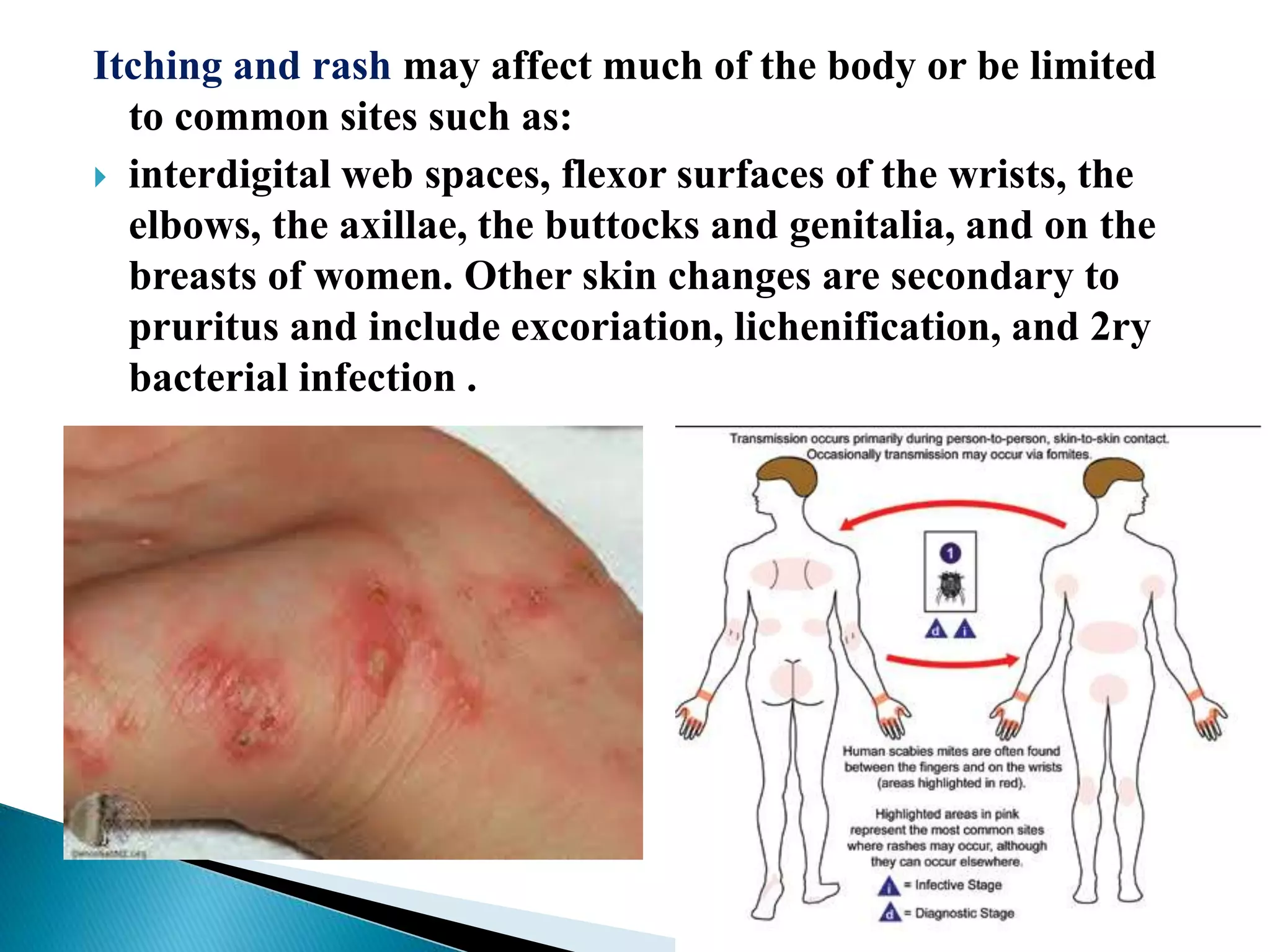
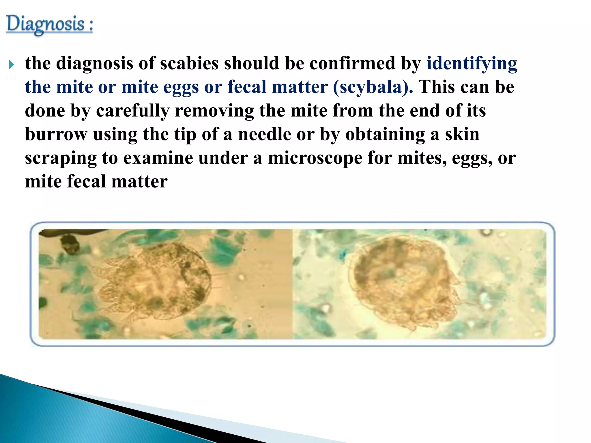
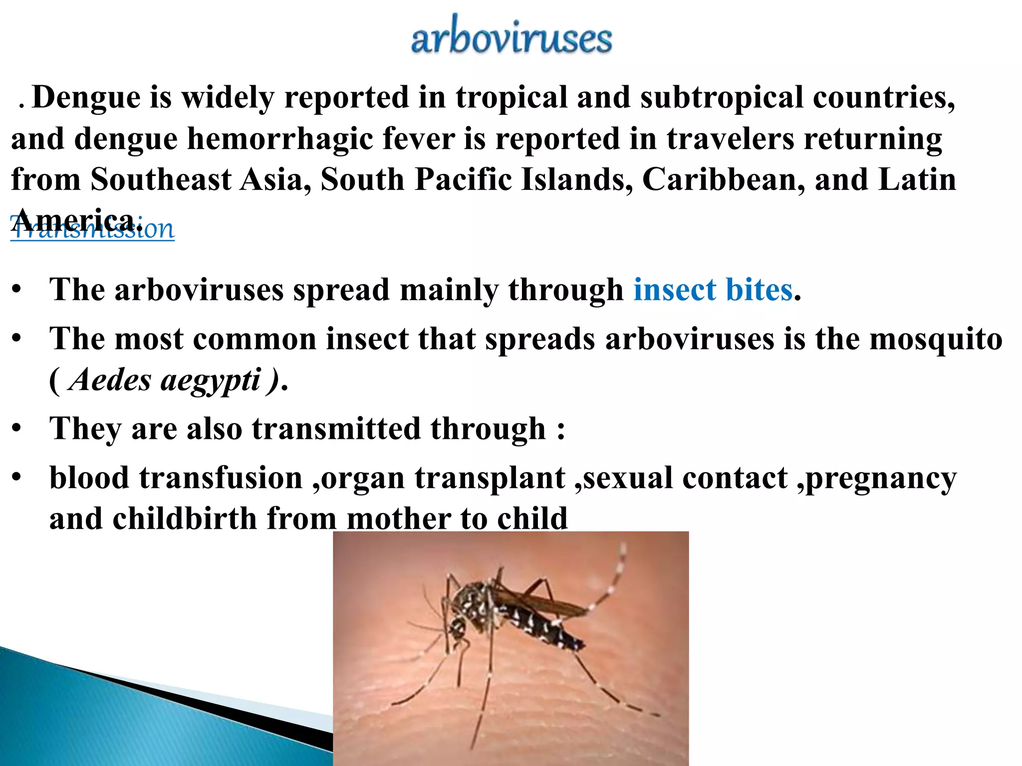
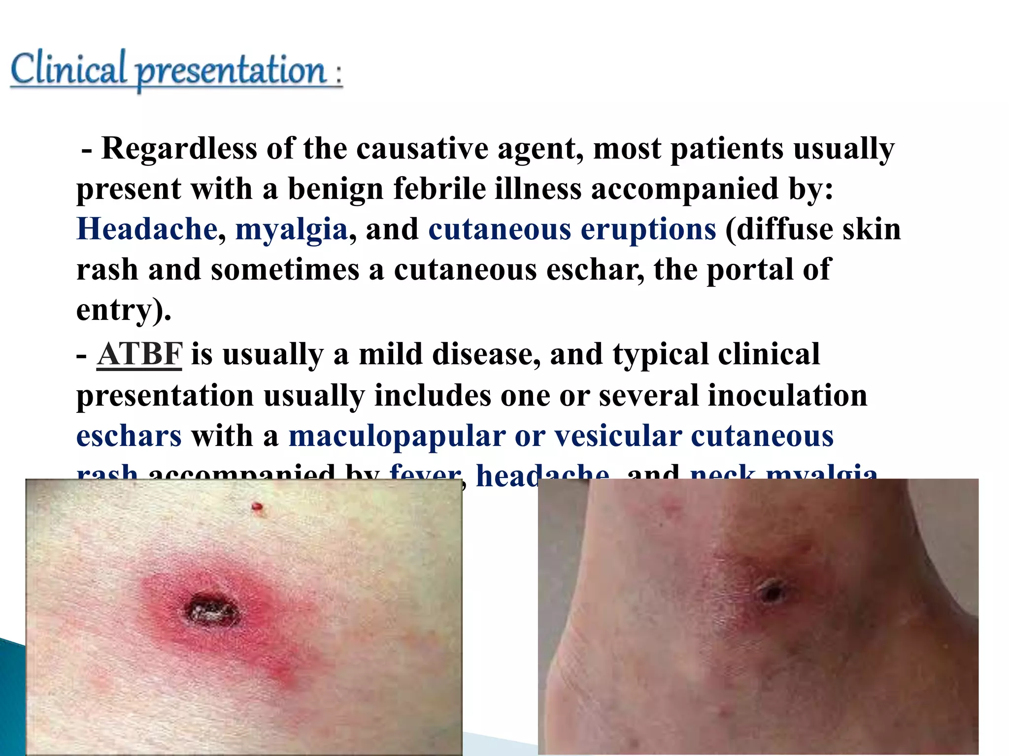
SlideShare a Scribd company logo
Common Skin Infections in Travelers
Group 3 Members
1- Epidemiology - Ahmed Yousef Mohamed 118
2- Bacterial skin infection - Ayman Hassan Hamed
302
3- HrCLM - Bashayer Michel Farag 349
4- Leishmaniasis - Ahmed Mohamed Mohamed Aly
105
5- Myiasis- Al shaimaa ahmed 211
6- Tungiasis- Alaa Gamal Soliman 258
7- Scabies - Dalia hassan attia 411
8-Arboviral infection - Amira Fetiany 243
 In Nepal, three studies showed that dermatoses were the third to the fourth
most frequent presenting illness among tourists
 in the Maldives and Fiji, dermatoses were the most frequent presenting
illnesses in tourists, with sunburns, superficial injuries (including those due to
contact with marine creatures), and skin infections documented most often
 South African tick typhus, or African tick-bite fever (Rickettsia africae) is the
most frequent cause of fever and rash in southern Africa. Transmitted by ticks
 In an international study concerning 17,353 returning travelers, dermatologic
disorders were the third most common cause of health problems after systemic
febrile illness and acute diarrhea. The most common causes of dermatologic
problems were insect bites (with or without secondary infection), cutaneous
larva migrans, allergic reactions, and skin abscesses.
“GeoSentinel Surveillance Network”
SKIN LESION
PERCENTAGE OF ALL DERMATOLOGIC
DIAGNOSES (N = 4,742)
Cutaneous larvae migrans 9.8
Insect bite 8.2
Skin abscess 7.7
Superinfected insect bite 6.8
Allergic rash 5.5
Rash, unknown origin 5.5
Dog bite 4.3
Superficial fungal infection 4.0
Dengue 3.4
Leishmaniasis 3.3
Myiasis 2.7
Spotted-fever group rickettsiae 1.5
Scabies 1.5
1-According to the presence of fever: with e.g cellulitis,
lymphangitis, bacteremia, toxin-mediated. Without e.g Most of
minor skin infection
2-According to the causative agent : e.g viral , bacterial , fungal ,
parasitic .
3-According to the dermatologic gross pathology :
Ulcers , papules , nodules , rash..etc
4-According to the spread of infection :
Localized (or) systemic with skin involvement
 Insect bites probably act as a frequent portal of entry of bacterial
skin infections in travelers.
 Lesions usually appear while the patient is still abroad but are
also a leading cause of consultation in returning travelers.
 The clinical spectrum ranges from impetigo and ecthyma to
erysipelas, abscess, and necrotizing cellulitis.
 Bacterial analysis and susceptibility testing should be widely
recommended considering the risk of antibiotic resistance and the
possibility of highly pathogenic S aureus strains , Streptococcus
pyogenes and also Gram‐negative bacilli such as Vibrio vulnificus
( swimming or seafood ) .
Treatment :
Penicillinase‐resistant penicillins.
first‐generation cephalosporins, clindamycin, or vancomycin for
patients with life‐threatening penicillin allergies.
 Is the most frequent travel‐associated skin disease of tropical
origin.
 Caused by : the penetration of the skin by cat or dog nematode
larvae usually while landing or walking on contaminated lake e.g
ancylostoma braziliense .
Signs and symptoms
- The striking symptom: is pruritus localized at
the site of the eruption .
- most frequent and characteristic sign: is
“creeping dermatitis,”
- Two other major clinical signs: are edema and
vesiculobullous lesions along the course of the
larva.
- Hookworm folliculitis
- Local complications are : secondary bacterial
infection
Without any treatment, the eruption usually
lasts between 2 and 8 weeks.
Diagnosis:
usually a clinical diagnosis based on the typical clinical presentation in
the context of recent travel to a tropical country and beach exposure.
Treatment:
thiabendazole ointment remains the first‐choice treatment.
First‐line treatments: Oral ivermectin and albendazole are the
Taken in a single dose.
In the case of hookworm folliculitis: Treatment may necessitate
repeated courses of oral anthelmintic agents.
** When oral ivermectin and albendazole are contraindicated (eg,
very young children): then the application of a 10% albendazole
ointment, twice a day for 10 days, is a safe and effective alternative
treatment.
 Localized cutaneous leishmaniasis (LCL) occurs in tropical and
warm temperate countries and is transmitted by sandflies.
 Old World : Leishmania major and Leishmania tropica )
(sub‐Saharan and North Africa, the Mediterranean basin, and the
Middle East) .
 New World LCL : Leishmania braziliensis and Leishmania
mexicana (Amazon Forest of South America) .
- The lesion may be papule, nodule, ulcer, and nodular
lymphangitis .
- Usual features of LCL include the anatomic location on exposed
skin (face, arms, and legs), absence of pain, chronicity (more than
15 d duration), and failure of antibiotics (which are often
prescribed, given that it often looks like pyoderma).
Clinical picture
- direct examination of a slit‐skin smear with Giemsa stain .
- culture with identification of subsequent species.
- Skin biopsy from the edge of the ulcer may reveal the
characteristic amastigotes within macrophages but is less sensitive
than culture.
- PCR
Treatment
PCR guided treatment
-antimony sodium gluconate (pentostam) Injected
intralesionally
- Amphotericin B
 Myiasis is defined as the infestation of human tissues by larvae or maggots of
flies usually occurring in tropical and subtropical areas e.g Central America,
South America, Africa, and the Caribbean Islands.
 The most common form of human myiasis reported in travelers is furuncular
myiasis, which is often caused by Cordylobia anthropophaga and Dermatobia
hominis
Signs and symptoms:
The cutaneous lesion is a
furuncle‐like lesion with a central
punctum through which
serosanguineous or purulent fluid
discharges. Importantly, the patient
complains of a crawling sensation
within the lesion, and movements of
the larvae may be seen within the
central punctum.
 Careful skin covering , window screens and mosquito nets
in endemic areas .
 insect repellant .
 In tropical areas, iron any clothes that were put on the line
to dry.
Treatment:
• The larvae need to be surgically removed by
a medical professional either by extraction
or lateral expression .
• Myiasis wounds should be disinfected in
addition to the provision of tetanus
prophylaxis and antibiotic treatment for
secondary bacterial infections
•Tungiasis is caused by penetration of the gravid female sand flea
Tunga penetrans that burrows into the skin of its host, usually on the
feet to feed on blood while producing and extruding eggs
Distribution:
Tunga penetrans is distributed in tropical and
subtropical regions of the world.
specially In South America, tungiasis has
been reported from Columbia to Argentina.
Symptoms:
 itching .
 Irritation.
 Inflammation and ulceration .
 difficulty in walking.
Signs:
The acute cutaneous lesion is a papule with a
central black dot at the site of penetration that
develops into a wart‐like nodule through which eggs
of the flea are expelled. There is a limited number
of nodules (most commonly one), which are usually
located on the feet (subungual, soles, tips or toes,
and web spaces) and lower extremities.
 Diagnosis:
Identification is made by the finding of adult fleas and
their eggs in lesions.
 Treatment:
 Complete sterile excision .
 Administration of tetanus prophylaxis, and oral
antibiotics if there are signs of secondary bacterial
infection.33
 Human scabies is caused by an infestation of the skin by the
human itch mite (Sarcoptes scabiei var. hominis).
 The adult female scabies mites burrow into the upper layer of
the skin (epidermis) where they live and deposit their eggs.
 scabies is passed by direct skin-to-skin contact with a infected
person.
 Humans are the source of infection ; animals do not spread
human scabies
Scabies
 The most common symptoms of scabies, itching and a skin
rash which are caused by allergic reaction to the proteins
and feces of the parasite.
* Severe itching (pruritus), especially at night.
Itching and rash may affect much of the body or be limited
to common sites such as:
 interdigital web spaces, flexor surfaces of the wrists, the
elbows, the axillae, the buttocks and genitalia, and on the
breasts of women. Other skin changes are secondary to
pruritus and include excoriation, lichenification, and 2ry
bacterial infection .
 the diagnosis of scabies should be confirmed by identifying
the mite or mite eggs or fecal matter (scybala). This can be
done by carefully removing the mite from the end of its
burrow using the tip of a needle or by obtaining a skin
scraping to examine under a microscope for mites, eggs, or
mite fecal matter
one or more of the following may be used
 Permethrin cream 5 %.
 Crotamiton lotion 10% and Crotamiton cream
10%.
 Sulfur ointment (5%-10%)
 Lindane lotion 1% .
 Ivermectin
 Bedding and clothing must be laundered or
removed from contact for at least 3 days. Personal
and household contacts must also be treated
• The arboviruses spread mainly through insect bites.
• The most common insect that spreads arboviruses is the mosquito
( Aedes aegypti ).
• They are also transmitted through :
• blood transfusion ,organ transplant ,sexual contact ,pregnancy
and childbirth from mother to child
Transmission
. Dengue is widely reported in tropical and subtropical countries,
and dengue hemorrhagic fever is reported in travelers returning
from Southeast Asia, South Pacific Islands, Caribbean, and Latin
America.
 The most frequent arboviral infections that give rise to a
cutaneous eruption in travelers are caused by dengue and
chikungunya viruses.
 Typical presentation of classic dengue fever includes the sudden
onset of fever, headache, retroorbital pain, fatigue, arthralgia ,
myalgia and an exanthem that usually appears when the fever
decreases. The exanthema is typically macular or maculopapular,
confluent with the sparing of small islands of normal skin. Other
dermatologic signs include pruritus, flushed facies, and
hemorrhagic manifestations such as petechiae and purpuraN.B : Manifestations of chickungunya virus is
similar to that of dengue fever so they must be
differentiated through PCR techniques because
they have different complications :
Dengue fever …. Shock and GIT Hge
Chikungunya ….. Long-lasting arthralgia
 CBC : thrombocytopenia, leukopenia, lymphopenia, low
hematocrit may indicate hemorrhagic dengue fever
 serology or PCR.
 evidence of plasma leakage (eg, pleural effusion, ascites, or
hypoproteinemia).
Treatment
- treatment of the acute phase is only symptomatic (eg,
antipyretic agents such as paracetamol and bed rest).
- Rehydration ( ORS or IV )
- Blood transfusion when needed
- Rickettsioses are zoonotic bacterial infections transmitted to
humans by arthropods :
- African tick bite fever (ATBF) :
ATBF is currently the leading rickettsiosis reported in
travelers. ATBF is endemic in large parts of rural sub‐Saharan
Africa and the eastern Caribbean.
- causative agent:
- It is caused by Rickettsia africae and transmitted by cattle ticks of
the Amblyomma genus
Rickettsioses
- Regardless of the causative agent, most patients usually
present with a benign febrile illness accompanied by:
Headache, myalgia, and cutaneous eruptions (diffuse skin
rash and sometimes a cutaneous eschar, the portal of
entry).
- ATBF is usually a mild disease, and typical clinical
presentation usually includes one or several inoculation
eschars with a maculopapular or vesicular cutaneous
rash accompanied by fever, headache, and neck myalgia .
 Diagnosis:
An early diagnosis may be possible using PCR testing
of skin biopsies performed on the eschar .
 Treatment:
- Doxycycline is recommended whenever a case of
rickettsiosis is suspected allowing for rapid recovery and
prevention of complications.
- The usual dosage of doxycycline is 200 mg/d.
- Symptoms usually resolve within 24 to 48 hours after the
onset of treatment, which may also help with the diagnosis
of rickettsiosis.
Reference :
Journal of Travel Medicine, Volume 15,
Issue 4, 1 July 2008

More Related Content

What's hot

24. fungal infections
24. fungal infections24. fungal infections
24. fungal infections
Ahmad Hamadi
 
Viral infections-Human papillomaviruses infection
Viral infections-Human papillomaviruses infectionViral infections-Human papillomaviruses infection
Viral infections-Human papillomaviruses infection
Hima Farag
 
Dermatology 5th year, 5th lecture (Dr. Darseem)
Dermatology 5th year, 5th lecture (Dr. Darseem)Dermatology 5th year, 5th lecture (Dr. Darseem)
Dermatology 5th year, 5th lecture (Dr. Darseem)
College of Medicine, Sulaymaniyah
 
Viral skin-diseases
Viral skin-diseasesViral skin-diseases
Viral skin-diseases
Saif Jawad
 
7. Fungal infections
7. Fungal infections7. Fungal infections
7. Fungal infections
Dr.Bijay Yadav
 
VIRAL SKIN INFECTIONS - WARTS (VERRUCA),KETUAT (PURU)
VIRAL SKIN INFECTIONS - WARTS (VERRUCA),KETUAT (PURU)VIRAL SKIN INFECTIONS - WARTS (VERRUCA),KETUAT (PURU)
VIRAL SKIN INFECTIONS - WARTS (VERRUCA),KETUAT (PURU)
Muhammad Nasrullah
 
Common Viral Skin Diseases
Common Viral Skin DiseasesCommon Viral Skin Diseases
Common Viral Skin Diseases
Ali Gargoom
 
Fungal infections
Fungal infectionsFungal infections
Fungal infections
sadaf ahsan
 
Fungal diseases in children
Fungal diseases in childrenFungal diseases in children
Fungal diseases in children
Shikha Khare
 
Cutaneous mycoses.ppt
Cutaneous mycoses.pptCutaneous mycoses.ppt
Cutaneous mycoses.ppt
Sk. Mizanur Rahman
 
Superficial mycosis
Superficial mycosisSuperficial mycosis
Superficial mycosis
Dr.Dinesh Jain
 
dermatophytes infections
 dermatophytes infections  dermatophytes infections
dermatophytes infections
شذى الزرد
 
common Skin infections
common Skin infectionscommon Skin infections
common Skin infections
Doha Rasheedy
 
Fungal infections part II
Fungal infections part IIFungal infections part II
Fungal infections part II
Ibrahim Farag
 
Fungal infection
Fungal infection Fungal infection
Fungal infection
dralinazari
 
Final ppt on fungal diseases
Final ppt on fungal diseasesFinal ppt on fungal diseases
Final ppt on fungal diseases
Safeena Sidiq
 
Bacterial skin infection
Bacterial skin infectionBacterial skin infection
Bacterial skin infection
Habrol Afzam
 
Viral infections
Viral infectionsViral infections
Viral infections
Dr Subodh Shah
 
Viral infection ppt
Viral infection pptViral infection ppt
Viral infection ppt
Nuhafadhil
 
Viral warts
Viral wartsViral warts
Viral warts
Hatlan Al Hatlan
 

What's hot (20)

24. fungal infections
24. fungal infections24. fungal infections
24. fungal infections
 
Viral infections-Human papillomaviruses infection
Viral infections-Human papillomaviruses infectionViral infections-Human papillomaviruses infection
Viral infections-Human papillomaviruses infection
 
Dermatology 5th year, 5th lecture (Dr. Darseem)
Dermatology 5th year, 5th lecture (Dr. Darseem)Dermatology 5th year, 5th lecture (Dr. Darseem)
Dermatology 5th year, 5th lecture (Dr. Darseem)
 
Viral skin-diseases
Viral skin-diseasesViral skin-diseases
Viral skin-diseases
 
7. Fungal infections
7. Fungal infections7. Fungal infections
7. Fungal infections
 
VIRAL SKIN INFECTIONS - WARTS (VERRUCA),KETUAT (PURU)
VIRAL SKIN INFECTIONS - WARTS (VERRUCA),KETUAT (PURU)VIRAL SKIN INFECTIONS - WARTS (VERRUCA),KETUAT (PURU)
VIRAL SKIN INFECTIONS - WARTS (VERRUCA),KETUAT (PURU)
 
Common Viral Skin Diseases
Common Viral Skin DiseasesCommon Viral Skin Diseases
Common Viral Skin Diseases
 
Fungal infections
Fungal infectionsFungal infections
Fungal infections
 
Fungal diseases in children
Fungal diseases in childrenFungal diseases in children
Fungal diseases in children
 
Cutaneous mycoses.ppt
Cutaneous mycoses.pptCutaneous mycoses.ppt
Cutaneous mycoses.ppt
 
Superficial mycosis
Superficial mycosisSuperficial mycosis
Superficial mycosis
 
dermatophytes infections
 dermatophytes infections  dermatophytes infections
dermatophytes infections
 
common Skin infections
common Skin infectionscommon Skin infections
common Skin infections
 
Fungal infections part II
Fungal infections part IIFungal infections part II
Fungal infections part II
 
Fungal infection
Fungal infection Fungal infection
Fungal infection
 
Final ppt on fungal diseases
Final ppt on fungal diseasesFinal ppt on fungal diseases
Final ppt on fungal diseases
 
Bacterial skin infection
Bacterial skin infectionBacterial skin infection
Bacterial skin infection
 
Viral infections
Viral infectionsViral infections
Viral infections
 
Viral infection ppt
Viral infection pptViral infection ppt
Viral infection ppt
 
Viral warts
Viral wartsViral warts
Viral warts
 

Similar to Final

FUNGAL ZOONOSES.pptx
FUNGAL ZOONOSES.pptx FUNGAL ZOONOSES.pptx
FUNGAL ZOONOSES.pptx
Zubair238676
 
Arthropods of medical importance 003.pdf
Arthropods of medical importance 003.pdfArthropods of medical importance 003.pdf
Arthropods of medical importance 003.pdf
DarHanan
 
Onchiocerciasis in ethiopia
Onchiocerciasis in ethiopia Onchiocerciasis in ethiopia
Onchiocerciasis in ethiopia
Edom Muluberhan
 
Lecture 3. epid. charact. of vector borne infections
Lecture 3. epid. charact. of vector borne infectionsLecture 3. epid. charact. of vector borne infections
Lecture 3. epid. charact. of vector borne infections
Vasyl Sorokhan
 
Neglected Tropical Diseases
Neglected Tropical DiseasesNeglected Tropical Diseases
Neglected Tropical Diseases
Sindhu Ravichandran
 
ashe.s.pptx
ashe.s.pptxashe.s.pptx
ashe.s.pptx
BarentuShemsu
 
HCM 124 lecture 6.pptx
HCM 124 lecture 6.pptxHCM 124 lecture 6.pptx
HCM 124 lecture 6.pptx
elphaswalela
 
Onchocerciasis.pptx
Onchocerciasis.pptxOnchocerciasis.pptx
Onchocerciasis.pptx
Omdurman Islamic University
 
Epidemiology and control of filariasis-
 Epidemiology and control of filariasis- Epidemiology and control of filariasis-
Epidemiology and control of filariasis-
Ubaida Fazaa
 
Filariasis 2
Filariasis 2Filariasis 2
Filariasis 2
ROMAN BAJRANG
 
100004568.ppt
100004568.ppt100004568.ppt
100004568.ppt
AvaB3
 
leishmania 4.pptx
leishmania 4.pptxleishmania 4.pptx
leishmania 4.pptx
agho john
 
2.3. TISSUE NEMATODE BY ADERAW.doc
2.3. TISSUE NEMATODE BY ADERAW.doc2.3. TISSUE NEMATODE BY ADERAW.doc
2.3. TISSUE NEMATODE BY ADERAW.doc
AderawAlemie
 
Viral diseases of the skin (Other)
Viral diseases of the skin (Other)Viral diseases of the skin (Other)
Viral diseases of the skin (Other)
Hima Farag
 
Cutaneous Larva Migrans: A Case Report in a Traveler Child
Cutaneous Larva Migrans: A Case Report in a Traveler ChildCutaneous Larva Migrans: A Case Report in a Traveler Child
Cutaneous Larva Migrans: A Case Report in a Traveler Child
pateldrona
 
Cutaneous Larva Migrans: A Case Report in a Traveler Child
Cutaneous Larva Migrans: A Case Report in a Traveler ChildCutaneous Larva Migrans: A Case Report in a Traveler Child
Cutaneous Larva Migrans: A Case Report in a Traveler Child
navasreni
 
Cutaneous Larva Migrans: A Case Report in a Traveler Child
Cutaneous Larva Migrans: A Case Report in a Traveler ChildCutaneous Larva Migrans: A Case Report in a Traveler Child
Cutaneous Larva Migrans: A Case Report in a Traveler Child
SarkarRenon
 
Cutaneous Larva Migrans: A Case Report in a Traveler Child
Cutaneous Larva Migrans: A Case Report in a Traveler ChildCutaneous Larva Migrans: A Case Report in a Traveler Child
Cutaneous Larva Migrans: A Case Report in a Traveler Child
clinicsoncology
 
Cutaneous Larva Migrans: A Case Report in a Traveler Child
Cutaneous Larva Migrans: A Case Report in a Traveler ChildCutaneous Larva Migrans: A Case Report in a Traveler Child
Cutaneous Larva Migrans: A Case Report in a Traveler Child
komalicarol
 
Cutaneous Larva Migrans: A Case Report in a Traveler Child
Cutaneous Larva Migrans: A Case Report in a Traveler ChildCutaneous Larva Migrans: A Case Report in a Traveler Child
Cutaneous Larva Migrans: A Case Report in a Traveler Child
AnonIshanvi
 

Similar to Final (20)

FUNGAL ZOONOSES.pptx
FUNGAL ZOONOSES.pptx FUNGAL ZOONOSES.pptx
FUNGAL ZOONOSES.pptx
 
Arthropods of medical importance 003.pdf
Arthropods of medical importance 003.pdfArthropods of medical importance 003.pdf
Arthropods of medical importance 003.pdf
 
Onchiocerciasis in ethiopia
Onchiocerciasis in ethiopia Onchiocerciasis in ethiopia
Onchiocerciasis in ethiopia
 
Lecture 3. epid. charact. of vector borne infections
Lecture 3. epid. charact. of vector borne infectionsLecture 3. epid. charact. of vector borne infections
Lecture 3. epid. charact. of vector borne infections
 
Neglected Tropical Diseases
Neglected Tropical DiseasesNeglected Tropical Diseases
Neglected Tropical Diseases
 
ashe.s.pptx
ashe.s.pptxashe.s.pptx
ashe.s.pptx
 
HCM 124 lecture 6.pptx
HCM 124 lecture 6.pptxHCM 124 lecture 6.pptx
HCM 124 lecture 6.pptx
 
Onchocerciasis.pptx
Onchocerciasis.pptxOnchocerciasis.pptx
Onchocerciasis.pptx
 
Epidemiology and control of filariasis-
 Epidemiology and control of filariasis- Epidemiology and control of filariasis-
Epidemiology and control of filariasis-
 
Filariasis 2
Filariasis 2Filariasis 2
Filariasis 2
 
100004568.ppt
100004568.ppt100004568.ppt
100004568.ppt
 
leishmania 4.pptx
leishmania 4.pptxleishmania 4.pptx
leishmania 4.pptx
 
2.3. TISSUE NEMATODE BY ADERAW.doc
2.3. TISSUE NEMATODE BY ADERAW.doc2.3. TISSUE NEMATODE BY ADERAW.doc
2.3. TISSUE NEMATODE BY ADERAW.doc
 
Viral diseases of the skin (Other)
Viral diseases of the skin (Other)Viral diseases of the skin (Other)
Viral diseases of the skin (Other)
 
Cutaneous Larva Migrans: A Case Report in a Traveler Child
Cutaneous Larva Migrans: A Case Report in a Traveler ChildCutaneous Larva Migrans: A Case Report in a Traveler Child
Cutaneous Larva Migrans: A Case Report in a Traveler Child
 
Cutaneous Larva Migrans: A Case Report in a Traveler Child
Cutaneous Larva Migrans: A Case Report in a Traveler ChildCutaneous Larva Migrans: A Case Report in a Traveler Child
Cutaneous Larva Migrans: A Case Report in a Traveler Child
 
Cutaneous Larva Migrans: A Case Report in a Traveler Child
Cutaneous Larva Migrans: A Case Report in a Traveler ChildCutaneous Larva Migrans: A Case Report in a Traveler Child
Cutaneous Larva Migrans: A Case Report in a Traveler Child
 
Cutaneous Larva Migrans: A Case Report in a Traveler Child
Cutaneous Larva Migrans: A Case Report in a Traveler ChildCutaneous Larva Migrans: A Case Report in a Traveler Child
Cutaneous Larva Migrans: A Case Report in a Traveler Child
 
Cutaneous Larva Migrans: A Case Report in a Traveler Child
Cutaneous Larva Migrans: A Case Report in a Traveler ChildCutaneous Larva Migrans: A Case Report in a Traveler Child
Cutaneous Larva Migrans: A Case Report in a Traveler Child
 
Cutaneous Larva Migrans: A Case Report in a Traveler Child
Cutaneous Larva Migrans: A Case Report in a Traveler ChildCutaneous Larva Migrans: A Case Report in a Traveler Child
Cutaneous Larva Migrans: A Case Report in a Traveler Child
 

Recently uploaded

The Best Ayurvedic Antacid Tablets in India
The Best Ayurvedic Antacid Tablets in IndiaThe Best Ayurvedic Antacid Tablets in India
The Best Ayurvedic Antacid Tablets in India
Swastik Ayurveda
 
Muscles of Mastication by Dr. Rabia Inam Gandapore.pptx
Muscles of Mastication by Dr. Rabia Inam Gandapore.pptxMuscles of Mastication by Dr. Rabia Inam Gandapore.pptx
Muscles of Mastication by Dr. Rabia Inam Gandapore.pptx
Dr. Rabia Inam Gandapore
 
Chapter 11 Nutrition and Chronic Diseases.pptx
Chapter 11 Nutrition and Chronic Diseases.pptxChapter 11 Nutrition and Chronic Diseases.pptx
Chapter 11 Nutrition and Chronic Diseases.pptx
Earlene McNair
 
Promoting Wellbeing - Applied Social Psychology - Psychology SuperNotes
Promoting Wellbeing - Applied Social Psychology - Psychology SuperNotesPromoting Wellbeing - Applied Social Psychology - Psychology SuperNotes
Promoting Wellbeing - Applied Social Psychology - Psychology SuperNotes
PsychoTech Services
 
Diabetic nephropathy diagnosis treatment
Diabetic nephropathy diagnosis treatmentDiabetic nephropathy diagnosis treatment
Diabetic nephropathy diagnosis treatment
arahmanzai5
 
Journal Article Review on Rasamanikya
Journal Article Review on RasamanikyaJournal Article Review on Rasamanikya
Journal Article Review on Rasamanikya
Dr. Jyothirmai Paindla
 
Role of Mukta Pishti in the Management of Hyperthyroidism
Role of Mukta Pishti in the Management of HyperthyroidismRole of Mukta Pishti in the Management of Hyperthyroidism
Role of Mukta Pishti in the Management of Hyperthyroidism
Dr. Jyothirmai Paindla
 
Netter's Atlas of Human Anatomy 7.ed.pdf
Netter's Atlas of Human Anatomy 7.ed.pdfNetter's Atlas of Human Anatomy 7.ed.pdf
Netter's Atlas of Human Anatomy 7.ed.pdf
BrissaOrtiz3
 
Artificial Intelligence Symposium (THAIS)
Artificial Intelligence Symposium (THAIS)Artificial Intelligence Symposium (THAIS)
Artificial Intelligence Symposium (THAIS)
Josep Vidal-Alaball
 
8 Surprising Reasons To Meditate 40 Minutes A Day That Can Change Your Life.pptx
8 Surprising Reasons To Meditate 40 Minutes A Day That Can Change Your Life.pptx8 Surprising Reasons To Meditate 40 Minutes A Day That Can Change Your Life.pptx
8 Surprising Reasons To Meditate 40 Minutes A Day That Can Change Your Life.pptx
Holistified Wellness
 
Efficacy of Avartana Sneha in Ayurveda
Efficacy of Avartana Sneha in AyurvedaEfficacy of Avartana Sneha in Ayurveda
Efficacy of Avartana Sneha in Ayurveda
Dr. Jyothirmai Paindla
 
Cell Therapy Expansion and Challenges in Autoimmune Disease
Cell Therapy Expansion and Challenges in Autoimmune DiseaseCell Therapy Expansion and Challenges in Autoimmune Disease
Cell Therapy Expansion and Challenges in Autoimmune Disease
Health Advances
 
CHEMOTHERAPY_RDP_CHAPTER 1_ANTI TB DRUGS.pdf
CHEMOTHERAPY_RDP_CHAPTER 1_ANTI TB DRUGS.pdfCHEMOTHERAPY_RDP_CHAPTER 1_ANTI TB DRUGS.pdf
CHEMOTHERAPY_RDP_CHAPTER 1_ANTI TB DRUGS.pdf
rishi2789
 
A Classical Text Review on Basavarajeeyam
A Classical Text Review on BasavarajeeyamA Classical Text Review on Basavarajeeyam
A Classical Text Review on Basavarajeeyam
Dr. Jyothirmai Paindla
 
Top 10 Best Ayurvedic Kidney Stone Syrups in India
Top 10 Best Ayurvedic Kidney Stone Syrups in IndiaTop 10 Best Ayurvedic Kidney Stone Syrups in India
Top 10 Best Ayurvedic Kidney Stone Syrups in India
Swastik Ayurveda
 
Post-Menstrual Smell- When to Suspect Vaginitis.pptx
Post-Menstrual Smell- When to Suspect Vaginitis.pptxPost-Menstrual Smell- When to Suspect Vaginitis.pptx
Post-Menstrual Smell- When to Suspect Vaginitis.pptx
FFragrant
 
OCT Training Course for clinical practice Part 1
OCT Training Course for clinical practice Part 1OCT Training Course for clinical practice Part 1
OCT Training Course for clinical practice Part 1
KafrELShiekh University
 
Part II - Body Grief: Losing parts of ourselves and our identity before, duri...
Part II - Body Grief: Losing parts of ourselves and our identity before, duri...Part II - Body Grief: Losing parts of ourselves and our identity before, duri...
Part II - Body Grief: Losing parts of ourselves and our identity before, duri...
bkling
 
CHEMOTHERAPY_RDP_CHAPTER 3_ANTIFUNGAL AGENT.pdf
CHEMOTHERAPY_RDP_CHAPTER 3_ANTIFUNGAL AGENT.pdfCHEMOTHERAPY_RDP_CHAPTER 3_ANTIFUNGAL AGENT.pdf
CHEMOTHERAPY_RDP_CHAPTER 3_ANTIFUNGAL AGENT.pdf
rishi2789
 
Local Advanced Lung Cancer: Artificial Intelligence, Synergetics, Complex Sys...
Local Advanced Lung Cancer: Artificial Intelligence, Synergetics, Complex Sys...Local Advanced Lung Cancer: Artificial Intelligence, Synergetics, Complex Sys...
Local Advanced Lung Cancer: Artificial Intelligence, Synergetics, Complex Sys...
Oleg Kshivets
 

Recently uploaded (20)

The Best Ayurvedic Antacid Tablets in India
The Best Ayurvedic Antacid Tablets in IndiaThe Best Ayurvedic Antacid Tablets in India
The Best Ayurvedic Antacid Tablets in India
 
Muscles of Mastication by Dr. Rabia Inam Gandapore.pptx
Muscles of Mastication by Dr. Rabia Inam Gandapore.pptxMuscles of Mastication by Dr. Rabia Inam Gandapore.pptx
Muscles of Mastication by Dr. Rabia Inam Gandapore.pptx
 
Chapter 11 Nutrition and Chronic Diseases.pptx
Chapter 11 Nutrition and Chronic Diseases.pptxChapter 11 Nutrition and Chronic Diseases.pptx
Chapter 11 Nutrition and Chronic Diseases.pptx
 
Promoting Wellbeing - Applied Social Psychology - Psychology SuperNotes
Promoting Wellbeing - Applied Social Psychology - Psychology SuperNotesPromoting Wellbeing - Applied Social Psychology - Psychology SuperNotes
Promoting Wellbeing - Applied Social Psychology - Psychology SuperNotes
 
Diabetic nephropathy diagnosis treatment
Diabetic nephropathy diagnosis treatmentDiabetic nephropathy diagnosis treatment
Diabetic nephropathy diagnosis treatment
 
Journal Article Review on Rasamanikya
Journal Article Review on RasamanikyaJournal Article Review on Rasamanikya
Journal Article Review on Rasamanikya
 
Role of Mukta Pishti in the Management of Hyperthyroidism
Role of Mukta Pishti in the Management of HyperthyroidismRole of Mukta Pishti in the Management of Hyperthyroidism
Role of Mukta Pishti in the Management of Hyperthyroidism
 
Netter's Atlas of Human Anatomy 7.ed.pdf
Netter's Atlas of Human Anatomy 7.ed.pdfNetter's Atlas of Human Anatomy 7.ed.pdf
Netter's Atlas of Human Anatomy 7.ed.pdf
 
Artificial Intelligence Symposium (THAIS)
Artificial Intelligence Symposium (THAIS)Artificial Intelligence Symposium (THAIS)
Artificial Intelligence Symposium (THAIS)
 
8 Surprising Reasons To Meditate 40 Minutes A Day That Can Change Your Life.pptx
8 Surprising Reasons To Meditate 40 Minutes A Day That Can Change Your Life.pptx8 Surprising Reasons To Meditate 40 Minutes A Day That Can Change Your Life.pptx
8 Surprising Reasons To Meditate 40 Minutes A Day That Can Change Your Life.pptx
 
Efficacy of Avartana Sneha in Ayurveda
Efficacy of Avartana Sneha in AyurvedaEfficacy of Avartana Sneha in Ayurveda
Efficacy of Avartana Sneha in Ayurveda
 
Cell Therapy Expansion and Challenges in Autoimmune Disease
Cell Therapy Expansion and Challenges in Autoimmune DiseaseCell Therapy Expansion and Challenges in Autoimmune Disease
Cell Therapy Expansion and Challenges in Autoimmune Disease
 
CHEMOTHERAPY_RDP_CHAPTER 1_ANTI TB DRUGS.pdf
CHEMOTHERAPY_RDP_CHAPTER 1_ANTI TB DRUGS.pdfCHEMOTHERAPY_RDP_CHAPTER 1_ANTI TB DRUGS.pdf
CHEMOTHERAPY_RDP_CHAPTER 1_ANTI TB DRUGS.pdf
 
A Classical Text Review on Basavarajeeyam
A Classical Text Review on BasavarajeeyamA Classical Text Review on Basavarajeeyam
A Classical Text Review on Basavarajeeyam
 
Top 10 Best Ayurvedic Kidney Stone Syrups in India
Top 10 Best Ayurvedic Kidney Stone Syrups in IndiaTop 10 Best Ayurvedic Kidney Stone Syrups in India
Top 10 Best Ayurvedic Kidney Stone Syrups in India
 
Post-Menstrual Smell- When to Suspect Vaginitis.pptx
Post-Menstrual Smell- When to Suspect Vaginitis.pptxPost-Menstrual Smell- When to Suspect Vaginitis.pptx
Post-Menstrual Smell- When to Suspect Vaginitis.pptx
 
OCT Training Course for clinical practice Part 1
OCT Training Course for clinical practice Part 1OCT Training Course for clinical practice Part 1
OCT Training Course for clinical practice Part 1
 
Part II - Body Grief: Losing parts of ourselves and our identity before, duri...
Part II - Body Grief: Losing parts of ourselves and our identity before, duri...Part II - Body Grief: Losing parts of ourselves and our identity before, duri...
Part II - Body Grief: Losing parts of ourselves and our identity before, duri...
 
CHEMOTHERAPY_RDP_CHAPTER 3_ANTIFUNGAL AGENT.pdf
CHEMOTHERAPY_RDP_CHAPTER 3_ANTIFUNGAL AGENT.pdfCHEMOTHERAPY_RDP_CHAPTER 3_ANTIFUNGAL AGENT.pdf
CHEMOTHERAPY_RDP_CHAPTER 3_ANTIFUNGAL AGENT.pdf
 
Local Advanced Lung Cancer: Artificial Intelligence, Synergetics, Complex Sys...
Local Advanced Lung Cancer: Artificial Intelligence, Synergetics, Complex Sys...Local Advanced Lung Cancer: Artificial Intelligence, Synergetics, Complex Sys...
Local Advanced Lung Cancer: Artificial Intelligence, Synergetics, Complex Sys...
 

Final

  • 1. Common Skin Infections in Travelers Group 3 Members 1- Epidemiology - Ahmed Yousef Mohamed 118 2- Bacterial skin infection - Ayman Hassan Hamed 302 3- HrCLM - Bashayer Michel Farag 349 4- Leishmaniasis - Ahmed Mohamed Mohamed Aly 105 5- Myiasis- Al shaimaa ahmed 211 6- Tungiasis- Alaa Gamal Soliman 258 7- Scabies - Dalia hassan attia 411 8-Arboviral infection - Amira Fetiany 243
  • 2.  In Nepal, three studies showed that dermatoses were the third to the fourth most frequent presenting illness among tourists  in the Maldives and Fiji, dermatoses were the most frequent presenting illnesses in tourists, with sunburns, superficial injuries (including those due to contact with marine creatures), and skin infections documented most often  South African tick typhus, or African tick-bite fever (Rickettsia africae) is the most frequent cause of fever and rash in southern Africa. Transmitted by ticks  In an international study concerning 17,353 returning travelers, dermatologic disorders were the third most common cause of health problems after systemic febrile illness and acute diarrhea. The most common causes of dermatologic problems were insect bites (with or without secondary infection), cutaneous larva migrans, allergic reactions, and skin abscesses. “GeoSentinel Surveillance Network”
  • 3. SKIN LESION PERCENTAGE OF ALL DERMATOLOGIC DIAGNOSES (N = 4,742) Cutaneous larvae migrans 9.8 Insect bite 8.2 Skin abscess 7.7 Superinfected insect bite 6.8 Allergic rash 5.5 Rash, unknown origin 5.5 Dog bite 4.3 Superficial fungal infection 4.0 Dengue 3.4 Leishmaniasis 3.3 Myiasis 2.7 Spotted-fever group rickettsiae 1.5 Scabies 1.5
  • 4. 1-According to the presence of fever: with e.g cellulitis, lymphangitis, bacteremia, toxin-mediated. Without e.g Most of minor skin infection 2-According to the causative agent : e.g viral , bacterial , fungal , parasitic . 3-According to the dermatologic gross pathology : Ulcers , papules , nodules , rash..etc 4-According to the spread of infection : Localized (or) systemic with skin involvement
  • 5.  Insect bites probably act as a frequent portal of entry of bacterial skin infections in travelers.  Lesions usually appear while the patient is still abroad but are also a leading cause of consultation in returning travelers.  The clinical spectrum ranges from impetigo and ecthyma to erysipelas, abscess, and necrotizing cellulitis.
  • 6.  Bacterial analysis and susceptibility testing should be widely recommended considering the risk of antibiotic resistance and the possibility of highly pathogenic S aureus strains , Streptococcus pyogenes and also Gram‐negative bacilli such as Vibrio vulnificus ( swimming or seafood ) . Treatment : Penicillinase‐resistant penicillins. first‐generation cephalosporins, clindamycin, or vancomycin for patients with life‐threatening penicillin allergies.
  • 7.  Is the most frequent travel‐associated skin disease of tropical origin.  Caused by : the penetration of the skin by cat or dog nematode larvae usually while landing or walking on contaminated lake e.g ancylostoma braziliense .
  • 8. Signs and symptoms - The striking symptom: is pruritus localized at the site of the eruption . - most frequent and characteristic sign: is “creeping dermatitis,” - Two other major clinical signs: are edema and vesiculobullous lesions along the course of the larva. - Hookworm folliculitis - Local complications are : secondary bacterial infection Without any treatment, the eruption usually lasts between 2 and 8 weeks.
  • 9. Diagnosis: usually a clinical diagnosis based on the typical clinical presentation in the context of recent travel to a tropical country and beach exposure. Treatment: thiabendazole ointment remains the first‐choice treatment. First‐line treatments: Oral ivermectin and albendazole are the Taken in a single dose. In the case of hookworm folliculitis: Treatment may necessitate repeated courses of oral anthelmintic agents. ** When oral ivermectin and albendazole are contraindicated (eg, very young children): then the application of a 10% albendazole ointment, twice a day for 10 days, is a safe and effective alternative treatment.
  • 10.  Localized cutaneous leishmaniasis (LCL) occurs in tropical and warm temperate countries and is transmitted by sandflies.  Old World : Leishmania major and Leishmania tropica ) (sub‐Saharan and North Africa, the Mediterranean basin, and the Middle East) .  New World LCL : Leishmania braziliensis and Leishmania mexicana (Amazon Forest of South America) . - The lesion may be papule, nodule, ulcer, and nodular lymphangitis . - Usual features of LCL include the anatomic location on exposed skin (face, arms, and legs), absence of pain, chronicity (more than 15 d duration), and failure of antibiotics (which are often prescribed, given that it often looks like pyoderma). Clinical picture
  • 11. - direct examination of a slit‐skin smear with Giemsa stain . - culture with identification of subsequent species. - Skin biopsy from the edge of the ulcer may reveal the characteristic amastigotes within macrophages but is less sensitive than culture. - PCR Treatment PCR guided treatment -antimony sodium gluconate (pentostam) Injected intralesionally - Amphotericin B
  • 12.  Myiasis is defined as the infestation of human tissues by larvae or maggots of flies usually occurring in tropical and subtropical areas e.g Central America, South America, Africa, and the Caribbean Islands.  The most common form of human myiasis reported in travelers is furuncular myiasis, which is often caused by Cordylobia anthropophaga and Dermatobia hominis Signs and symptoms: The cutaneous lesion is a furuncle‐like lesion with a central punctum through which serosanguineous or purulent fluid discharges. Importantly, the patient complains of a crawling sensation within the lesion, and movements of the larvae may be seen within the central punctum.
  • 13.  Careful skin covering , window screens and mosquito nets in endemic areas .  insect repellant .  In tropical areas, iron any clothes that were put on the line to dry. Treatment: • The larvae need to be surgically removed by a medical professional either by extraction or lateral expression . • Myiasis wounds should be disinfected in addition to the provision of tetanus prophylaxis and antibiotic treatment for secondary bacterial infections
  • 14. •Tungiasis is caused by penetration of the gravid female sand flea Tunga penetrans that burrows into the skin of its host, usually on the feet to feed on blood while producing and extruding eggs Distribution: Tunga penetrans is distributed in tropical and subtropical regions of the world. specially In South America, tungiasis has been reported from Columbia to Argentina.
  • 15. Symptoms:  itching .  Irritation.  Inflammation and ulceration .  difficulty in walking. Signs: The acute cutaneous lesion is a papule with a central black dot at the site of penetration that develops into a wart‐like nodule through which eggs of the flea are expelled. There is a limited number of nodules (most commonly one), which are usually located on the feet (subungual, soles, tips or toes, and web spaces) and lower extremities.
  • 16.  Diagnosis: Identification is made by the finding of adult fleas and their eggs in lesions.  Treatment:  Complete sterile excision .  Administration of tetanus prophylaxis, and oral antibiotics if there are signs of secondary bacterial infection.33
  • 17.  Human scabies is caused by an infestation of the skin by the human itch mite (Sarcoptes scabiei var. hominis).  The adult female scabies mites burrow into the upper layer of the skin (epidermis) where they live and deposit their eggs.  scabies is passed by direct skin-to-skin contact with a infected person.  Humans are the source of infection ; animals do not spread human scabies Scabies
  • 18.  The most common symptoms of scabies, itching and a skin rash which are caused by allergic reaction to the proteins and feces of the parasite. * Severe itching (pruritus), especially at night.
  • 19. Itching and rash may affect much of the body or be limited to common sites such as:  interdigital web spaces, flexor surfaces of the wrists, the elbows, the axillae, the buttocks and genitalia, and on the breasts of women. Other skin changes are secondary to pruritus and include excoriation, lichenification, and 2ry bacterial infection .
  • 20.  the diagnosis of scabies should be confirmed by identifying the mite or mite eggs or fecal matter (scybala). This can be done by carefully removing the mite from the end of its burrow using the tip of a needle or by obtaining a skin scraping to examine under a microscope for mites, eggs, or mite fecal matter
  • 21. one or more of the following may be used  Permethrin cream 5 %.  Crotamiton lotion 10% and Crotamiton cream 10%.  Sulfur ointment (5%-10%)  Lindane lotion 1% .  Ivermectin  Bedding and clothing must be laundered or removed from contact for at least 3 days. Personal and household contacts must also be treated
  • 22. • The arboviruses spread mainly through insect bites. • The most common insect that spreads arboviruses is the mosquito ( Aedes aegypti ). • They are also transmitted through : • blood transfusion ,organ transplant ,sexual contact ,pregnancy and childbirth from mother to child Transmission . Dengue is widely reported in tropical and subtropical countries, and dengue hemorrhagic fever is reported in travelers returning from Southeast Asia, South Pacific Islands, Caribbean, and Latin America.
  • 23.  The most frequent arboviral infections that give rise to a cutaneous eruption in travelers are caused by dengue and chikungunya viruses.  Typical presentation of classic dengue fever includes the sudden onset of fever, headache, retroorbital pain, fatigue, arthralgia , myalgia and an exanthem that usually appears when the fever decreases. The exanthema is typically macular or maculopapular, confluent with the sparing of small islands of normal skin. Other dermatologic signs include pruritus, flushed facies, and hemorrhagic manifestations such as petechiae and purpuraN.B : Manifestations of chickungunya virus is similar to that of dengue fever so they must be differentiated through PCR techniques because they have different complications : Dengue fever …. Shock and GIT Hge Chikungunya ….. Long-lasting arthralgia
  • 24.  CBC : thrombocytopenia, leukopenia, lymphopenia, low hematocrit may indicate hemorrhagic dengue fever  serology or PCR.  evidence of plasma leakage (eg, pleural effusion, ascites, or hypoproteinemia). Treatment - treatment of the acute phase is only symptomatic (eg, antipyretic agents such as paracetamol and bed rest). - Rehydration ( ORS or IV ) - Blood transfusion when needed
  • 25. - Rickettsioses are zoonotic bacterial infections transmitted to humans by arthropods : - African tick bite fever (ATBF) : ATBF is currently the leading rickettsiosis reported in travelers. ATBF is endemic in large parts of rural sub‐Saharan Africa and the eastern Caribbean. - causative agent: - It is caused by Rickettsia africae and transmitted by cattle ticks of the Amblyomma genus Rickettsioses
  • 26. - Regardless of the causative agent, most patients usually present with a benign febrile illness accompanied by: Headache, myalgia, and cutaneous eruptions (diffuse skin rash and sometimes a cutaneous eschar, the portal of entry). - ATBF is usually a mild disease, and typical clinical presentation usually includes one or several inoculation eschars with a maculopapular or vesicular cutaneous rash accompanied by fever, headache, and neck myalgia .
  • 27.  Diagnosis: An early diagnosis may be possible using PCR testing of skin biopsies performed on the eschar .  Treatment: - Doxycycline is recommended whenever a case of rickettsiosis is suspected allowing for rapid recovery and prevention of complications. - The usual dosage of doxycycline is 200 mg/d. - Symptoms usually resolve within 24 to 48 hours after the onset of treatment, which may also help with the diagnosis of rickettsiosis.
  • 28. Reference : Journal of Travel Medicine, Volume 15, Issue 4, 1 July 2008