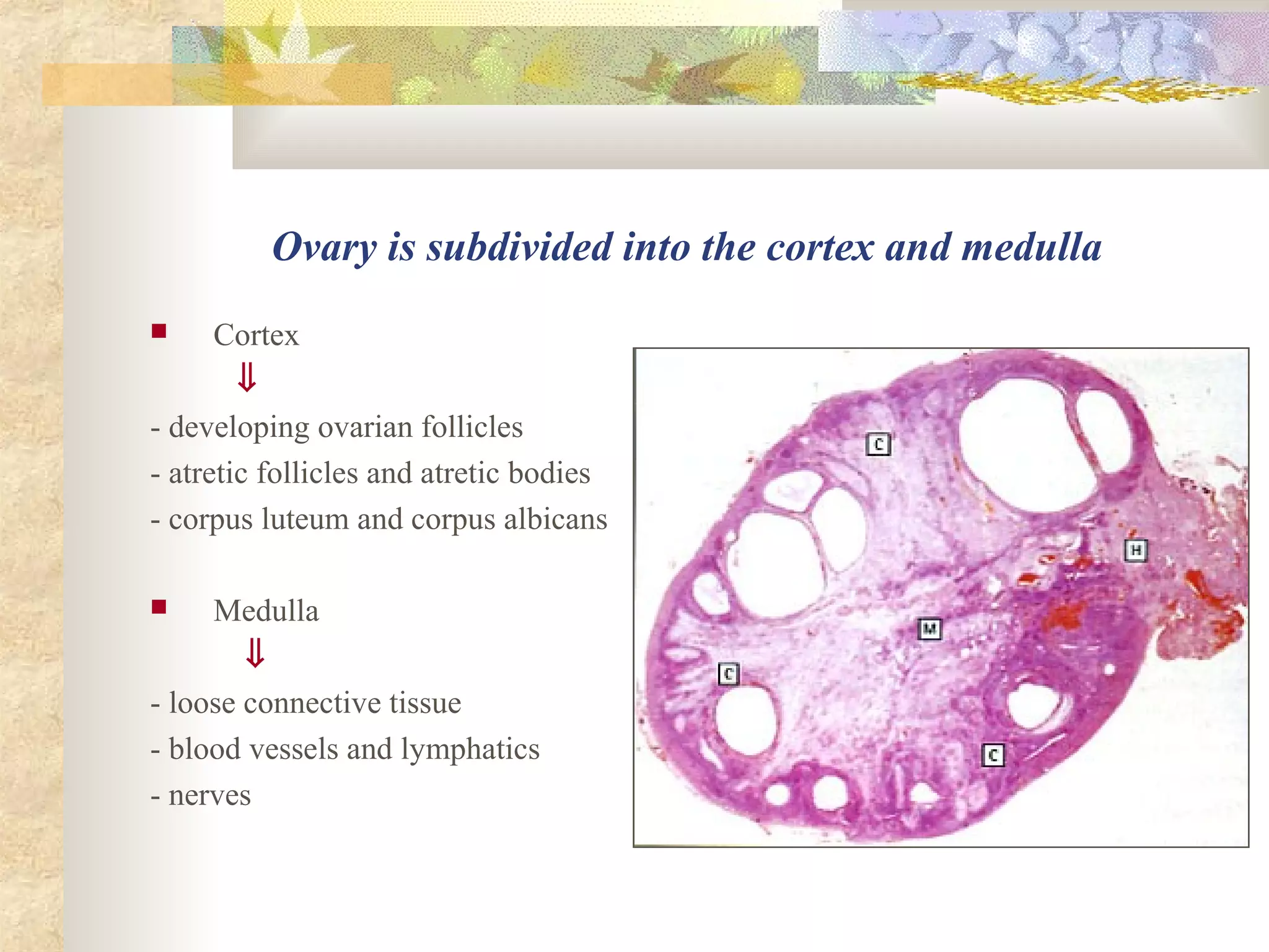
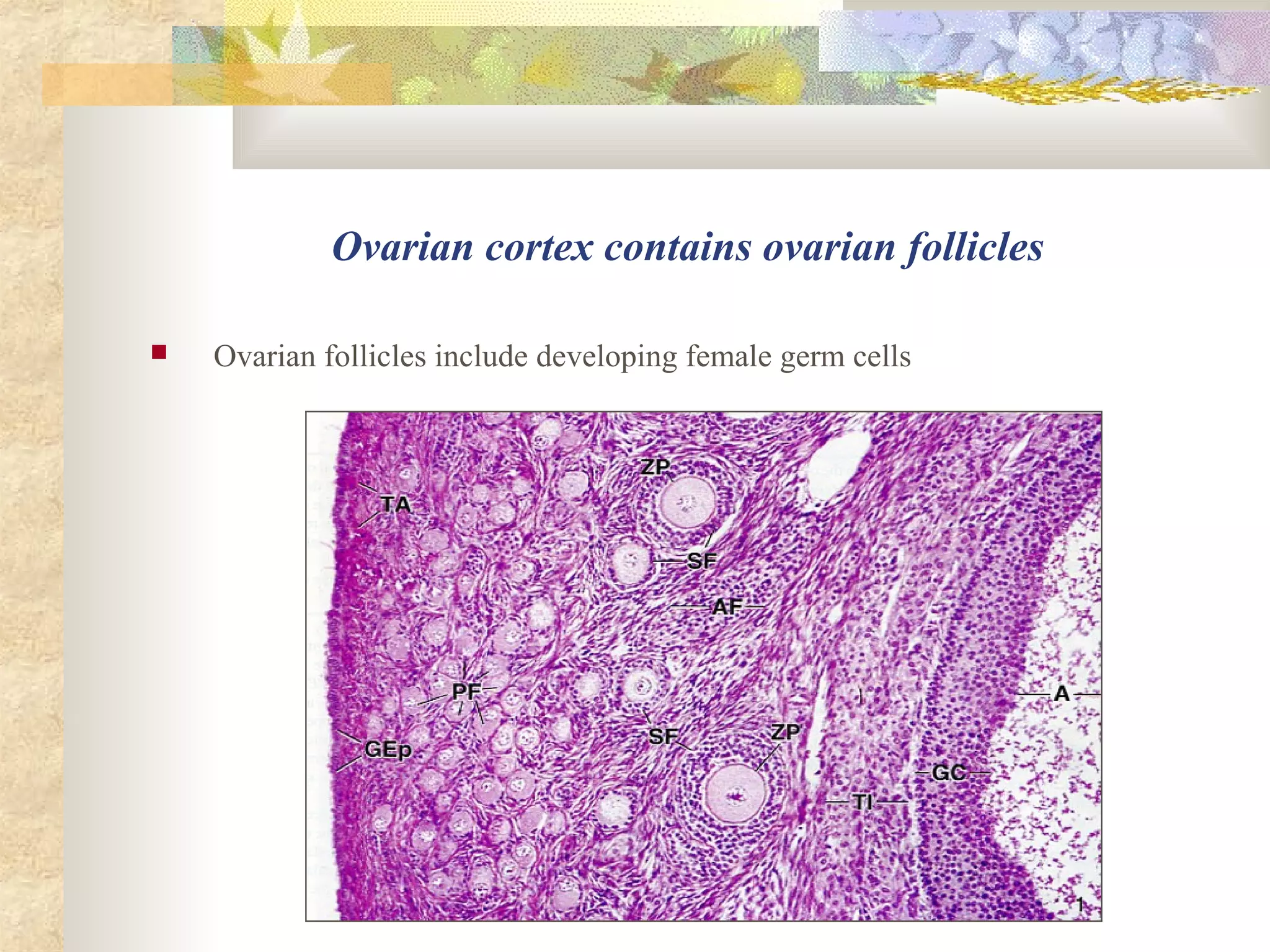
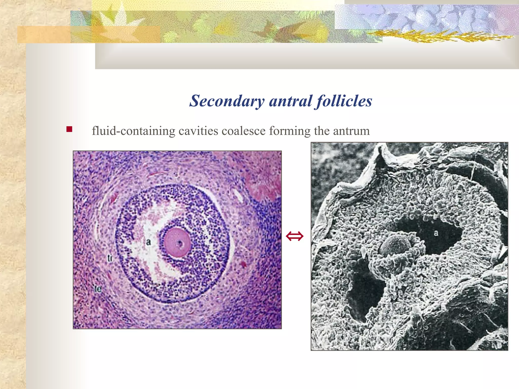
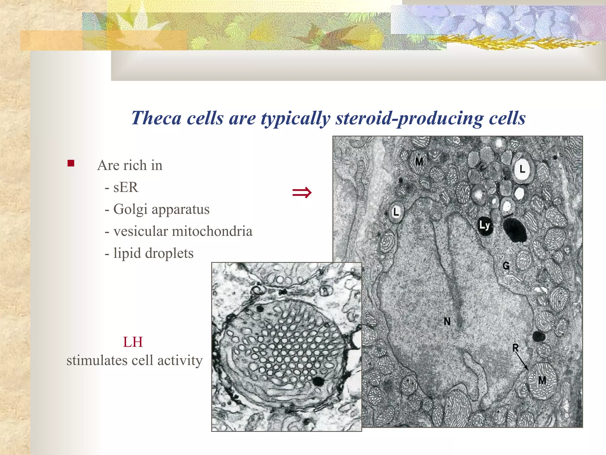
Female Reproductive System I
Female reproductive system
 Ovaries
 Oviducts
 Uterus
 Vagina
 External genitalia
Ovary (female sex gland, female gonad)
 Functions
- female gamete production – ovogenesis
- female sex hormone secretion
⇓
estrogens
progesterone
Ovary is almond-shaped organ
 is enclosed by the tunica albuginea
 is covered with the germinal epithelium
⇓
Ovary is subdivided into the cortex and medulla
 Cortex
⇓
- developing ovarian follicles
- atretic follicles and atretic bodies
- corpus luteum and corpus albicans
 Medulla
⇓
- loose connective tissue
- blood vessels and lymphatics
- nerves
Ovarian cortex contains ovarian follicles
 Ovarian follicles include developing female germ cells
Ovogenesis is a process of the ovum development
 Includes 3 stages
- stage of proliferation
- stage of growth
- stage of maturation
Ovogenesis stage of proliferation
 is represented by oogonia (2n, 2c) proliferating by mitotic divisions
⇑
oogonia in the embryonic ovary
Oogonium proliferation occurs only in the fetal period
 oogonia are converted to the primary oocytes before birth
⇔
Ovogenesis stage of growth
 is represented by primary oocytes (2n, 4c)
 is subdivided into
- small growth
- large growth
Small growth begins in the embryogenesis
 primary oocytes enter the prophase
of the first meiotic division
 prophase does not complete until puberty
 oocytes remain in suspended prophase
called the dictyotene for several years
Large growth begins at puberty
 lasts for two weeks until before ovulation
 oocytes considerably enlarge in size
⇓
from 30µm to 150 µm
 oocytes synthesize and accumulate
⇓
- organelles
- cortical granules
- yolk granules
⇒
Large growth is accompanied by folliculogenesis
⇒
⇒
0vogenesis stage of maturation includes meiosis
 primary oocyte (2n, 4c)
⇓
1st
meiotic division
⇓
 secondary oocyte (n, 2c)
⇓
2nd
meiotic division
⇓
 ootida (n, c)
First meiotic division completes before ovulation
 second meiotic division
⇓
- begins at ovulation
- is arrested at metaphase
- is completed in fertilization
Female meiotic division is unequal
 only one cell receives almost all the cytoplasm
 polar bodies receive minimal cytoplasm and are nonfunctional cells
Ovarian follicle
 is the oocyte surrounded by envelopes
 provides the microenvironment for developing oocyte
Folliculogenesis
 Includes stages
⇓
- primordial follicle
- primary follicle
- secondary follicle
- tertiary follicle
Primordial follicle
 Consists of
- primary oocyte in the small growth
- a single layer of flat follicular cells
Primordial follicle primary oocyte
 is arrested in the meiotic dictyotene
 measures about 30 µm in diameter
Primordial follicles predominate in the ovary
 are located in the cortex periphery just beneath the tunica albuginea
⇒
Primary follicle
 Consists of
- primary oocyte
- zona pellucida
- a single layer of cuboidal follicular cells
Primary follicle oocyte
 Primary oocyte
- enters the large growth
- becomes 50 to 80 µm in diameter
⇔
Zona pellucida
 is homogeneous acidophilic gel-like layer
 consists of glycosaminoglycans and glycoproteins
 is secreted by oocyte and follicular cells
⇔
Secondary (growing) follicle
 Consists of
- primary oocyte in the large growth
- zona pellucida
- several layers of follicular cells
- connective tissue theca folliculi
⇔
Membrana granulosa or stratum granulosum
 is stratified follicular epithelium
 follicular cells are identified as the granulosa cells
Stratum granulosum results from cell proliferation
estrogens stimulate granulosa cell proliferation
⇒
Follicular fluid or liquor folliculi
 appears when the stratum granulosum reaches
a thickness of 6 to 12-cell layers
 is secreted by the granulosa cells
FSH stimulates fluid secretion
Secondary antral follicles
 fluid-containing cavities coalesce forming the antrum
⇔
Granulosa cell endocrine function
 Granulosa cells
- convert androgens to estrogens under
FSH stimulation
- release estrogens to fluid and blood
Theca folliculi
 Is subdivided into
- theca interna – loose connective tissue
- theca externa – more dense connective tissue
Theca interna
 Contains
- rich network of small vessels
- theca cells
⇔
⇑
theca cells arise from mesenchyme
Theca cells are typically steroid-producing cells
 Are rich in
- sER
- Golgi apparatus
- vesicular mitochondria
- lipid droplets
⇒
LH
stimulates cell activity
Theca cells produce androgens under LH stimulation
 Androgens
are transported to the stratum granulosum
 Granulosa cells
- convert androgens into estrogens
- release estrogens into the fluid and blood
⇑
⇑
Growing follicles are estrogen-producing structures
 Estrogen blood level ⇒
- increases as the follicles grow
- attains the maximal size before ovulation
Hormonal regulation of ovarian follicle endocrine function
 LH stimulates the theca cells
- to produce androgens
 FSH stimulates the granulosa cells
- to convert androgens to estrogens
- to release estrogens into the blood
 Estrogens through feed-back loop
- inhibit FSH release
- activate LH production
Tertiary follicle (mature follicle or Graafian vesicle)
 represents the final stage of folliculogenesis
 is preovulatory follicle
 measures 10 mm and more in diameter
 extends through the full cortex thickness
 bulges on the ovarian surface
Tertiary follicle oocyte
 reaches its final size – about 150 µm in diameter
 resumes the first meiotic division
 becomes secondary oocyte
Tertiary follicle oocyte is acentrically positioned
 on the cumulus oophorus
 is surrounded by envelopes
- zona pellucida
- corona radiata
cumulus oophorus ⇑
Tertiary follicle envelopes
 Stratum granulosum
- becomes thin
- forms the cumulus oophorus
 Antrum
- increases in size
 Theca folliculi
- is more prominent
Ovulation
 is the release of the secondary oocyte
with zona pellucida and corona radiata
from the Graafian follicle ⇒
 occurs on the 14th
day of the large growth
Ovulating oocyte
 begins the second meiotic division
 progresses only to the metaphase
 division is arrested at the metaphase II
 secondary oocyte at the metaphase II
leaves the ovary in ovulation ⇒
Stigma formation
 Stigma ruptures, forming a small gap in
- germinal epithelium
- ovarian capsule
- wall of the Graafian follicle
⇓
secondary oocyte with envelopes leaves the ovary
Ovulation mechanisms
 follicular fluid increases in volume and pressure
 proteolytic enzymes
- lyse the stigma and follicular wall
- separate the oocyte-cumulus complex
 theca externa myofibroblasts contract
the oocyte leaves the ovary ⇒
Ovulation is hormonal-mediated process
 is induced by the gonadotropin maximal
blood concentration ⇒
⇓
LH peak (surge) – 12 hours before ovulation
FSH peak (surge) – 36 hours before ovulation
Ovulated oocyte enters the oviduct
 Oviduct infundibulum fimbriae
 cover the ovarian surface
 direct the oocyte into the oviduct
 prevent it to enter the peritoneal cavity
Corpus luteum or yellow body
 develops in the place of collapsed follicle
after ovulation
Bleeding from the capillaries into the follicular lumen
 leads to the corpus hemorrhagicum formation with a central clot
 connective tissue invades the former follicular cavity
⇔
Corpus luteum formation – luteinization
 Stages of
 vascularization and proliferation
 glandular metamorphosis
 secretion
 involution
Stage of vascularization and proliferation
 granulosa basement membrane is destroyed
 capillaries enter the stratum granulosum and form a rich vascular network
 granulosa cells and theca cells proliferate
Stage of glandular metamorphosis
 granulosa cells and theca-cells differentiate and become the lutein cells
 increase in size and accumulate
- sER
- mitochondria with vesicular cristae
- Golgi apparatus
- lipid droplets
- pigment lipochrome
 lipochrome imparts to lutein cells a yellow appearance in fresh preparation
Granulosa lutein cells
 are derived from the granulosa cells
 are pale and large – 30 µm in diameter
 are centrally located
⇔
Theca lutein cells
 are derived from the theca-cells
 are darker and smaller – 15 µm in diameter
 are peripherally located
⇔
Corpus luteum development
 lasts from 2 to 3 days after ovulation
 is controlled by LH
Stage of secretion
 Corpus luteum secretes progesterone
⇓
- prepares female organs (the endometrium, mammary glands) for pregnancy
- inhibits ovarian follicle development
 secretion is regulated by LTH (prolactin)
Corpus luteum types
 Corpus luteum of pregnancy
 develops if fertilization and implantation occur
 attains a size of 2 to 3 cm
 exists about 6 months
 Corpus luteum of menstruation
 develops if fertilization do not occur
 is small in size
 exists about 12 days
Stage of involution occurs after pregnancy or menstruation
 lutein cells die by apoptosis
 cell remnants are phagocytosed by macrophages
 connective tissue corpus albicans is formed
⇔
Atresia is a process of ovarian follicle degeneration
 primordial and primary follicles degenerate and disappear,
leaving no trace of their existence
Atresia of secondary and tertiary follicles
 results in the atretic body formation
⇒ ⇒ ⇒
⇑
Stratum granulosum changes appear in the first place
 granulosa is infiltrated by neutrophils and macrophages
 granulosa cells are sloughed into the antrum
 connective tissue and blood vessels invade
the granulosa and antrum
Oocyte degeneration occurs for the second time
 oocyte undergoes apoptosis
 zona pellucida becomes folded and is broken down
 their remnants are phagocytozed by macrophages
Theca-cells proliferate and differentiate
 become the atretic body cells
 produce steroid hormones, mainly estrogens
⇔
⇑
atretic body contains rich capillary network
Glassy membrane is a characteristic feature of the atretic bodies
 is the former granulosa basal membrane
 separates from follicular cells
 increases in thickness and becomes folded
the glassy membrane in an atretic follicle ⇒
The End
Thank you for attention!

Female reproductive system

  • 1.
  • 2.
    Female reproductive system Ovaries  Oviducts  Uterus  Vagina  External genitalia
  • 3.
    Ovary (female sexgland, female gonad)  Functions - female gamete production – ovogenesis - female sex hormone secretion ⇓ estrogens progesterone
  • 4.
    Ovary is almond-shapedorgan  is enclosed by the tunica albuginea  is covered with the germinal epithelium ⇓
  • 5.
    Ovary is subdividedinto the cortex and medulla  Cortex ⇓ - developing ovarian follicles - atretic follicles and atretic bodies - corpus luteum and corpus albicans  Medulla ⇓ - loose connective tissue - blood vessels and lymphatics - nerves
  • 6.
    Ovarian cortex containsovarian follicles  Ovarian follicles include developing female germ cells
  • 7.
    Ovogenesis is aprocess of the ovum development  Includes 3 stages - stage of proliferation - stage of growth - stage of maturation
  • 8.
    Ovogenesis stage ofproliferation  is represented by oogonia (2n, 2c) proliferating by mitotic divisions ⇑ oogonia in the embryonic ovary
  • 9.
    Oogonium proliferation occursonly in the fetal period  oogonia are converted to the primary oocytes before birth ⇔
  • 10.
    Ovogenesis stage ofgrowth  is represented by primary oocytes (2n, 4c)  is subdivided into - small growth - large growth
  • 11.
    Small growth beginsin the embryogenesis  primary oocytes enter the prophase of the first meiotic division  prophase does not complete until puberty  oocytes remain in suspended prophase called the dictyotene for several years
  • 12.
    Large growth beginsat puberty  lasts for two weeks until before ovulation  oocytes considerably enlarge in size ⇓ from 30µm to 150 µm  oocytes synthesize and accumulate ⇓ - organelles - cortical granules - yolk granules ⇒
  • 13.
    Large growth isaccompanied by folliculogenesis ⇒ ⇒
  • 14.
    0vogenesis stage ofmaturation includes meiosis  primary oocyte (2n, 4c) ⇓ 1st meiotic division ⇓  secondary oocyte (n, 2c) ⇓ 2nd meiotic division ⇓  ootida (n, c)
  • 15.
    First meiotic divisioncompletes before ovulation  second meiotic division ⇓ - begins at ovulation - is arrested at metaphase - is completed in fertilization
  • 16.
    Female meiotic divisionis unequal  only one cell receives almost all the cytoplasm  polar bodies receive minimal cytoplasm and are nonfunctional cells
  • 17.
    Ovarian follicle  isthe oocyte surrounded by envelopes  provides the microenvironment for developing oocyte
  • 18.
    Folliculogenesis  Includes stages ⇓ -primordial follicle - primary follicle - secondary follicle - tertiary follicle
  • 19.
    Primordial follicle  Consistsof - primary oocyte in the small growth - a single layer of flat follicular cells
  • 20.
    Primordial follicle primaryoocyte  is arrested in the meiotic dictyotene  measures about 30 µm in diameter
  • 21.
    Primordial follicles predominatein the ovary  are located in the cortex periphery just beneath the tunica albuginea ⇒
  • 22.
    Primary follicle  Consistsof - primary oocyte - zona pellucida - a single layer of cuboidal follicular cells
  • 23.
    Primary follicle oocyte Primary oocyte - enters the large growth - becomes 50 to 80 µm in diameter ⇔
  • 24.
    Zona pellucida  ishomogeneous acidophilic gel-like layer  consists of glycosaminoglycans and glycoproteins  is secreted by oocyte and follicular cells ⇔
  • 25.
    Secondary (growing) follicle Consists of - primary oocyte in the large growth - zona pellucida - several layers of follicular cells - connective tissue theca folliculi ⇔
  • 26.
    Membrana granulosa orstratum granulosum  is stratified follicular epithelium  follicular cells are identified as the granulosa cells
  • 27.
    Stratum granulosum resultsfrom cell proliferation estrogens stimulate granulosa cell proliferation ⇒
  • 28.
    Follicular fluid orliquor folliculi  appears when the stratum granulosum reaches a thickness of 6 to 12-cell layers  is secreted by the granulosa cells FSH stimulates fluid secretion
  • 29.
    Secondary antral follicles fluid-containing cavities coalesce forming the antrum ⇔
  • 30.
    Granulosa cell endocrinefunction  Granulosa cells - convert androgens to estrogens under FSH stimulation - release estrogens to fluid and blood
  • 31.
    Theca folliculi  Issubdivided into - theca interna – loose connective tissue - theca externa – more dense connective tissue
  • 32.
    Theca interna  Contains -rich network of small vessels - theca cells ⇔ ⇑ theca cells arise from mesenchyme
  • 33.
    Theca cells aretypically steroid-producing cells  Are rich in - sER - Golgi apparatus - vesicular mitochondria - lipid droplets ⇒ LH stimulates cell activity
  • 34.
    Theca cells produceandrogens under LH stimulation  Androgens are transported to the stratum granulosum  Granulosa cells - convert androgens into estrogens - release estrogens into the fluid and blood ⇑ ⇑
  • 35.
    Growing follicles areestrogen-producing structures  Estrogen blood level ⇒ - increases as the follicles grow - attains the maximal size before ovulation
  • 36.
    Hormonal regulation ofovarian follicle endocrine function  LH stimulates the theca cells - to produce androgens  FSH stimulates the granulosa cells - to convert androgens to estrogens - to release estrogens into the blood  Estrogens through feed-back loop - inhibit FSH release - activate LH production
  • 37.
    Tertiary follicle (maturefollicle or Graafian vesicle)  represents the final stage of folliculogenesis  is preovulatory follicle  measures 10 mm and more in diameter  extends through the full cortex thickness  bulges on the ovarian surface
  • 38.
    Tertiary follicle oocyte reaches its final size – about 150 µm in diameter  resumes the first meiotic division  becomes secondary oocyte
  • 39.
    Tertiary follicle oocyteis acentrically positioned  on the cumulus oophorus  is surrounded by envelopes - zona pellucida - corona radiata cumulus oophorus ⇑
  • 40.
    Tertiary follicle envelopes Stratum granulosum - becomes thin - forms the cumulus oophorus  Antrum - increases in size  Theca folliculi - is more prominent
  • 41.
    Ovulation  is therelease of the secondary oocyte with zona pellucida and corona radiata from the Graafian follicle ⇒  occurs on the 14th day of the large growth
  • 42.
    Ovulating oocyte  beginsthe second meiotic division  progresses only to the metaphase  division is arrested at the metaphase II  secondary oocyte at the metaphase II leaves the ovary in ovulation ⇒
  • 43.
    Stigma formation  Stigmaruptures, forming a small gap in - germinal epithelium - ovarian capsule - wall of the Graafian follicle ⇓ secondary oocyte with envelopes leaves the ovary
  • 44.
    Ovulation mechanisms  follicularfluid increases in volume and pressure  proteolytic enzymes - lyse the stigma and follicular wall - separate the oocyte-cumulus complex  theca externa myofibroblasts contract the oocyte leaves the ovary ⇒
  • 45.
    Ovulation is hormonal-mediatedprocess  is induced by the gonadotropin maximal blood concentration ⇒ ⇓ LH peak (surge) – 12 hours before ovulation FSH peak (surge) – 36 hours before ovulation
  • 46.
    Ovulated oocyte entersthe oviduct  Oviduct infundibulum fimbriae  cover the ovarian surface  direct the oocyte into the oviduct  prevent it to enter the peritoneal cavity
  • 47.
    Corpus luteum oryellow body  develops in the place of collapsed follicle after ovulation
  • 48.
    Bleeding from thecapillaries into the follicular lumen  leads to the corpus hemorrhagicum formation with a central clot  connective tissue invades the former follicular cavity ⇔
  • 49.
    Corpus luteum formation– luteinization  Stages of  vascularization and proliferation  glandular metamorphosis  secretion  involution
  • 50.
    Stage of vascularizationand proliferation  granulosa basement membrane is destroyed  capillaries enter the stratum granulosum and form a rich vascular network  granulosa cells and theca cells proliferate
  • 51.
    Stage of glandularmetamorphosis  granulosa cells and theca-cells differentiate and become the lutein cells  increase in size and accumulate - sER - mitochondria with vesicular cristae - Golgi apparatus - lipid droplets - pigment lipochrome  lipochrome imparts to lutein cells a yellow appearance in fresh preparation
  • 52.
    Granulosa lutein cells are derived from the granulosa cells  are pale and large – 30 µm in diameter  are centrally located ⇔
  • 53.
    Theca lutein cells are derived from the theca-cells  are darker and smaller – 15 µm in diameter  are peripherally located ⇔
  • 54.
    Corpus luteum development lasts from 2 to 3 days after ovulation  is controlled by LH
  • 55.
    Stage of secretion Corpus luteum secretes progesterone ⇓ - prepares female organs (the endometrium, mammary glands) for pregnancy - inhibits ovarian follicle development  secretion is regulated by LTH (prolactin)
  • 56.
    Corpus luteum types Corpus luteum of pregnancy  develops if fertilization and implantation occur  attains a size of 2 to 3 cm  exists about 6 months  Corpus luteum of menstruation  develops if fertilization do not occur  is small in size  exists about 12 days
  • 57.
    Stage of involutionoccurs after pregnancy or menstruation  lutein cells die by apoptosis  cell remnants are phagocytosed by macrophages  connective tissue corpus albicans is formed ⇔
  • 58.
    Atresia is aprocess of ovarian follicle degeneration  primordial and primary follicles degenerate and disappear, leaving no trace of their existence
  • 59.
    Atresia of secondaryand tertiary follicles  results in the atretic body formation ⇒ ⇒ ⇒ ⇑
  • 60.
    Stratum granulosum changesappear in the first place  granulosa is infiltrated by neutrophils and macrophages  granulosa cells are sloughed into the antrum  connective tissue and blood vessels invade the granulosa and antrum
  • 61.
    Oocyte degeneration occursfor the second time  oocyte undergoes apoptosis  zona pellucida becomes folded and is broken down  their remnants are phagocytozed by macrophages
  • 62.
    Theca-cells proliferate anddifferentiate  become the atretic body cells  produce steroid hormones, mainly estrogens ⇔ ⇑ atretic body contains rich capillary network
  • 63.
    Glassy membrane isa characteristic feature of the atretic bodies  is the former granulosa basal membrane  separates from follicular cells  increases in thickness and becomes folded the glassy membrane in an atretic follicle ⇒
  • 64.
    The End Thank youfor attention!