Downloaded 102 times


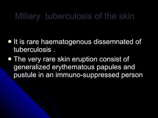


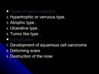


This document summarizes different types of cutaneous tuberculosis, their characteristics, and treatment approaches. The main types discussed are primary tuberculosis of the skin, miliary tuberculosis of the skin, lupus vulgaris, scrofuloderma, tuberculosis verrucosa cutis, tuberculosis cutis orificialis, and tuberculides. Lupus vulgaris is described as the most common form, presenting as reddish-brown plaques on the head and neck that can cause scarring and deformity. Treatment involves a combination of anti-tuberculosis medications like rifampicin, isoniazid, and ethambutol, as well as surgical excision for some small lesions.


![GRANULOMATOUS LESION OF SKIN2[299] - Read-Only (1).pptx](https://cdn.slidesharecdn.com/ss_thumbnails/granulomatouslesionofskin2299-read-only1-240725163335-974d6cd5-thumbnail.jpg?width=640&height=640&fit=bounds)


