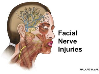
Facial nerve injuries symptoms, treatment
•Download as PPTX, PDF•
27 likes•5,811 views

The facial nerve carries motor, secretory and sensory fibers. It originates in the pons and has crossed and uncrossed fibers in the facial nucleus. Damage to the facial nerve can occur at different locations and causes varying symptoms depending on if it is an upper or lower motor neuron lesion. Treatment options depend on the severity and chronicity of the injury and include medications, physiotherapy, nerve grafting and transfers. Microsurgical techniques may allow for primary repair of acute injuries or cross facial nerve grafts for more longstanding paralysis.

Recommended
More Related Content
What's hot
What's hot (20)
Similar to Facial nerve injuries symptoms, treatment
Similar to Facial nerve injuries symptoms, treatment (20)
Recently uploaded
Recently uploaded (20)
Facial nerve injuries symptoms, treatment
- 2. The facial nerve - cranial nerve VII carries : (1) motor (2) secretory, (3) afferent fibers from the anterior two thirds of the tongue. It originates in the facial nucleus, which is located at the caudal pontine area. and the Corticobulbar fibers from the precentral gyrus (frontal lobe) project to the facial nucleus, with most crossing to the contralateral side. As a result, crossed and uncrossed fibers are found in the nucleus. the facial nucleus can be divided into two parts: (1) the upper part, which receives corticobulbar projections bilaterally and later courses to the upper parts of the face, including the forehead, (2) the lower part, the predominantly crossed projections of which supply innervation to lower facial muscles : stylohyoid, posterior belly of digastric, buccinator, and platysma “The facial nerve”
- 3. “Intra Cranial part” The portion of the nerve from the brainstem to the internal auditory canal Carries preganglionic parasympathetic fibers and special afferent sensory fibers Important branches of facial nerve in this part (1)Greater superfacial petrosal nerve : Carries parasympathetic fibers to lacrimal gland and glands of the nose and palate. (2)Nerve to Stapedius muscle (3)Chorda tympani : carries parasympathetics to the submandibular and sublingual glands & Taste to anterior 2/3 of the tongue . “Intra Temporal part”
- 4. Main trunk 15-20 mm : (1)Give branches to the posterior belly of the digastric and stylohyoid muscles. (2)Postauricular to occipitofrontalis muscles The facial nerve branches off to smaller nerves and muscles that go to 5 different parts of the face. Therefore, when the nerve is damaged those smaller veins are not supplied with enough blood for circulation which is necessary for muscles on the different areas of the face to move. Each nerve branch affects the movement of different muscles. “Extra Cranial part”
- 5. Branching of the extracranial segments in the parotid gland that splitting it into a superficial and deep lobe : 3. Buccal Branch - (Infraorbital Branches): This Nerve Branch affects the Cheek and Above the Mouth Muscles. 4. Marginal Mandibular Branch: This Nerve Branch affects the Chin Muscles. 5. Cervical Branch: This Nerve Branch some of the Neck Muscles. 1. Temporal Branch - (Frontal Branch): This Nerve Branch affects the muscles in the Forehead. 2. Zygomatic Branch - (Malar Branches): This Nerve Branch affects the Upper Cheek. 1&2. Temporal & Zygomatic Branch: Together these Nerve Branches affect the muscles control opening and closure of the Eye.
- 6. • Endoneurium – Surrounds each axon – Adherent to Schwann cell layer – Vital for regeneration • Perineurium – Encases endoneural tubules – Tensile strength – Barrier to infection • Epineurium (nerve sheath) – Outermost layer – Houses vasa nervosum for nutrition “component of nerve fibers”
- 7. • Nerve injury is most serious complication that may occur during oral surgical procedures especially when we are damaging large nerve branches such as during: • dental injections , RCT , insertion of dental implants , extraction of teeth & other surgical treatments ...etc. “Nerve Injuries”
- 8. “Sunderland Nerve Injury Classification” Class I (Neuropraxia): -Axon remain intact -Conduction block caused by cessation(stoping) of axoplasmic flow -Full recovery Class II (Axonotmesis): -Axons are disrupted -Endoneural tube still intact -Full recovery expected Class III (Neurotmesis): -Neural tube is disrupted Injury to endoneurium or myelin sheath -Poor prognosis -If regeneration occurs, high incidence of synkinesis (involuntary movement of muscles associated with voluntary movement other muscles ) Class IV (Partial transection) -Epineurium remains intact -Perineurium, endoneurium, and axon disrupted -Poor functional outcome with higher risk for synkinesis. Class V (Complete transection) -Complete disruption -Little chance of regeneration -Risk of neuroma formation
- 9. “Causes of Facial nerve paralysis” supranuclear lesions UMN : • Congenital abnormalities, stroke , malignancies, trauma , vascular conditions and other causes . • only lower part of the opposite side of the face is paralyzed. • The upper part with the frontalis and orbicularis oculi escapes due to bilateral representation in the cerebral cortex. infranuclear lesions LMN : • Malignancy (parotid gland as well as tumors of adjacent structures) , trauma, infections, Bell’s palsy, osteopetrosis and iatrogenic causes . • the whole of the face of the same side gets paralyzed.
- 10. LMN LESIONS UMN LESIONS Only lower 2/3 rd of the facial muscles are affected. Mid face is paralysed. Eye brow’s can move normally. Totally half side of the is affected. Half of the Mid face is only paralysed. Eye brow’s can’t move normally.
- 11. “Signs and symptoms of facial nerve paralysis” The symptoms according to the level of injury of facial nerve. At internal auditory meatus: loss of lacrimation, stapedial reflex, taste from most of anterior two-third of tongue, lack of salivation and paralysis of muscles of facial expression. Below geniculate ganglion: loss of stapedial reflex, taste from anterior two third of tongue, lack of salivation and paralayis of facial expression muscles. Region below stylomastoid foramen paralysis of facial expression muscles. Sign & symptoms : unilateral facial weakness loss of taste decreased salivation and tear secretion Hyperacusis-A heightened sensitivity to some sounds. Facial palsy : caused by trauma, infection, tumour to the facial nerve .
- 12. “Dental Etiology” 1. During nerve block of IAN & Mental nerve ( deep dental injection ) . 2. While creating incision extend to mental formen & lingual vestibuler fold 3. during incision at the alveolar ridge of edentolous pt whose mental foramen located superficially due to bone resoption 4. during excessive flap retraction 5. when bone near the nerve is excessively heated ,if the surgical handpiece used without coolent ( water or saline solution ) 6. in case of removel impacted tooth , root & root tips that are deep in the bone which is near the nerve. 7. during perforation & fracture of lingual cortical plate during sectioning of the roots and crown of impacted 3rd moler. 8. when a bur enters the mandibuler canal , during sectioning. 9. during displacement of a root tip inside the mandibuler canal during extraction attempt. 10. during cleaning of periapical lesion oa posterior teeth that are in direct contact with mand. canal . 11.or by chance Suturing of the Nerve . 12.during putting implant 13.during endodontic treatment because of proximity of the tooth to IAN by over-instrumentation or overfilling or irrigation.
- 13. “Facial nerve paralysis” BELL’S palsy : It is the commonest type of facial palsy. It is the major cause of the acute facial nerve paralysis. It affects totally half side of the face due to the LMN Lesion. Its idiopathic Its due to the inflammation of the facial nerve. The inflammation prevents nerve from sending correct signals to brain &facial muscles. Sign & symptoms : droopy eyelid, drooping corner of the mouth,unilateral facial,weakness loss of taste decreased salivation and tear secretion
- 14. “DIFFERENCE BETWEEN FACIAL PALSY&BELL’S PALSY” FACIAL PALSY BELL’S PALSY 1)Cause can be known (infection,trauma, tumour). 2)Permanent(lasts for years to life). 3)need surgical treatment. 4)Site of affection depends upon UMN&LMN Lesions. 1)It is idiopathic(may de velop suddenly). 2)Temporary(permanent cure with in 3 months in 90% of cases). 3)Without treatment or surgery regains facial function. 4)It is mainly due to LMN Lesions.half side of the face is totally affected.
- 15. Herpes zoster virus reactivation of virus within dorsal root ganglion of facial nerve is associated with vesicles affecting ear canal. Symptoms 1)ear pain 2)vesicles 3)hearing loss 4)vertigo Treatment 1)anti viral 2)steroids(corticosteroids) Otitis media inflammation of the middle ear due to infections can spread to facial nerve &inflame it causing compression. Symptoms 1)ottorrhoea(discharge). 2)otalgia(no ear pain). Treatment –myringotomy(incision to tympanic membrane) “INFECTION”
- 16. 1)fractures of temporal bone due to injury in accidents. 2)birth injury to the facial nerve at the time of delivery due to application of fore ceps. reason :it remains unprotected after its exit through stylomastoid foramen investigation CT Scan “TRAUMA” “TUMOR” The bells palsy may be due to tumour’s which compress the the nerve alo BELL’S PALSY DUE TO COMPRESSION investigation 1)Tomography. 2)MRI(to locate tumour) 3)CT Scan
- 17. Used to assess the degree of electrical dysfunction and to pinpoint the site of injury also it help to determine treatment to predict recovery of function – partial paralysis is a much better prognosis than total paralysis “Facial Nerve Testing and Diagnosis ” Topographictests Tests function of specific facial nerve branches Do not predict potential recovery of function Rarely utilized today Electrodiagnostictests Utilize electrical stimulation to assess function Most commonly used today Magnetic stimulation of intra-cranial facial nerve CT scan temporal bone: for progressive palsy MRI brain Surgical exploration Tests for faAsk the patient to show his teeth. Ask the patient to puff his cheeks. Ask the patient to close his eyes against resistance. Ask the patient to lift his eyebrows.cial palsy: Ask the patient to lift his eyebrows
- 18. Depends on the age of patient , type of the damage , time that elapsed till the management of injury , correct treatment “Prognosis” “Treatment” • No treatment required for type 1 & 2 unless if there foreign body or root tip compressing on the nerve we just prescribe analgesic & vitamin B to restore the sensation . • Treatment of the neaurotmesis is grafting to replace the injured part or suturing . • For bell’s palsy is often treated with the corticosteriod
- 19. “Treatment AND Medication” • Oral antivirals - Acyclovir • Corticosteroids • Eye protection • Follow progression with serial exams • Physiotherapy • If the patient is seen within 2 to 3 weeks of onset of symptoms-tab. Prednisolone in doses of 1mg/kg/d for 10 to 14 days has been recommended with a gradual tapering. • Vitamins B1, B6, B12 may be administered. • If pt is seen after 3-4 weeks, then steroid therapy is of no use. “SURGICAL TREATMENT MODALITIES” • Nerve decompression - Internally or externally • Nerve anastomosis • Nerve grafting
- 20. A. Acute (< 3 wks) 1. Nerve exploration/decompression 2. Nerve repair a. Primary anastomosis b. Cable grafting i. Great auricular nerve ii. Sural nerve B. Intermediate (3 wks- 2 yrs) 1. Nerve transfer a. Hypoglossal-facial b. Spinal accessory-facial c. Masseteric-facial 2. Cross face nerve grafting using sural nerve C. Chronic (>2 yrs) 1. Muscle transfers a. Temporalis b. Masseter c. Digastrics 2. Free muscle flaps/ microneurovascular transfer a. Gracilis b. Latissimus dorsi c. Serratus anterior d. Pectoralis minor D. Static procedures/ancillary procedures (can be performed at any time period listed above) 1. Gold weight/spring implants 2. Slings 3. Lid procedures “SURGICAL TREATMENT MODALITIES”
- 21. Facial nerve repair is the most effective procedure to restore facial function in patients who have suffered nerve damage from an accident or during surgery. It involves microscopic repair of a nerve that has been cut. “Micro-neurological Surgery”
- 22. End-to-end anastomosis preferred No tension Extratemporal repair performed < 72 hrs of injury Most common methods Group fascicular repair Epineural repair “PRIMARY NERVE REPAIR” Group fascicular repair
- 23. 1. Parotidectomy incision extended into cervical crease ~ 2-3 cm below inferior border of mandible 2. Facial nerve identified and dissected distal to pes anserinus 3. Identify hypoglossal nerve a. SCM retracted posteriorly b. Dissect superiorly until posterior belly of digastic is identified c. Retract digastric superiorly and CN XII is found inferiorly. d. Hypoglossal is within 2-3 c m of main trunk of the facial nerve 4. Hypoglossal nerve is dissected anteriorly and medially into the tongue. 1. Transect distal to ansa hypoglossis 5. Facial nerve transected at the stylomastoid foramen 6. Anastomose nerves using 9-0 epineural suture. Hypoglossal-Facial Technique Hypoglossal Facial Nerve Transfer
- 24. “Treatment” Brow ptosis correction – direct brow lift,endoscopic brow lift. Eye lid weight placement – occuloplastic management for lagopthalmus. Static facial suspension – by using facial slings from zygomatic/temporalis arch to nasolabial fold & oral commisure. Extra nasal valve repair – facia lata sling from alar base to temporalis facia to open extra nasal valve. Cross Face Nerve Transplant(CFNT) – It is most advanced. It is making continuity between paralysed&normal facial nerves by means of bridge grafts.
- 25. 😇 😇 😇😇 😇