Mind Maps forMedicine 181
Neurology
Chapter 6
06
Bell’s palsy.......................................................................................................................................................................182
Delirium...........................................................................................................................................................................186
Epilepsy............................................................................................................................................................................190
Extradural haematoma (epidural haemorrhage)...................................................................................194
Guillain–Barré syndrome......................................................................................................................................196
Migraine and other causes of headache....................................................................................................198
Motor neurone disease..........................................................................................................................................202
Multiple sclerosis.......................................................................................................................................................204
Myasthenia gravis......................................................................................................................................................206
Neurofibromatosis....................................................................................................................................................208
Parkinsonism................................................................................................................................................................210
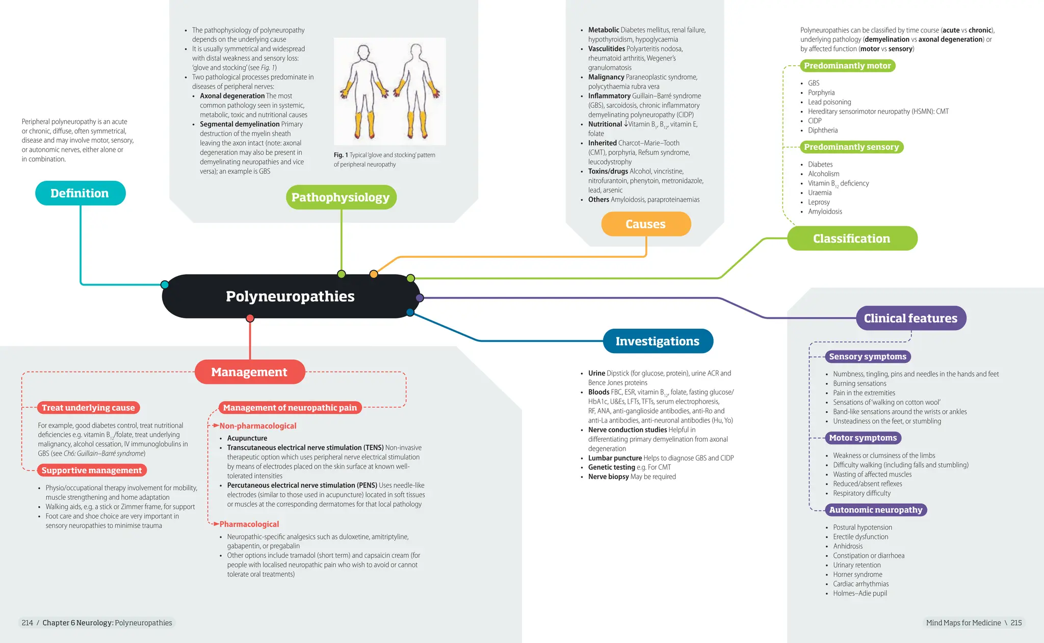
Polyneuropathies.......................................................................................................................................................214
Transient ischaemic attack...................................................................................................................................216
Stroke................................................................................................................................................................................218
Subarachnoid haemorrhage..............................................................................................................................222
Subdural haematoma.............................................................................................................................................224
2.
Notes
Bell’s palsy
Mind Mapsfor Medicine 183
182 / Chapter 6 Neurology: Bell’s palsy
Bell’s palsy
The diagnosis of Bell’s palsy is clinical and investigations
are not usually required but may be done to rule out other
causes or determine the severity of palsy:
• Serology Raised borrelia antibodies (in Lyme disease)
or varicella zoster virus antibodies (in Ramsay Hunt
syndrome)
• MRI brain To rule out stroke, space-occupying lesion
or MS
• Nerve conduction studies Predict delayed recovery
by showing axonal degeneration
• Schirmer’s tear test Shows reduced flow of tears on
the side of the palsy
• Stapedial reflex (audiological test) Absent if stapedius
muscle is affected
• Most people with Bell’s palsy begin to
recover, even without treatment, within
2–3 weeks (approx. 85%); complete recovery
usually occurs within 3–4 months
• If untreated, around 15% of patients have
permanent moderate to severe weakness
• Poor prognostic features include:
• Complete palsy or severe degeneration
(on electrophysiology)
• No signs of recovery by 3 weeks
• Age >60 years
• Severe pain
• Ramsay Hunt syndrome
• Associated with hypertension, diabetes or
pregnancy
Eye care
• ↓Tear production and difficulty closing
the eye puts people at risk of corneal
dryness, ulceration and even blindness;
appropriate eye care is vital
• Prescription of artificial tears and eye
lubricants should be considered
• Taping the eyelids shut at night is
recommended
• If the cornea remains exposed after
attempting to close the eyelid, urgent
referral to ophthalmology should be
considered
Steroids
• Should be prescribed for patients within
72h of onset of Bell’s palsy
• Prednisolone 1mg/kg or 60mg OD for
5 days then reducing by 10mg each day
for a further 5 days (10 days in total)
• Antivirals such as aciclovir are NO LONGER
recommended
Physiotherapy
‘Facial re-training’to improve facial motor
function may help but evidence of its
effectiveness is lacking.
Botulinum
Botulinum toxin can augment facial
symmetry.
Surgery
• Facial nerve decompression is an option for
patients with facial palsy not responding to
medical treatment
• If residual paralysis after 6–9 months,
referral to plastic surgery should be
considered for surgical nerve grafting
An acute, unilateral, partial or
complete paralysis of the face
due to idiopathic unilateral
lower motor neurone facial
nerve palsy. It is the most
common cause of facial nerve
palsy but it is often a diagnosis
of exclusion as other important
diagnoses such as stroke,
infections, parotid tumours,
middle ear disease should be
excluded first.
Definition
• Likely cause is ischaemic compression of
the facial nerve within the facial canal due
to inflammation
• Inflammation is most likely caused by a
viral infection (with herpes simplex virus
and varicella zoster virus being the likely
culprits) although the exact pathogenesis
remains unclear
• There may be a familial component
in recurrent cases, possibly due to
anatomical abnormality of the facial canal
Pathophysiology
Prognosis Management
Rapid onset (<72h):
• Unilateral sagging of the
mouth
• One-sided facial paralysis
(see Fig. 1)
• Drooling of saliva
• Speech difficulty
• Hyperacusis
• Altered taste (loss of taste
sensation in the anterior
two-thirds of tongue)
• Failure of eye closure: may
cause watery or dry eyes
Clinical features
Investigations
Epidemiology/risk factors
• Overall, it is relatively uncommon with
an incidence of approx. 20–30 people
per 100 000 each year
• Most commonly seen at age 15–60 years
• It is more common in people who are
diabetic, immunocompromised, obese,
hypertensive or have upper respiratory
conditions, or in pregnant women
Idiopathic
• Bell’s palsy (most common)
(see Fig. 1)
Infectious
• Ramsay Hunt syndrome
• Lyme disease
• Meningitis
• TB
• Other viruses: HIV, EBV, CMV
Intracranial lesions
• Stroke
• Brain tumours
• Multiple sclerosis (MS)
Systemic disease
• Diabetes mellitus
• Sarcoidosis
• Guillain–Barré syndrome (usually
bilateral facial nerve palsy)
• Rheumatoid arthritis and Sjögren
syndrome
• Vasculitides
ENT
• Acoustic neuroma
• Otitis media
• Parotid tumours
• Cholesteatoma
Trauma
• Basal skull fracture
• Forceps delivery
• Post acupuncture haematoma
• Diving (barotrauma)
Causes of facial nerve palsy
Fig. 1 Bell’s palsy showing right
facial paralysis when the patient
was asked to smile
3.
Notes
Bell’s palsy
Mind Mapsfor Medicine 185
184 / Chapter 6 Neurology: Bell’s palsy notes
Ramsay Hunt syndrome
Definition
Ramsay Hunt syndrome (herpes zoster oticus) is caused by the reactivation of
the varicella zoster virus in the geniculate ganglion of the 7th cranial nerve.
Epidemiology
It most commonly occurs in elderly people
(over 60 years) although it can affect all ages.
Clinical features
• Auricular pain (often first feature)
• Facial nerve palsy on affected side (as above)
• Vesicular rash around the ear (see Fig. 3)
• Vertigo and tinnitus
Management
• Oral corticosteroids: prednisolone regime as above
• Oral antivirals e.g. aciclovir
• Analgesia: paracetamol (± codeine) and NSAIDs are 1st line; tricyclic
antidepressants, gabapentin, pregabalin, and opioids are other options
• Eye care as above
Prognosis
• Recovery of facial nerve function is less likely than in Bell’s palsy
• The prognosis is excellent for younger and otherwise healthy patients
• Elderly people have increased risk of post-herpetic neuralgia, bacterial
infections and scarring
Neuroanatomy of the facial nerve
(7th cranial nerve)
• The facial nerve is largely motor in function, supplying the muscles
of facial expression
• In addition it has two major branches which arise during its intracranial
course through the facial canal of the petrous temporal bone:
• The chorda tympani, which carries taste from the anterior
two-thirds of the tongue
• Nerve to stapedius muscle, which has a damping effect to
protect the ear from loud noise
• Therefore damage to the facial nerve in the temporal bone
(e.g. in Bell’s palsy) causes hyperacusis and taste disturbance to the
anterior two-thirds of the tongue
• Within the parotid gland, the nerve terminates by splitting into five
extracranial branches which can be remembered by the mnemonic
‘Two Zebras Bit My Coccyx’: Temporal, Zygomatic, Buccal, Mandibular
and Cervical
Clinically distinguishing upper vs
lower motor neurone lesions that
cause facial weakness
• It is vital to be able to distinguish clinically facial weakness caused
by an upper (central) vs a lower (peripheral) motor neurone lesion
as this will significantly alter the management plan
• Upper motor neurone lesions, such as a stroke, cause contralateral face
weakness sparing the forehead, while lower motor neurone lesions,
such as a facial nerve injury, typically cause weakness involving the
whole ipsilateral face (see Fig. 2)
Fig. 2 The innervation to the muscles of the upper face originates on both sides of the brain, whereas the innervation to the muscles of the lower face is from the contralateral side of
the brain only; when the cortex is injured, e.g. in a stroke, there is weakness in the contralateral lower face only, therefore‘forehead sparing’; when the facial nerve is injured, there is
weakness in the ipsilateral upper and lower face
Loss of forehead &
brow movements
Preservation of
forehead & brow
movements
Loss of nasolabial
folds & drooping
of lower lip
Peripheral facial palsy Central facial palsy
Inability to close
eyes & drooping
of eyelids
Loss of nasolabial
folds & drooping
of lower lip
Fig. 3 Vesicular rash in Ramsay
Hunt syndrome
Bell’s palsy notes
4.
Notes
Delirium
Mind Maps forMedicine 187
186 / Chapter 6 Neurology: Delirium
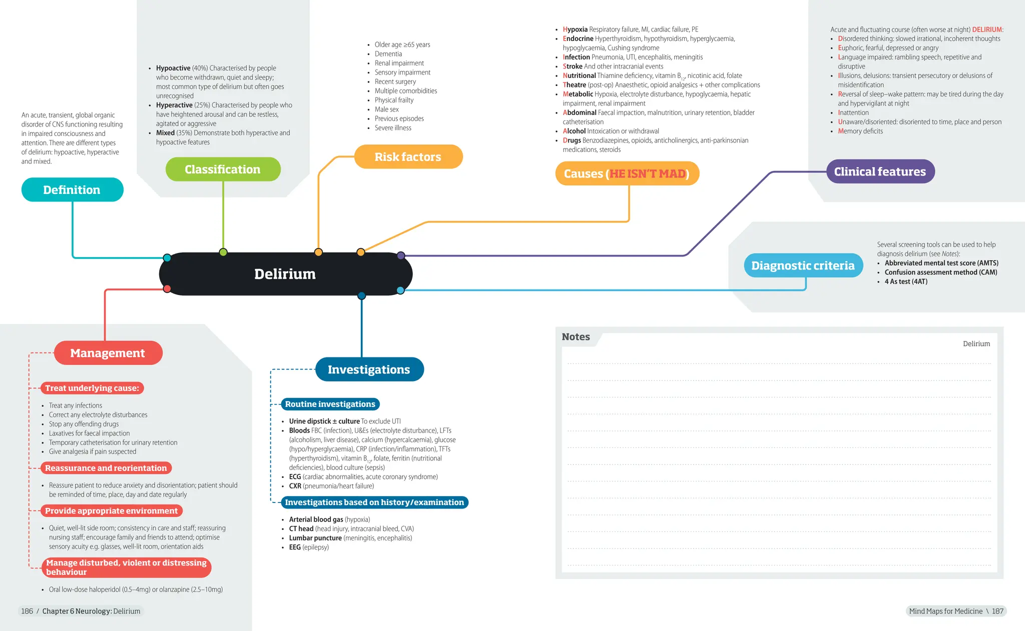
Delirium
An acute, transient, global organic
disorder of CNS functioning resulting
in impaired consciousness and
attention. There are different types
of delirium: hypoactive, hyperactive
and mixed.
• Hypoactive (40%) Characterised by people
who become withdrawn, quiet and sleepy;
most common type of delirium but often goes
unrecognised
• Hyperactive (25%) Characterised by people who
have heightened arousal and can be restless,
agitated or aggressive
• Mixed (35%) Demonstrate both hyperactive and
hypoactive features
Several screening tools can be used to help
diagnosis delirium (see Notes):
• Abbreviated mental test score (AMTS)
• Confusion assessment method (CAM)
• 4 As test (4AT)
Routine investigations
• Urine dipstick ± culture To exclude UTI
• Bloods FBC (infection), U&Es (electrolyte disturbance), LFTs
(alcoholism, liver disease), calcium (hypercalcaemia), glucose
(hypo/hyperglycaemia), CRP (infection/inflammation), TFTs
(hyperthyroidism), vitamin B12
, folate, ferritin (nutritional
deficiencies), blood culture (sepsis)
• ECG (cardiac abnormalities, acute coronary syndrome)
• CXR (pneumonia/heart failure)
Investigations based on history/examination
• Arterial blood gas (hypoxia)
• CT head (head injury, intracranial bleed, CVA)
• Lumbar puncture (meningitis, encephalitis)
• EEG (epilepsy)
Treat underlying cause:
• Treat any infections
• Correct any electrolyte disturbances
• Stop any offending drugs
• Laxatives for faecal impaction
• Temporary catheterisation for urinary retention
• Give analgesia if pain suspected
Reassurance and reorientation
• Reassure patient to reduce anxiety and disorientation; patient should
be reminded of time, place, day and date regularly
Provide appropriate environment
• Quiet, well-lit side room; consistency in care and staff; reassuring
nursing staff; encourage family and friends to attend; optimise
sensory acuity e.g. glasses, well-lit room, orientation aids
Manage disturbed, violent or distressing
behaviour
• Oral low-dose haloperidol (0.5–4mg) or olanzapine (2.5–10mg)
Definition
Classification
Acute and fluctuating course (often worse at night) DELIRIUM:
• Disordered thinking: slowed irrational, incoherent thoughts
• Euphoric, fearful, depressed or angry
• Language impaired: rambling speech, repetitive and
disruptive
• Illusions, delusions: transient persecutory or delusions of
misidentification
• Reversal of sleep–wake pattern: may be tired during the day
and hypervigilant at night
• Inattention
• Unaware/disoriented: disoriented to time, place and person
• Memory deficits
Clinical features
Diagnostic criteria
Management
Investigations
Risk factors
• Older age ≥65 years
• Dementia
• Renal impairment
• Sensory impairment
• Recent surgery
• Multiple comorbidities
• Physical frailty
• Male sex
• Previous episodes
• Severe illness
• Hypoxia Respiratory failure, MI, cardiac failure, PE
• Endocrine Hyperthyroidism, hypothyroidism, hyperglycaemia,
hypoglycaemia, Cushing syndrome
• Infection Pneumonia, UTI, encephalitis, meningitis
• Stroke And other intracranial events
• Nutritional Thiamine deficiency, vitamin B12
, nicotinic acid, folate
• Theatre (post-op) Anaesthetic, opioid analgesics + other complications
• Metabolic Hypoxia, electrolyte disturbance, hypoglycaemia, hepatic
impairment, renal impairment
• Abdominal Faecal impaction, malnutrition, urinary retention, bladder
catheterisation
• Alcohol Intoxication or withdrawal
• Drugs Benzodiazepines, opioids, anticholinergics, anti-parkinsonian
medications, steroids
Causes (HE ISN’T MAD)
5.
Notes
Delirium
Mind Maps forMedicine 189
188 / Chapter 6 Neurology: Delirium notes
Delirium notes
Delirium vs dementia
Delirium Dementia
Sleep–wake cycle Disrupted Usually normal
Attention Markedly reduced Normal/reduced
Arousal Increased/decreased Usually normal
Autonomic features Abnormal Normal
Duration Hours to weeks Months to years
Delusions Fleeting Complex
Course Fluctuating Stable/slow/
progressive
Conscious level Impaired Not impaired
Hallucinations Common Less common
Onset Acute/subacute Chronic
Psychomotor activity Usually abnormal Usually normal
Confusion assessment method (CAM)
The confusion assessment method (CAM) involves assessing a patient for 4
features; diagnosis involves the presence of 1 and 2 + either 3 or 4:
1. Acute onset and fluctuating course
2. Inattention
3. Disorganised thinking
4. Altered consciousness
4AT
1. Alertness
This includes patients who may be markedly drowsy (e.g. difficult
to rouse and/or obviously sleepy during assessment) or agitated/
hyperactive. Observe the patient. If asleep, attempt to wake with speech
or gentle touch on shoulder. Ask the patient to state their name and
address to assist rating.
Normal (fully alert, but not agitated, throughout assessment) 0
Mild sleepiness for <10sec after waking, then normal 0
Clearly abnormal 4
2. AMT4
Age, date of birth, place (name of the hospital or building), current year
No mistakes 0
1 mistake 1
2 or more mistakes/untestable 2
3. Attention
Months of the year backwards
Achieves 7 months or more correctly 0
Starts but scores <7 months/refuses to start 1
Untestable (cannot start because unwell, drowsy, inattentive) 2
4. Acute change or fluctuating course
Evidence of significant change or fluctuation in: alertness, cognition,
other mental function (e.g. paranoia, hallucinations) arising over the
last 2 weeks and still evident in last 24h
No 0
Yes 4
4 or above Possible delirium ± cognitive impairment
1–3 Possible cognitive impairment
0 Delirium or cognitive impairment unlikely
Abbreviated mental test score (AMTS)
1. Age? (1)
2. Time to the nearest hour? (1)
3. Recall address at end:‘42 West Street’(1)
4. ‘What year it is?’(1)
5. ‘Where are you right now?’(1)
6. Identify two people (e.g. doctor, nurse) (1)
7. ‘What is your date of birth?’(1)
8. ‘When did the Second World War end?’(1)
9. ‘Who is the current monarch?’(1)
10. Count backwards from 20 to 1 (1)
(<8 correct Cognitive impairment likely)
6.
Mind Maps forMedicine 191
190 / Chapter 6 Neurology: Epilepsy
Epilepsy
Epilepsy is a neurological condition
characterised by recurrent seizures
unprovoked by any immediately
identifiable cause. An epileptic
seizure is the sudden transient
attack of symptoms and signs due
to abnormal electrical activity in the
brain, leading to a disturbance of
consciousness, behaviour, emotion,
motor function or sensation.
Focal seizures
• Previously termed partial seizures
• These start in a specific area, on one side of the brain
• The level of awareness can be used to further classify
focal seizures: focal aware (previously termed‘simple
partial’), focal impaired awareness (previously
termed‘complex partial’) and awareness unknown
• Focal seizures can also be classified as being motor,
non-motor (e.g. déjà vu, jamais vu) or having other
features such as aura
• Idiopathic (most common)
• Cerebrovascular disease: cerebral infarction, cerebral haemorrhage and
venous thrombosis
• Head injury
• Post cranial surgery
• CNS infections: meningitis or encephalitis
• Neurodegenerative diseases: Alzheimer’s and multi-infarct dementia
are risk factors for epilepsy
• Autoimmune disease: e.g. anti-NMDA receptor encephalitis and
anti-LG11 encephalitis
• Brain neoplasm
• Genetic diseases: e.g. Dravet syndrome
• Drugs: e.g. phenothiazines, isoniazid, tricyclic antidepressants,
benzodiazepines, binge alcohol drinking or alcohol withdrawal
• Metabolic medical disorders: uraemia, hypoglycaemia, hyponatraemia,
hypernatraemia, hypercalcaemia and hypocalcaemia
• Aura Subjective symptoms at the start of the seizure
(the patient is aware of this) – suggestive of focal
epilepsy e.g. strange feeling in the gut, déjà vu, strange
smells or flashing lights
• Potential triggers Sleep deprivation, stress, light
sensitivity or alcohol use
• Specific features of the seizure:
• Tonic Short-lived, abrupt, generalised muscle
stiffening (may cause a fall) with rapid recovery –
suggestive of tonic seizure
• Generalised tonic–clonic Generalised stiffening
and subsequent rhythmic jerking of the limbs,
urinary incontinence and tongue biting
• Absence seizure Brief pauses, e.g. suddenly stop
talking full sentence then carrying on where left off
(presents in childhood)
• Atonic seizure Sudden onset of loss of muscle tone
causing falls
• Myoclonic seizure Brief,‘shock-like’involuntary
single or multiple jerks
• Post-ictal phenomena (residual symptoms after
the attack) e.g. Drowsiness, headaches, amnesia or
confusion (usually occur only after generalised tonic
and/or clonic seizures).
• Bloods e.g. Glucose, Ca2+
, LFTs to identify potential causes
• EEG Supports the diagnosis of epilepsy and may be used to help to
determine seizure type and epilepsy syndrome; it is however often normal
in between attacks (therefore normal EEG does not rule out epilepsy) but
during a seizure it almost always shows an abnormal pattern (typically
showing a cortical spike or generalised spike activity); long-term video or
ambulatory EEG may be used if diagnostic uncertainty remains after clinical
assessment and standard EEG
• ECG In all those with altered consciousness, particularly those in older age
groups, when disorders of cardiac rhythm may simulate epilepsy; 24-h
ambulatory ECG and other cardiovascular tests e.g. implantable loop devices
may also be helpful
• Neuroimaging (used to identify structural abnormalities):
• MRI brain Imaging investigation of choice and particularly important in
those with a focal onset on history (unless examination or EEG suggests
evidence of benign focal epilepsy), and in those who do not respond to
1st-line medication
• CT brain To identify gross pathology if MRI is not available or is
contraindicated
• Polysomnography May be used to confirm a diagnosis of sleep-related
epilepsy
• Handheld video recordings Asking family members or friends to video
record events should be considered in patients with uncertain diagnosis
(after consent from patient)
Definition Classification
Clinical features
Investigations
Causes
Generalised seizures
• These involve both sides of the
brain at the onset
• Consciousness is lost immediately
• Can be further subdivided into
motor (e.g. tonic–clonic) and
non-motor (e.g. absence)
• Specific types include tonic–
clonic (grand mal), tonic, clonic,
absence (petit mal) and atonic
• A seizure results when a sudden imbalance occurs between the
excitatory and inhibitory forces within the network of cortical neurones
in favour of a sudden-onset net excitation
• This imbalance can result from an alteration at many levels of brain
function, from genes and subcellular signalling cascades to widespread
neuronal circuits
• If the affected cortical network is in the visual cortex, the clinical
manifestations are visual phenomena; other affected areas of primary
cortex give rise to sensory, gustatory or motor manifestations; the psychic
phenomenon of déjà vu occurs when the temporal lobe is involved
Pathophysiology
• Injuries sustained during seizures
• Social stigmatisation and occupational issues
• Anxiety/depression
• Status epilepticus
• Sudden unexplained death in epilepsy (SUDEP)
• Increased mortality rate from SUDEP, deaths due to
accidents during seizures, deaths due to status epilepticus
Complications
General advice
• Take precautions e.g. avoid swimming alone, avoid dangerous sports, e.g.
rock climbing, leave door open when having a bath
• Driving:
• All patients must not drive and must inform the DVLA
• For first unprovoked seizure: 6 months off if there are no relevant
structural abnormalities on brain imaging and no definite epileptiform
activity on EEG (if not met then this is increased to 12 months)
• For patients with established epilepsy or those with multiple unprovoked
seizures: may qualify for a driving licence if they have been free from any
seizure for 12 months
Anti-epileptics (see Table 1)
Anti-epileptics are usually started following a second epileptic seizure; NICE
guidelines suggest starting anti-epileptics after the first seizure if any of the
following are present:
• the patient has a neurological deficit
• brain imaging shows a structural abnormality
• the EEG shows unequivocal epileptic activity
• the patient or their family or carers consider the risk of having a further
seizure unacceptable
Neurosurgical treatment
• Neurosurgical treatment has particular benefit for selected people
with refractory focal epilepsy
• Some neurosurgical procedures involve resection of part of the brain
and the aim is to obtain complete seizure freedom
• For the most commonly performed procedures, involving anterior
and medial temporal lobe resection, about 70% of patients will
become seizure-free
Management
7.
Mind Maps forMedicine 193
192 / Chapter 6 Neurology: Epilepsy notes
Anti-epileptics
Table 1 Anti-epileptics
Sodium
valproate
• Indication 1st-line treatment for patients with generalised seizures including generalised tonic–clonic, absence and
myoclonic seizures
• Mechanism of action Blockage of voltage-gated Na+
channels and increased brain levels of gamma-aminobutyric acid
(GABA)
• Side effects Include nausea/vomiting, weight gain, hair loss, confusion, drowsiness, hepatotoxicity, thrombocytopenia,
teratogenicity, encephalopathy, oedema, SIADH
• Cautions/contraindications Pregnancy, acute porphyrias, known or suspected mitochondrial disorders, liver failure,
urea cycle disorders
Carbamazepine • Indications 1st-line treatment for focal seizures
• Mechanism of action Preferentially binds to voltage-gated Na+
channels in their inactive form
• Side effects Rash, vomiting, drowsiness, hyponatraemia, leucopenia, thrombocytopenia, vision disturbance, movement
disorders
• Cautions/contraindications Pregnancy, acute porphyrias, AV conduction abnormalities, bone marrow depression
Lamotrigine • Indications 1st-line treatment for focal seizures and 2nd-line for generalised tonic–clonic seizures
• Mechanism of action Blocks Na+
channels and suppresses the release of glutamate and aspartate
• Side effects Aggression, agitation, diarrhoea, dizziness, drowsiness, sleep disorders, tremor, vomiting, aplastic anaemia
• Cautions/contraindications Myoclonic seizures (may be exacerbated), Parkinson’s disease (may be exacerbated),
caution in hepatic and renal impairment
Levetiracetam • Indications 2nd-line treatment for patients with focal seizures
• Mechanism of action Exact mechanism unclear but binding to synaptic vesicle protein 2A (SV2A) appears to be the key driver
• Side effects Depression/anxiety, diarrhoea/vomiting, dyspepsia, insomnia, vertigo, blood dyscrasias
• Cautions/contraindications Caution in severe hepatic impairment and dose adjustment in renal impairment
Ethosuximide • Indications 1st-line treatment for people with absent seizures
• Mechanism of action Binds to T-type voltage-sensitive calcium channels
• Side effects Aggression, agranulocytosis, reduced appetite, concentration impaired, generalised tonic–clonic seizure,
headache, bone marrow disorders
• Cautions/contraindications Acute porphyrias, pregnancy, caution in hepatic failure and renal failure
Phenytoin • Indications Used for generalised, focal seizures and status epilepticus but not used 1st line in generalised and focal
seizures due to side effects and narrow therapeutic index
• Mechanism of action Blocks voltage-dependent Na+
channels
• Side effects Gingival hyperplasia, hirsutism, coarsening of facial features, drowsiness, megaloblastic anaemia, peripheral
neuropathy, lymphadenopathy, dyskinesia, teratogenicity; toxicity causes dizziness, diplopia, nystagmus, slurred speech,
ataxia, confusion, seizures
• Monitoring Phenytoin levels do not need to be monitored routinely but trough levels, immediately before dose, should
be checked if: adjustment of phenytoin dose, suspected toxicity or detection of non-adherence to the prescribed
medication
• Contraindications Pregnancy, second- and third-degree heart block, sino-atrial block, sinus bradycardia, Stokes–Adams
syndrome; caution in hepatic impairment
Phenobarbital • Indications All seizures (including status epilepticus) except absent seizures
• Mechanism of action Acts on GABAA
receptors enhancing synaptic inhibition
• Side effects Rash, sedation, bone disorders, depression, ataxia
• Cautions/contraindications Pregnancy, history of porphyria, severe hepatic impairment, caution in renal impairment
Status epilepticus (Fig. 1)
Convulsive status epilepticus is defined as a convulsive seizure which continues for a prolonged period (e.g. longer than 5 min), or when convulsive
seizures occur one after the other with no recovery between. Convulsive status epilepticus is an emergency and requires immediate medical attention
Management
Secure IV access in a large vein and take venous blood gas and venous bloods
for glucose, electrolytes, calcium, U&Es, LFTs and toxicology screen and
anticonvulsant levels (if indicated)
IV dextrose if hypoglycaemia suspected/confirmed;
IV thiamine if alcoholism/malnourishment suspected
Monitor ECG, oxygen saturations, temperature,
pulse rate and blood pressure
IV lorazepam up to 4mg as 1st-line treatment; buccal midazolam
10mg or rectal diazepam 10–20mg should be given if
there is no IV access
If seizures continue, contact ITU; IV phenobarbital or phenytoin is 2nd-line
treatment; fosphenytoin (a prodrug of phenytoin) can be given more rapidly
and causes fewer injection-site reactions than phenytoin
For refractory convulsive status epilepticus summon anaesthetist and
ITU; administer IV midazolam, propofol or thiopental sodium for general
anaesthesia with intubation and ventilation
Secure airway, high-flow oxygen
Fig. 1 Management of status epilepticus
Epilepsy notes
8.
Notes
Acute respiratory distresssyndrome
Extradural haematoma (epidural haemorrhage)
Mind Maps for Medicine 195
194 / Chapter 6 Neurology: Extradural haematoma (epidural haemorrhage)
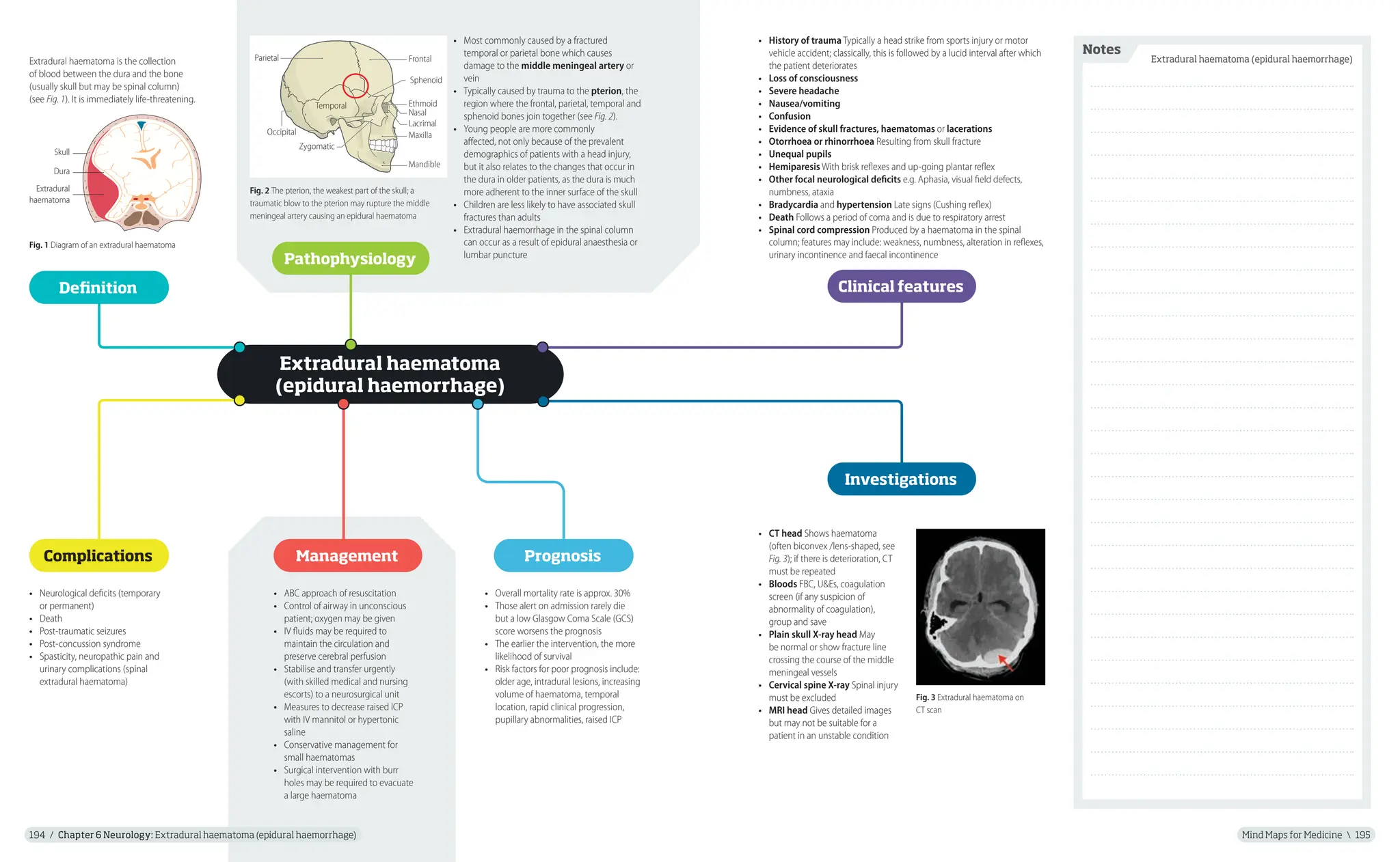
Extradural haematoma
(epidural haemorrhage)
Extradural haematoma is the collection
of blood between the dura and the bone
(usually skull but may be spinal column)
(see Fig. 1). It is immediately life-threatening.
• ABC approach of resuscitation
• Control of airway in unconscious
patient; oxygen may be given
• IV fluids may be required to
maintain the circulation and
preserve cerebral perfusion
• Stabilise and transfer urgently
(with skilled medical and nursing
escorts) to a neurosurgical unit
• Measures to decrease raised ICP
with IV mannitol or hypertonic
saline
• Conservative management for
small haematomas
• Surgical intervention with burr
holes may be required to evacuate
a large haematoma
Definition
Pathophysiology
Investigations
Management
• Most commonly caused by a fractured
temporal or parietal bone which causes
damage to the middle meningeal artery or
vein
• Typically caused by trauma to the pterion, the
region where the frontal, parietal, temporal and
sphenoid bones join together (see Fig. 2).
• Young people are more commonly
affected, not only because of the prevalent
demographics of patients with a head injury,
but it also relates to the changes that occur in
the dura in older patients, as the dura is much
more adherent to the inner surface of the skull
• Children are less likely to have associated skull
fractures than adults
• Extradural haemorrhage in the spinal column
can occur as a result of epidural anaesthesia or
lumbar puncture
Dura
Extradural
haematoma
Skull
Fig. 1 Diagram of an extradural haematoma
Fig. 3 Extradural haematoma on
CT scan
• CT head Shows haematoma
(often biconvex /lens-shaped, see
Fig. 3); if there is deterioration, CT
must be repeated
• Bloods FBC, U&Es, coagulation
screen (if any suspicion of
abnormality of coagulation),
group and save
• Plain skull X-ray head May
be normal or show fracture line
crossing the course of the middle
meningeal vessels
• Cervical spine X-ray Spinal injury
must be excluded
• MRI head Gives detailed images
but may not be suitable for a
patient in an unstable condition
• Neurological deficits (temporary
or permanent)
• Death
• Post-traumatic seizures
• Post-concussion syndrome
• Spasticity, neuropathic pain and
urinary complications (spinal
extradural haematoma)
Complications
• Overall mortality rate is approx. 30%
• Those alert on admission rarely die
but a low Glasgow Coma Scale (GCS)
score worsens the prognosis
• The earlier the intervention, the more
likelihood of survival
• Risk factors for poor prognosis include:
older age, intradural lesions, increasing
volume of haematoma, temporal
location, rapid clinical progression,
pupillary abnormalities, raised ICP
Prognosis
Fig. 2 The pterion, the weakest part of the skull; a
traumatic blow to the pterion may rupture the middle
meningeal artery causing an epidural haematoma
Parietal
Zygomatic
Temporal
Mandible
Maxilla
Sphenoid
Ethmoid
Lacrimal
Nasal
Occipital
Frontal
Clinical features
• History of trauma Typically a head strike from sports injury or motor
vehicle accident; classically, this is followed by a lucid interval after which
the patient deteriorates
• Loss of consciousness
• Severe headache
• Nausea/vomiting
• Confusion
• Evidence of skull fractures, haematomas or lacerations
• Otorrhoea or rhinorrhoea Resulting from skull fracture
• Unequal pupils
• Hemiparesis With brisk reflexes and up-going plantar reflex
• Other focal neurological deficits e.g. Aphasia, visual field defects,
numbness, ataxia
• Bradycardia and hypertension Late signs (Cushing reflex)
• Death Follows a period of coma and is due to respiratory arrest
• Spinal cord compression Produced by a haematoma in the spinal
column; features may include: weakness, numbness, alteration in reflexes,
urinary incontinence and faecal incontinence
9.
Notes
Guillain–Barré syndrome
Mind Mapsfor Medicine 197
196 / Chapter 6 Neurology: Guillain–Barré syndrome
Guillain–Barré syndrome
Guillain–Barré syndrome (GBS)
describes an immune-mediated
demyelination of the peripheral
nervous system often triggered
by an infection. It can lead to
life-threatening respiratory
failure.
• Reflexes May be reduced or absent
• Sensory symptoms These can include paraesthesia
and sensory loss, starting in the lower extremities
• Autonomic symptoms Involvement of the autonomic
system may present, with reduced sweating, reduced
heat tolerance, paralytic ileus and urinary hesitancy;
severe autonomic dysfunction may occur
• Miller Fisher syndrome (variant of GBS):
• Associated with ophthalmoplegia, areflexia and ataxia
• Usually presents as a descending paralysis rather
than ascending as seen in other forms of GBS
• Anti-GQ1b antibodies are present in 90% of cases
Definition
Clinical features
• Weakness:
• In 60% of cases, onset occurs approximately 3 weeks
after an infection
• Usually presents with an ascending pattern of progressive
symmetrical weakness, starting in the lower extremities
• This reaches a level of maximum severity 2 weeks after
initial onset of symptoms and usually stops progressing
after 5 weeks
• Facial weakness, dysphasia, diplopia or dysarthria may
develop
• In severe cases, muscle weakness may lead to
respiratory failure
• Pain Neuropathic pain may develop, particularly in the legs;
back pain may also occur
Risk factors
Investigations
• History of gastrointestinal or
respiratory infection from 1 to
3 weeks prior to the onset of
weakness
• Zika virus
• Vaccinations: live and dead vaccines
have been implicated
• Malignancy: e.g. lymphomas,
especially Hodgkin’s disease
• Pregnancy: incidence decreases
during pregnancy but increases in
the months after delivery
• GBS is usually triggered by an infection: Campylobacter
jejuni, Epstein–Barr virus (EBV) and cytomegalovirus have all
been linked
• It is thought that the infectious organism shares epitopes
with an antigen in the peripheral nervous system leading to
autoantibody-mediated cell damage
• The suppressor T-cell response is reduced, suggesting a cell-
mediated immunological reaction directed at the peripheral
nerves; occasionally, serum antibodies to myelin components
are detected; nerve damage occurs segmentally; lymphocytes
infiltrate the nerve roots and release cytotoxic substances that
damage the Schwann cells and myelin
• Correlation between anti-ganglioside antibody (e.g. anti-GM1)
and clinical features has been demonstrated; anti-GM1
antibodies are present in approx. 25% of patients
• Plasma exchange Leads to shorter
periods of ventilation and a shorter
period until patients are able to walk
unaided
• IV immunoglobulin If started within
2 weeks from the onset of illness
it accelerates recovery as much as
plasma exchange
• DVT prophylaxis DVT due to
immobility; prevent with gradient
compression stockings and
subcutaneous low molecular weight
heparin
• Admission to ICU Intubation and
assisted ventilation may be required
• Pain relief May be required for
neuropathic pain
Management
• Lumbar puncture Most patients have an elevated
level of CSF protein, with no elevation in CSF cell
counts (the gamma-globulin fraction is usually
raised); note: the rise in the CSF protein may not be
seen until 1–2 weeks after the onset of weakness
• Antibody screen In Miller Fisher syndrome there
are often antibodies against GQ1b
• Spirometry Forced vital capacity is a major
determinant of the need for admission to ICU
and then the need for intubation
• Nerve conduction studies Abnormal in 85% of
patients, even early on in the disease; they show
prolonged conduction velocities; repeat after
2 weeks if initially normal
• ECG Many different abnormalities may be seen:
2nd-degree and 3rd-degree AV block, T-wave
abnormalities, ST depression, QRS widening and
a variety of rhythm disturbances
• Campylobacter serology Should be performed;
positive titres identify a group with a poorer
prognosis
Pathophysiology
Complications
• Persistent paralysis
• Respiratory failure requiring
mechanical ventilation
• Aspiration pneumonia
• Hypotension or hypertension
• Thromboembolism, pneumonia,
skin breakdown
• Cardiac arrhythmia
• Urinary retention
• Ileus
• Psychiatric problems e.g. depression,
anxiety
10.
Notes
Migraine
Mind Maps forMedicine 199
198 / Chapter 6 Neurology: Migraine
Migraine
Migraine is a primary headache
disorder which is characterised by
episodic severe headaches (often
but not always unilateral), with
commonly associated symptoms
such as photophobia, phonophobia
and nausea/vomiting.
• Severe, often unilateral, throbbing headache
• Aura:‘classic’migraine attacks are precipitated
by an aura; these occur in around one-third of
migraine patients; typical aura are visual, progressive,
last 5–60min and are characterised by transient
hemianopic disturbance or a spreading scintillating
scotoma
• Nausea/vomiting
• Photophobia
• Phonophobia
• Attacks may last up to 72h
The diagnosis of migraine is a clinical diagnosis.
Table 1 International Headache Society diagnostic criteria
Point Criteria
A At least 5 attacks fulfilling criteria B–D
B Headache attacks lasting 4–72h (untreated or unsuccessfully
treated)
C Headache has at least 2 of the following characteristics:
• Unilateral location
• Pulsating quality (i.e. varying with the heartbeat)
• Moderate or severe pain intensity
• Aggravation by or causing avoidance of routine physical
activity (e.g. walking or climbing stairs)
D During headache at least one of the following:
• Nausea and/or vomiting
• Photophobia and phonophobia
E Not attributed to another disorder (history and examination
do not suggest a secondary headache disorder or, if they do, it
is ruled out by appropriate investigations or headache attacks
do not occur for the first time in close temporal relation to the
other disorder)
Definition Common triggers
Clinical features
Management
• Tiredness, stress
• Alcohol
• Combined oral contraceptive pill
• Lack of food or dehydration
• Cheese, chocolate, red wines, citrus fruits
• Menstruation
• Bright lights
Acute
• Paracetamol e.g. 1g oral QDS; 1st-line treatment for pregnant
women as considered safe
• NSAIDs e.g. Soluble aspirin 600–900mg (not in children)
or ibuprofen 400–600mg
• Triptans e.g. Sumatriptan; should be taken as soon as possible
after the onset of headache; oral, orodispersible, nasal spray and SC
injections are available
• Anti-emetics For nausea/vomiting e.g. buccal prochlorperazine or
metoclopramide
• Opiate-containing medication, e.g. codeine, should be avoided
Prevention
• Avoid triggers (if possible)
• Prophylaxis should be given if patients are experiencing 2 or more
attacks per month
• Topiramate or propranolol 1st-line for prophylaxis according to
the person’s preference, comorbidities and risk of adverse events;
propranolol should be used in preference to topiramate in women
of childbearing age as topiramate may be teratogenic and can
reduce the effectiveness of hormonal contraceptives
• Acupuncture If above measures fail, NICE recommend a course of
up to 10 sessions of acupuncture over 5–8 weeks or gabapentin
• Riboflavin May be effective in reducing migraine frequency and
intensity for some people
• Triptans Frovatriptan or zolmitriptan can be used as a type of
‘mini-prophylaxis’for women with predictable menstrual migraine
International Headache Society
Diagnostic criteria
11.
Mind Maps forMedicine 201
200 / Chapter 6 Neurology: Migraine notes
Table 2 Causes of headaches
Condition Notes
Chronic
headache
Tension headache • Recurrent, non-disabling, bilateral headache, often described as a‘tight band’
• Not aggravated by routine activities of daily living
• Not associated with aura, nausea/vomiting or aggravated by routine physical activity
• May be associated with stress
• Acute treatment: aspirin, paracetamol or an NSAID is 1st-line
• Prophylaxis: NICE recommend up to 10 sessions of acupuncture over 5–8 weeks; low-dose
amitriptyline is widely used in the UK for prophylaxis against tension-type headache
Medication overuse
headache
• Present for ≥15 days per month
• Developed or worsened while taking regular symptomatic medication specifically for headaches
• Most common offending drugs are opioids and triptans
• There may be a psychiatric comorbidity
• Simple analgesics and triptans should be withdrawn abruptly (may initially worsen headaches)
• Opioid analgesics should be gradually withdrawn
Raised intracranial
pressure
• e.g. Tumour, idiopathic intracranial hypertension
• Typically worse on waking, lying down or bending forward, or with coughing
• Associated with vomiting, papilloedema, fits and neurological signs
• CT or MRI scan is investigation of choice
• Lumbar puncture contraindicated until after imaging
Recurrent
acute
attacks
of
headache
Migraine See Mind Map
Cluster headache • Intense pain around one eye; recurrent attacks‘always’affect same side
• Patient is often restless during an attack due to severity of pain
• Pain typically occurs once or twice a day, each episode lasting 15 min to 2 hours with clusters typically
lasting 4–12 weeks
• Associated eye symptoms include redness, lacrimation, lid swelling
• More common in men and smokers
• Acute: 100% oxygen, subcutaneous triptan
• Prophylaxis: verapamil is 1st-line; a tapering dose of prednisolone can also be considered
Trigeminal neuralgia • A unilateral disorder characterised by transient electric shock-like pains, abrupt in onset and
termination, limited to one or more divisions of the trigeminal nerve
• The pain is commonly triggered by light touch e.g. washing, shaving, talking and brushing the teeth,
and frequently occurs spontaneously
• Carbamazepine is 1st-line treatment
Subacute
onset
Giant cell arteritis (GCA) • Typically patient >60 years old
• Usually rapid onset (e.g. <1 month) of unilateral headache
• Often associated jaw or tongue claudication
• Tender, palpable temporal artery
• Raised ESR
• See Ch7: Giant cell arteritis for further information
Condition Notes
Acute
single
episode
Subarachnoid
haemorrhage
• Sudden onset severe‘thunderclap’headache
• Nausea and vomiting
• Meningism (photophobia, neck stiffness)
• See Ch6: Subarachnoid haemorrhage for further information
Head injury • Headache is common at the site of head injury but it may also be more generalised
• Serious head injury can lead to intracranial bleeds; a CT scan should be done if subdural or extradural
haematoma is suspected
• See Ch6: Subdural haematoma and Extradural haematoma for further information
Sinusitis • Facial pain: typically frontal pressure pain which is worse on bending forward
• Associated symptoms include nasal discharge and nasal obstruction
• Postnasal drip may produce chronic cough
• Acute sinusitis can be treated with analgesia, intranasal decongestants and antibiotics for severe
presentations; intranasal corticosteroids are often useful for recurrent or chronic sinusitis
Acute glaucoma • Severe pain which may be ocular or headache
• Decreased visual acuity, red eye, haloes, semi-dilated non-reacting pupil, hazy cornea
• Symptoms worse with mydriasis e.g. watching TV in a dark room
• Systemic upset may be seen, such as nausea and vomiting and even abdominal pain
• Requires urgent ophthalmology assessment
Meningitis/encephalitis • Meningitis: classically presents with fever, photophobia, neck stiffness, headache, purpuric rash
• Encephalitis: classically presents with headache, fever, confusion/odd behaviour, seizures, reduced
consciousness
• Requires CT scan followed by lumbar puncture for diagnosis (in absence of raised intracranial
pressure)
Central venous sinus
thrombosis
• Headache which may be sudden onset
• There may be associated nausea and vomiting, cranial nerve palsies, vision disturbance, seizures
• The diagnosis is usually made with the aid of CT or MRI scan
• Specific treatment involves anticoagulation or thrombolytic treatments
Red flags for headaches
• Immunocompromised e.g. HIV or on immunosuppressive drugs
• Age <20 years and a history of malignancy
• History of malignancy known to metastasise to the brain
• Sudden-onset headache reaching maximum intensity within 5min
• Vomiting without other obvious cause
• Recent (typically within the past 3 months) head trauma
• Worsening headache with fever
• New-onset neurological deficit
• New-onset cognitive dysfunction
• Change in personality
• Impaired level of consciousness
• Headache exacerbated by cough, Valsalva (trying to breathe out with
nose and mouth blocked), sneeze or exercise
• Orthostatic headache (headache that changes with posture)
• Symptoms suggestive of GCA (see Table 2)
• Symptoms suggestive of acute narrow-angle glaucoma (see Table 2)
• A substantial change in the characteristics of patient’s headache
Migrainenotes
Table 2 (continued)
12.
Notes
Motor neurone disease
MindMaps for Medicine 203
202 / Chapter 6 Neurology: Motor neurone disease
Motor neurone disease
Motor neurone disease (MND) is a
rare but devastating neurological
condition of unknown cause which
can present with both upper and
lower motor neurone signs. It leads
to progressive paralysis and eventual
death from respiratory failure.
• MND is a degenerative condition that affects motor
neurones, specifically the anterior horn cells of the spinal
cord and the motor cranial nuclei
• The cause of the disease is unknown, although 5% of
those affected have a familial form of the disease due to a
mutation in the superoxide dismutase-1 gene
• It may be caused by abnormality of mitochondrial function
causing oxidative stress in motor neurones for which there
may be several causes
• It results in lower motor neurone (LMN) and upper motor
neurone (UMN) dysfunction, leading to a mixed picture of
muscular paralysis, typically with LMN signs predominating
• Amyotrophic lateral sclerosis (ALS)
The most common form of MND; it is
a combination of disease of the lateral
corticospinal tracts and anterior horn cells
producing a progressive spastic tetraparesis
and paraparesis with added lower motor
neurone signs (muscle wasting and
fasciculation)
• Progressive bulbar palsy and
pseudobulbar palsy Approximately 20%
of people with MND have this type; it
results from the destruction of the upper
(pseudobulbar) and lower (bulbar palsy)
motor neurones in the lower cranial nerves;
this results in dysarthria, dysphagia with
wasting, and fasciculation of the tongue
• Progressive muscular atrophy An
uncommon form of MND; predominantly
lower motor neurone lesion of the spinal cord;
the small muscles of the hands and feet are
usually first affected, but muscle spasticity is
absent
• Primary lateral sclerosis Another rare type
of MND; it mainly causes weakness in the leg
muscles and there is progressive tetraparesis
Riluzole
• A neuroprotective glutamate-release inhibitor;
the only drug of proven disease-modifying efficacy
• It is used mainly in ALS
• Prolongs life by approximately 3 months
Other symptomatic treatment
• Dysarthria Speech assessment and communication aids
• Dysphagia Feeding gastrostomy; cricopharyngeal
myotomy
• Dysphonia Speech therapists can give expert advice on
speech and swallowing difficulties
• Saliva problems Consider a trial of antimuscarinic
medicine as the 1st-line treatment for sialorrhoea in
people with MND
• Muscle weakness Physiotherapy, walking aids, splints
• Muscle cramps Consider quinine as 1st-line and
baclofen as 2nd-line treatment
• Muscle stiffness, spasticity or increased tone
Consider baclofen, tizanidine, dantrolene or gabapentin
• Non-invasive ventilation (usually BIPAP) Used at
night; studies have shown a survival benefit of around
7 months
There are no specific investigations
that will confirm a diagnosis of MND;
a range of investigations are carried
out to confirm consistent features and
exclude other possible pathologies:
• Nerve conduction studies Show
normal motor conduction and can
help exclude a neuropathy
• EMG Shows a reduced number of
action potentials with an increased
amplitude
• MRI Usually performed to exclude the
differential diagnosis of cervical cord
compression and myelopathy
Definition
Pathophysiology
Investigations
Management
Clinical features/types
• MND is relatively uncommon
with annual incidence of
around 2 cases per 100 000
population
• It can occur at any age but
is more common in people
aged >50 years
• The male to female ratio is 2:1
• Approximately 5–10% of
cases are inherited
Epidemiology
Prognosis
• Prognosis is poor with 50%
of patients dying within
3 years
• Most patients die of
respiratory failure
13.
Mind Maps forMedicine 205
204 / Chapter 6 Neurology: Multiple sclerosis
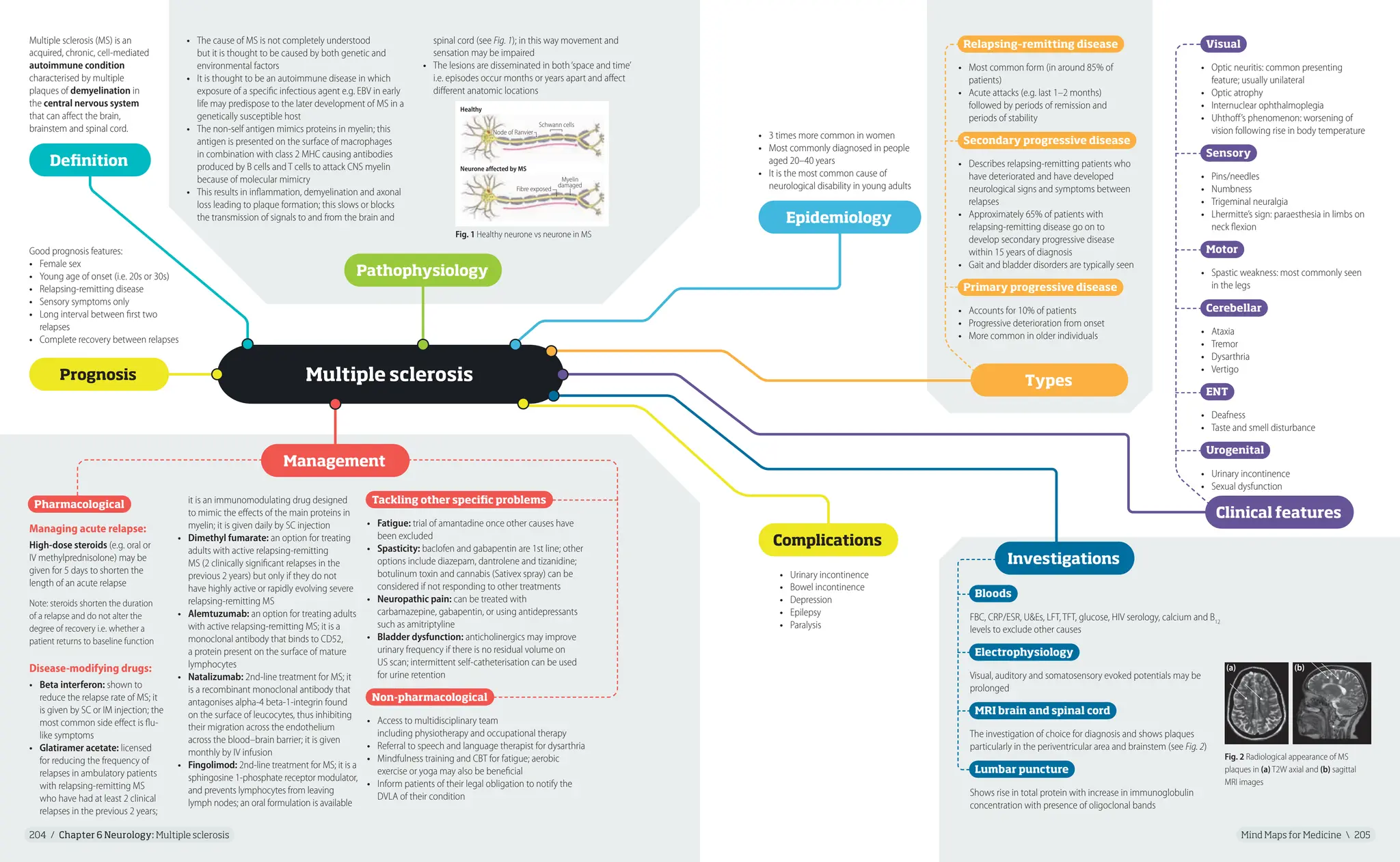
Multiple sclerosis
Multiple sclerosis (MS) is an
acquired, chronic, cell-mediated
autoimmune condition
characterised by multiple
plaques of demyelination in
the central nervous system
that can affect the brain,
brainstem and spinal cord.
• The cause of MS is not completely understood
but it is thought to be caused by both genetic and
environmental factors
• It is thought to be an autoimmune disease in which
exposure of a specific infectious agent e.g. EBV in early
life may predispose to the later development of MS in a
genetically susceptible host
• The non-self antigen mimics proteins in myelin; this
antigen is presented on the surface of macrophages
in combination with class 2 MHC causing antibodies
produced by B cells and T cells to attack CNS myelin
because of molecular mimicry
• This results in inflammation, demyelination and axonal
loss leading to plaque formation; this slows or blocks
the transmission of signals to and from the brain and
Bloods
FBC, CRP/ESR, U&Es, LFT, TFT, glucose, HIV serology, calcium and B12
levels to exclude other causes
Electrophysiology
Visual, auditory and somatosensory evoked potentials may be
prolonged
MRI brain and spinal cord
The investigation of choice for diagnosis and shows plaques
particularly in the periventricular area and brainstem (see Fig. 2)
Lumbar puncture
Shows rise in total protein with increase in immunoglobulin
concentration with presence of oligoclonal bands
• 3 times more common in women
• Most commonly diagnosed in people
aged 20–40 years
• It is the most common cause of
neurological disability in young adults
Good prognosis features:
• Female sex
• Young age of onset (i.e. 20s or 30s)
• Relapsing-remitting disease
• Sensory symptoms only
• Long interval between first two
relapses
• Complete recovery between relapses
Definition
Pathophysiology
Types
Prognosis
Epidemiology
• Urinary incontinence
• Bowel incontinence
• Depression
• Epilepsy
• Paralysis
Complications
Management
spinal cord (see Fig. 1); in this way movement and
sensation may be impaired
• The lesions are disseminated in both‘space and time’
i.e. episodes occur months or years apart and affect
different anatomic locations
Visual
• Optic neuritis: common presenting
feature; usually unilateral
• Optic atrophy
• Internuclear ophthalmoplegia
• Uhthoff’s phenomenon: worsening of
vision following rise in body temperature
Sensory
• Pins/needles
• Numbness
• Trigeminal neuralgia
• Lhermitte’s sign: paraesthesia in limbs on
neck flexion
Motor
• Spastic weakness: most commonly seen
in the legs
Cerebellar
• Ataxia
• Tremor
• Dysarthria
• Vertigo
ENT
• Deafness
• Taste and smell disturbance
Urogenital
• Urinary incontinence
• Sexual dysfunction
Pharmacological
Managing acute relapse:
High-dose steroids (e.g. oral or
IV methylprednisolone) may be
given for 5 days to shorten the
length of an acute relapse
Note: steroids shorten the duration
of a relapse and do not alter the
degree of recovery i.e. whether a
patient returns to baseline function
Disease-modifying drugs:
• Beta interferon: shown to
reduce the relapse rate of MS; it
is given by SC or IM injection; the
most common side effect is flu-
like symptoms
• Glatiramer acetate: licensed
for reducing the frequency of
relapses in ambulatory patients
with relapsing-remitting MS
who have had at least 2 clinical
relapses in the previous 2 years;
it is an immunomodulating drug designed
to mimic the effects of the main proteins in
myelin; it is given daily by SC injection
• Dimethyl fumarate: an option for treating
adults with active relapsing-remitting
MS (2 clinically significant relapses in the
previous 2 years) but only if they do not
have highly active or rapidly evolving severe
relapsing-remitting MS
• Alemtuzumab: an option for treating adults
with active relapsing-remitting MS; it is a
monoclonal antibody that binds to CD52,
a protein present on the surface of mature
lymphocytes
• Natalizumab: 2nd-line treatment for MS; it
is a recombinant monoclonal antibody that
antagonises alpha-4 beta-1-integrin found
on the surface of leucocytes, thus inhibiting
their migration across the endothelium
across the blood–brain barrier; it is given
monthly by IV infusion
• Fingolimod: 2nd-line treatment for MS; it is a
sphingosine 1-phosphate receptor modulator,
and prevents lymphocytes from leaving
lymph nodes; an oral formulation is available
Tackling other specific problems
• Fatigue: trial of amantadine once other causes have
been excluded
• Spasticity: baclofen and gabapentin are 1st line; other
options include diazepam, dantrolene and tizanidine;
botulinum toxin and cannabis (Sativex spray) can be
considered if not responding to other treatments
• Neuropathic pain: can be treated with
carbamazepine, gabapentin, or using antidepressants
such as amitriptyline
• Bladder dysfunction: anticholinergics may improve
urinary frequency if there is no residual volume on
US scan; intermittent self-catheterisation can be used
for urine retention
Non-pharmacological
• Access to multidisciplinary team
including physiotherapy and occupational therapy
• Referral to speech and language therapist for dysarthria
• Mindfulness training and CBT for fatigue; aerobic
exercise or yoga may also be beneficial
• Inform patients of their legal obligation to notify the
DVLA of their condition
Clinical features
Investigations
Fig. 1 Healthy neurone vs neurone in MS
Healthy
Schwann cells
Node of Ranvier
Neurone affected by MS
Fibre exposed
Myelin
damaged
Fig. 2 Radiological appearance of MS
plaques in (a) T2W axial and (b) sagittal
MRI images
(a) (b)
Relapsing–remitting disease
• Most common form (in around 85% of
patients)
• Acute attacks (e.g. last 1–2 months)
followed by periods of remission and
periods of stability
Secondary progressive disease
• Describes relapsing-remitting patients who
have deteriorated and have developed
neurological signs and symptoms between
relapses
• Approximately 65% of patients with
relapsing-remitting disease go on to
develop secondary progressive disease
within 15 years of diagnosis
• Gait and bladder disorders are typically seen
Primary progressive disease
• Accounts for 10% of patients
• Progressive deterioration from onset
• More common in older individuals
14.
Notes
Myasthenia gravis
The keyfeature is muscle fatigability – muscles become progressively weaker
during periods of activity and slowly improve after periods of rest:
• Extraocular muscle weakness: diplopia
• Proximal muscle weakness: face, neck, limb girdle
• Ptosis (see Fig. 1)
• Bulbar involvement: dysphagia, dysphonia, dysarthria
Clinical features
Fig. 1 Myasthenia gravis illustration showing
left-sided ptosis
Mind Maps for Medicine 207
206 / Chapter 6 Neurology: Myasthenia gravis
Myasthenia gravis
Myasthenia gravis (MG) is an
acquired autoimmune disorder
resulting in insufficient functioning
acetylcholine receptors. It is
characterised by weakness, typically
of the periocular, facial, bulbar and
girdle muscles.
• MG is more common in women (2:1)
• It can occur in any age but peaks occur
in the 20–30s and 60–80s
• Thymomas in 15%
• Autoimmune disorders: pernicious
anaemia, autoimmune thyroid
disorders, rheumatoid, SLE
• Thymic hyperplasia in 50–70%
• MG is an autoimmune disease in which
antibodies result in a loss of muscle
acetylcholine receptors (AChRs)
• In 85% of cases the antibodies bind to the
AChRs themselves and in the remaining
cases the antibodies bind to a different
muscle membrane target
• There are associations between MG and
thymic hyperplasia (75% of cases) and
thymoma (15%)
• Autoantibodies Around 85–90% of patients have antibodies to
acetylcholine receptors; in the remaining patients, about 40% are
positive for anti-muscle-specific tyrosine kinase antibodies
• CK Normal
• EMG (single fibre) High sensitivity (92–100%)
• CT or MRI thorax To exclude thymoma
• Tensilon test IV edrophonium reduces muscle weakness
temporarily (rarely used any more due to the risk of cardiac
arrhythmia)
• A reversible life-threatening neurological emergency that affects
20–30% of myasthenic patients, usually within the first year of illness; it
may be the first indication of the disease
• Results in weakness of respiratory muscles; facial muscles may be slack,
and face may be expressionless; patient may be unable to support the
head, which will fall onto the chest while the patient is seated; jaw is
slack; voice has a nasal quality; body is limp
• Often triggered by medications e.g. aminoglycosides, beta blockers
• Gag reflex is often absent, and such patients are at risk for aspiration
of oral secretions
• Management: monitor forced vital capacity, ventilatory support,
plasmapheresis or IV immunoglobulins
• Emotional stress
• Pregnancy
• Menses
• Secondary illness
• Thyroid dysfunction
• Trauma
• Temperature extremes
• Hypokalaemia
• Drugs: aminoglycosides,
beta blockers, calcium
channel blockers, quinidine,
procainamide, chloroquine,
lithium, macrolides, tetracycline,
penicillamine, succinylcholine,
magnesium,
ACE inhibitor
• Surgery
• Long-acting anticholinesterase
inhibitors Pyridostigmine is the
preferred symptomatic treatment
• Immunosuppression
Prednisolone initially
• Plasmapheresis and IV
immunoglobulin Used for
myasthenic crisis
• Thymectomy Important if a
thymoma is present but may be
beneficial even without one
Definition Epidemiology
Pathophysiology
Triggers Myasthenic crisis
Associations
Management
Investigations
Complications
• Aspiration pneumonia due
to throat muscle weakness
• Acute respiratory
failure during an
exacerbation
15.
Notes
Neurofibromatosis
Mind Maps forMedicine 209
208 / Chapter 6 Neurology: Neurofibromatosis
Neurofibromatosis
Neurofibromatosis (NF) refers to
a group of genetic disorders that
primarily affect the cell growth of
neural tissues. There are two types:
• Neurofibromatosis type 1
(NF1), also known as von
Recklinghausen’s disease
• Neurofibromatosis type 2 (NF2)
NF1
• NF1 is a dominantly inherited genetic disorder that results
from a germline mutation in the NF1 tumour-suppressor gene,
neurofibromin, which is located on chromosome 17q11.2
• About 50% of individuals with NF1 have no family history of the
disease and the disease is due to de novo mutations
NF2
• NF2 is caused by a mutation in the gene encoding for the
protein merlin or schwannomin on chromosome 22
• It is also autosomal dominant although around 50% are
de novo with mosaicism in some
NF1
• Mild learning disability
• Nerve root compressions caused by
neurofibromas
• GI bleeds/obstruction
• MSK complications: bone-cystic
lesions, scoliosis, pseudarthrosis
• Hypertension (from renal artery
stenosis)
• Phaeochromocytoma
• Malignancy
• Optic glioma
• Increased risk of epilepsy
• Carcinoid syndrome (rare)
NF2
• Partial/total deafness and tinnitus
• Facial nerve damage
• Visual disturbance
• Schwannomas
• Weakness or numbness in the
extremities
• Multiple benign brain tumours
Definition Pathophysiology
Management
NF1
• Multidisciplinary team involvement including geneticist, neurologist,
surgeon and physiotherapist, coordinated by a GP
• Comprehensive examination each year in children e.g. detailed skin
examination, eye tests, and assessment of bone, behaviour, blood
pressure, physical ability and progress at school
• Neurofibromas should not be excised unless they show evidence
of malignancy or are causing symptoms
• Other options include chemotherapy or radiation if a tumour has turned
malignant or cancerous
• Plexiform neurofibromas may be treated with plastic surgery but there is
risk of paralysis especially if the cranial nerves are involved superficially
• Cranial and spinal neurofibromas are amenable to corrective surgery
• Any gliomas or meningiomas should usually be extirpated, partially
or completely, once intracranial pressure is raised
• Genetic counselling
NF2
• Annual monitoring usually involving hearing tests, an MRI brain scan
and eye testing
• Hearing aids and management of tinnitus may be required
• Surgery and radiotherapy (less common) are options for brain tumours
depending on size
• Genetic counselling
Complications
Fig. 1 Café-au-lait macules
Fig. 2 Neurofibromas
Fig. 3 Lisch nodules
NF1
Diagnosis is made if at least 2 of the following are
found (in the absence of
alternative diagnoses):
1. ≥6 Café-au-lait spots or
hyperpigmented macules
>5mm in diameter in
prepubertal children and
>15mm postpubertal
(see Fig. 1)
2. Axillary or inguinal
freckles
3. ≥2 Typical neurofibromas
(see Fig. 2) or 1 plexiform
neurofibroma
4. Optic nerve glioma
5. ≥2 Iris hamartomas
(Lisch nodules): often
only through slit-lamp
examination by an
ophthalmologist (see Fig. 3)
6. Sphenoid dysplasia
or typical long-bone
abnormalities such as
pseudarthrosis
7. 1st-degree relative
(e.g. mother, father, sister,
brother) with NF1
Clinical features/diagnostic criteria
NF2
Diagnosis requires at least 1 of the following clinical scenarios:
1. Bilateral vestibular schwannomas
2. A 1st-degree relative with NF2 and
• Unilateral vestibular schwannoma or
• Any 2 of: meningioma, schwannoma, glioma, neurofibroma,
posterior subcapsular lenticular opacities
3. Unilateral vestibular schwannoma and
• Any 2 of: meningioma, schwannoma, glioma, neurofibroma,
posterior subcapsular lenticular opacities
4. Multiple meningiomas and
• Unilateral vestibular schwannoma or
• Any 2 of: schwannoma, glioma, neurofibroma, cataract
16.
Mind Maps forMedicine 211
210 / Chapter 6 Neurology: Parkinsonism
Parkinsonism
Parkinsonism is an umbrella term for the
clinical syndrome involving bradykinesia
plus at least one of tremor, rigidity
and/or postural instability. Parkinson’s
disease (PD) is an idiopathic progressive
neurodegenerative condition caused by
degeneration of dopaminergic neurones
in the substantia nigra of the basal
ganglia.
• PD (most common cause)
• Drug induced: antipsychotics,
metoclopramide, phenothiazines e.g.
chlorpromazine
• Progressive supranuclear palsy
or Steele–Richardson–Olszewski
syndrome
• Multiple system atrophy (previously
Shy–Drager syndrome)
• Wilson’s disease
• Post encephalitis
• Dementia pugilistica or chronic
traumatic encephalopathy (secondary
to chronic head trauma e.g. boxing)
• Toxins: carbon monoxide, MPTP,
copper
• Infections, most commonly aspiration
pneumonia
• Bed sores
• Poor nutrition
• Falls
• Contractures
• Bowel and bladder disorders
• Acute akinesia
Conservative
• Education and support, notifying DVLA, carer support,
access to a multidisciplinary team
Pharmacological (see Notes)
• Dopamine receptor agonists
• Levodopa
• MAO-B (monoamine oxidase B) inhibitors
• COMT (catechol-O-methyltransferase) inhibitors
• Amantadine
• Antimuscarinics
Deep brain stimulation/surgery
• Considered for people with advanced PD who fail to
be controlled by medical therapy, are biologically fit,
are levodopa responsive and have no mental health
problems
Definition Causes
Complications Management
Clinical features
Note: diagnosis is clinical, and investigations mainly focus on
excluding other causes of the presentation:
• CT or MRI brain For patients who do not respond to
levodopa; MRI can be used to exclude secondary causes of
parkinsonism e.g. tumours
• PET, SPECT, DaT scan Used to measure basal ganglia
dopaminergic function where diagnosis is unclear
• Genetic testing e.g. Huntington’s gene; <5% of all PD
cases are caused by known single-gene mutations
• Olfactory testing
• Caeruloplasmin levels (rule out Wilson’s disease) and
syphilis serology (rule out syphilis) For young-onset or
atypical disease
Investigations
• The 2 major neuropathological
findings in PD:
• Loss of pigmented dopaminergic
neurones in the pars compacta of
the substantia nigra
• The presence of Lewy bodies and
Lewy neurites
• Approximately 60–80% of
dopaminergic neurones are lost before
the motor signs of PD emerge
Pathophysiology
Parkinson-plus syndromes
A group of neurodegenerative diseases featuring the classical
features of PD with additional features that distinguish them
from simple idiopathic PD:
• Progressive supranuclear palsy Impairment of vertical
gaze (patients may complain of difficulty reading or
descending stairs), early postural instability, symmetrical
onset, speech and swallowing problems, little tremor
• Multiple system atrophy Early autonomic disturbance
(postural hypotension, impotence/incontinence) and
cerebellar signs
• Lewy body dementia Fluctuating cognition with visual
hallucinations and early dementia
• Corticobasal degeneration Akinetic rigidity affecting one
limb, cortical sensory loss, apraxia
• Vascular parkinsonism Pyramidal signs (legs), e.g.
in diabetic/hypertensive patients who fall or have gait
problems
Blank facial
expression
Trembling of
extremities
Forward tilt
to posture
Reduced
arm
swinging
Rigidity
Short, shuffling gait
Slow, monotonous,
slurred speech
Rigidity and tremor
of extremities
and head
Fig. 1 Typical appearance of parkinsonism
Motor (see Fig. 1)
• Tremor Worse at rest and usually improves with
movement; asymmetrical onset; often‘pill rolling’of
thumb over fingers (4–6 cycles/sec)
• Rigidity/↑tone Lead-pipe and cog-wheel rigidity
• Bradykinesia/hypokinesia Slow to initiate
movements, expressionless face
• Postural instability May cause falls
• Gait disorder ↓Arm swing, festinating (shuffling steps
difficult to stop with flexed trunk), freezing
Non-motor
• Sense of smell reduced
• Constipation
• Psychosis: complex visual hallucinations and paranoid
ideation
• Frequency/urgency
• Dribbling of saliva
• Sweating
• Sleep disorders
• Swallowing difficulties
• Depression
• Dementia
.
17.
Notes
Parkinsonism
Mind Maps forMedicine 213
212 / Chapter 6 Neurology: Parkinsonism notes
Pharmacological management
MAO-B (monoamine
oxidase B) inhibitors
COMT (catechol-O-
methyltransferase)
inhibitors
Amantadine
Antimuscarinics
Apomorphine
Levodopa • Levodopa (l-dopa) should be offered to people in the early stages of PD whose motor symptoms impact on
their quality of life
• It crosses the blood–brain barrier where it is converted to dopamine
• Usually combined with a decarboxylase inhibitor (e.g. carbidopa or benserazide) to prevent peripheral
metabolism of l-dopa to dopamine
• Effectiveness reduces with time (usually by 2 years)
• Side effects include: dyskinesia, dry mouth,‘on-off’effect, drowsiness, anorexia, palpitations, postural
hypotension, psychosis
Dopamine receptor
agonists
• Dopamine agonists may be used 1st line as a symptomatic treatment for people with early PD
• Can also be used in advanced disease in conjunction with l-dopa to control fluctuations in response
• May be ergot derived or non-ergot derived
• Non-ergot-derived dopamine agonists are preferred (pramipexole and ropinirole) due to fewer adverse effects
• Ergot-derived drugs (e.g. bromocriptine, cabergoline, lisuride) should not be offered as 1st-line treatment for
PD because of the risk of pulmonary, retroperitoneal and cardiac fibrosis; echocardiogram, ESR, creatinine and
CXR should be obtained prior to treatment and patients should be closely monitored
• Side effects include: impulse control disorders, excessive daytime somnolence, hallucinations, postural
hypotension, nasal congestion
• e.g. Selegiline, rasagiline
• Inhibit the breakdown of dopamine secreted by the dopaminergic neurones
• May be used as a symptomatic treatment for people with early PD or in advanced Parkinson’s to reduce motor
fluctuations
• e.g. Entacapone, tolcapone
• Used as a 2nd-line treatment for PD
• Used as an adjunct to l-dopa as increases half-life of the drug
• May be used as a treatment for people with early PD but should not be used 1st line as response rate is low and
tolerance occurs
• Mechanism of action is not entirely understood; it probably increases dopamine release and inhibits its uptake
at dopaminergic synapses
• Side effects include: ataxia, slurred speech, confusion, dizziness and livedo reticularis
• e.g. Procyclidine, benzatropine, trihexyphenidyl
• Block cholinergic receptors
• Used more to treat drug-induced parkinsonism rather than idiopathic PD; help with tremor and rigidity
• Side effects include: dry mouth, constipation, urinary retention, blurred vision
• A potent dopamine agonist
• Useful in patients with severe motor complications to decrease‘off’periods and dyskinesia
• Two main treatment options: intermittent subcutaneous rescue injections or via a continuous infusion
Beta blockers • Propranolol may be used as an adjunct for the symptomatic treatment of postural tremor in PD
Parkinsonism notes
18.
Mind Maps forMedicine 215
214 / Chapter 6 Neurology: Polyneuropathies
Polyneuropathies
Peripheral polyneuropathy is an acute
or chronic, diffuse, often symmetrical,
disease and may involve motor, sensory,
or autonomic nerves, either alone or
in combination.
• Metabolic Diabetes mellitus, renal failure,
hypothyroidism, hypoglycaemia
• Vasculitides Polyarteritis nodosa,
rheumatoid arthritis, Wegener’s
granulomatosis
• Malignancy Paraneoplastic syndrome,
polycythaemia rubra vera
• Inflammatory Guillain–Barré syndrome
(GBS), sarcoidosis, chronic inflammatory
demyelinating polyneuropathy (CIDP)
• Nutritional ↓Vitamin B1
, B12
, vitamin E,
folate
• Inherited Charcot–Marie–Tooth
(CMT), porphyria, Refsum syndrome,
leucodystrophy
• Toxins/drugs Alcohol, vincristine,
nitrofurantoin, phenytoin, metronidazole,
lead, arsenic
• Others Amyloidosis, paraproteinaemias
Treat underlying cause
For example, good diabetes control, treat nutritional
deficiencies e.g. vitamin B12
/folate, treat underlying
malignancy, alcohol cessation, IV immunoglobulins in
GBS (see Ch6: Guillain–Barré syndrome)
Supportive management
• Physio/occupational therapy involvement for mobility,
muscle strengthening and home adaptation
• Walking aids, e.g. a stick or Zimmer frame, for support
• Foot care and shoe choice are very important in
sensory neuropathies to minimise trauma
Definition
Causes
Management
• The pathophysiology of polyneuropathy
depends on the underlying cause
• It is usually symmetrical and widespread
with distal weakness and sensory loss:
‘glove and stocking’(see Fig. 1)
• Two pathological processes predominate in
diseases of peripheral nerves:
• Axonal degeneration The most
common pathology seen in systemic,
metabolic, toxic and nutritional causes
• Segmental demyelination Primary
destruction of the myelin sheath
leaving the axon intact (note: axonal
degeneration may also be present in
demyelinating neuropathies and vice
versa); an example is GBS
Pathophysiology
Classification
Polyneuropathies can be classified by time course (acute vs chronic),
underlying pathology (demyelination vs axonal degeneration) or
by affected function (motor vs sensory)
Predominantly motor
• GBS
• Porphyria
• Lead poisoning
• Hereditary sensorimotor neuropathy (HSMN): CMT
• CIDP
• Diphtheria
Predominantly sensory
• Diabetes
• Alcoholism
• Vitamin B12
deficiency
• Uraemia
• Leprosy
• Amyloidosis
• Urine Dipstick (for glucose, protein), urine ACR and
Bence Jones proteins
• Bloods FBC, ESR, vitamin B12
, folate, fasting glucose/
HbA1c, U&Es, LFTs, TFTs, serum electrophoresis,
RF, ANA, anti-ganglioside antibodies, anti-Ro and
anti-La antibodies, anti-neuronal antibodies (Hu,Yo)
• Nerve conduction studies Helpful in
differentiating primary demyelination from axonal
degeneration
• Lumbar puncture Helps to diagnose GBS and CIDP
• Genetic testing e.g. For CMT
• Nerve biopsy May be required
Investigations
Sensory symptoms
• Numbness, tingling, pins and needles in the hands and feet
• Burning sensations
• Pain in the extremities
• Sensations of‘walking on cotton wool’
• Band-like sensations around the wrists or ankles
• Unsteadiness on the feet, or stumbling
Motor symptoms
• Weakness or clumsiness of the limbs
• Difficulty walking (including falls and stumbling)
• Wasting of affected muscles
• Reduced/absent reflexes
• Respiratory difficulty
Autonomic neuropathy
• Postural hypotension
• Erectile dysfunction
• Anhidrosis
• Constipation or diarrhoea
• Urinary retention
• Horner syndrome
• Cardiac arrhythmias
• Holmes–Adie pupil
Clinical features
Management of neuropathic pain
Non-pharmacological
• Acupuncture
• Transcutaneous electrical nerve stimulation (TENS) Non-invasive
therapeutic option which uses peripheral nerve electrical stimulation
by means of electrodes placed on the skin surface at known well-
tolerated intensities
• Percutaneous electrical nerve stimulation (PENS) Uses needle-like
electrodes (similar to those used in acupuncture) located in soft tissues
or muscles at the corresponding dermatomes for that local pathology
Pharmacological
• Neuropathic-specific analgesics such as duloxetine, amitriptyline,
gabapentin, or pregabalin
• Other options include tramadol (short term) and capsaicin cream (for
people with localised neuropathic pain who wish to avoid or cannot
tolerate oral treatments)
Fig. 1 Typical‘glove and stocking’pattern
of peripheral neuropathy
19.
Mind Maps forMedicine 217
216 / Chapter 6 Neurology: Transient ischaemic attack
Transient ischaemic attack
Transient ischaemic attack, sometimes
referred to as‘mini stroke’, refers to
transient (<24h) neurological dysfunction
caused by focal brain, spinal cord or
retinal ischaemia, without evidence
of acute infarction.
• Stroke
• Hypoglycaemia
• Migraine with aura
• Focal epilepsy
• Intracranial lesion e.g. tumour or haemorrhage
• Hyperventilation
• Retinal or vitreous haemorrhage
• Labyrinth disorder
• Malignant hypertension
Initial
• Aspirin 300mg Unless
contraindicated should be given
immediately; if the patient is
already taking low-dose aspirin
regularly it should be continued
on the current dose until reviewed
by a specialist
• Specialist assessment by
stroke physician Within
24h if the patient has had a
suspected TIA <7 days ago and
within 7 days if they have had
suspected TIA >7 days ago
Long-term
• Control risk factors e.g. HTN and diabetes,
obesity, smoking cessation
• Antiplatelet long term Clopidogrel
is recommended 1st line 75mg OD;
aspirin + dipyridamole should be given to
patients who cannot tolerate clopidogrel
• Anticoagulate In AF (see Ch1: Atrial fibrillation)
• Long-term statin e.g. Atorvastatin
20–80mg daily
• Carotid endarterectomy Consider if carotid
stenosis >70% according to ECST criteria or
> 50% according to NASCET criteria
• Driving Must not drive for 1 month following a
TIA; if multiple TIAs over a short period, require
3 months free from further attacks before
resuming driving, and DVLA should be notified
Definition Causes
Clinical features
Investigations
Risk factors
Management
• The cause is usually embolic; it may
be thrombotic, and occasionally
haemorrhagic
• The most common source of emboli is the
carotids, usually at the bifurcation; they
may originate in the heart (particularly
in AF); the vertebrobasilar arteries may
also be a source
Carotid territory symptoms
• Amaurosis fugax
• Aphasia
• Hemiparesis
• Hemisensory loss
• Hemianopic visual loss
Vertebrobasilar territory symptoms
• Diplopia
• Vertigo
• Vomiting
• Choking and dysarthria
• Ataxia
• Hemisensory loss
• Hemianopic or bilateral visual loss
• Tetraparesis
• Loss of consciousness
Estimates stroke risk after suspected TIA but
should not be used any more for deciding
management:
• Age ≥60 years (1 point)
• Blood pressure ≥140/90mmHg (1 point)
• Clinical features Unilateral weakness
(2 points), speech disturbance without
weakness (1 point)
• Duration of symptoms ≥60 min (2 points),
10–59 min (1 point)
• Diabetes Presence of diabetes (1 point)
(low risk: 1–3, moderate risk: 4–5,
high risk: 6–7)
ABCD2
score
Differential diagnosis
• Bloods FBC, ESR/CRP, U&Es, glucose, LFTs, TFTs,
cholesterol, clotting and antiphospholipid
antibodies
• ECG/24-h ECG Look for arrhythmias
e.g. AF/paroxysmal AF
• CT brain To primarily rule out haemorrhage
• MRI brain To determine the region of ischaemia,
rule out haemorrhage or other pathologies
• Carotid imaging Carotid Doppler and duplex
US to look for atheroma and stenosis; MRI or CT
angiography may be required
• Transoesophageal/transthoracic ECHO To rule
out transmural thrombus or valvular heart disease
Lifestyle factors
• Smoking
• Alcohol misuse and drug abuse e.g. cocaine,
methamphetamine
• Physical inactivity
• Poor diet
Established cardiovascular disease
• Hypertension
• AF (causes more than 20% of ischaemic strokes)
• Ischaemic heart disease
• Infective endocarditis
• Valvular heart disease
• Congestive heart failure
• Congenital or structural heart disease
Other factors
• Increasing age
• Gender: men, ↑risk at younger
age; women, ↑stroke risk linked
to oral contraceptives, migraine
with aura, the immediate
postpartum period and
pre-eclampsia
• Peripheral vascular disease
• Hyperlipidaemia
• Diabetes mellitus
• Sickle cell disease
• Antiphospholipid syndrome and
other hypercoagulable disorders
• Chronic kidney disease
• Obstructive sleep apnoea
20.
Mind Maps forMedicine 219
218 / Chapter 6 Neurology: Stroke
Stroke
A clinical syndrome characterised
by sudden onset of rapidly
developing focal or global
neurological disturbance which
lasts more than 24h or leads to
death.
Ischaemic stroke (85%)
Occurs when large arteries (such as the
extracranial carotid or vertebral arteries),
intracranial arteries or small penetrating
arteries (lacunar) are occluded by:
• Thrombus due to atherosclerosis or
• Embolus of fatty material from an
atherosclerotic plaque or a clot in a
larger artery or the heart (often due
to AF or atherosclerosis of the carotid
arteries)
Haemorrhagic stroke (15%)
• Intracerebral haemorrhage (main
cause is hypertension)
• Subarachnoid haemorrhage
Same as TIA (see Ch6:
Transient ischaemic attack)
Cerebral hemisphere infarcts (approx. 50%)
Most commonly due to infarction of the internal capsule due to
occlusion of a branch of the middle cerebral artery:
• Contralateral hemiplegia
• Contralateral hemisensory loss
• Homonymous hemianopia
• Dysphasia: if the site of the lesion is in the left (usually the
dominant cerebral hemisphere) then language functions will be
affected
• Upper motor neurone signs, including facial weakness
• In right-sided cerebral infarcts there is likely to be neglect of
the contralateral limbs, constructional or dressing apraxia, and
topographical agnosia
Lacunar infarcts (approx. 25%)
Caused by small infarcts that produce localised deficits:
• Pure sensory, pure motor or mixed motor and sensory deficit
• Sudden-onset unilateral cerebellar ataxia and sudden dysarthria
with a clumsy hand are typically caused by single lacunar infarct
Acute stroke management
• Arrange urgent CT head (see Fig. 1)
or MRI scan
• Blood glucose, hydration, oxygen
saturations and temperature should
be maintained within normal limits;
(hypertension and labile BP are common
on presentation and should not be acutely
corrected)
• Give aspirin 300mg orally or rectally (if
unable to swallow) ASAP if a haemorrhagic
stroke has been excluded
• Admit to stroke unit
• NBM if swallowing deemed unsafe with
SALT assessment and support with feeding
• Prevention of DVT with early mobilisation
and TED stockings
Thrombolysis
Alteplase should only be given if administered <4.5h from
onset of stroke symptoms and haemorrhage has been
definitively excluded (see Notes: Thrombolysis contraindications).
Thrombectomy
Mechanical thrombectomy is a relatively new treatment option
for patients with an acute ischaemic stroke. All decisions about
thrombectomy should take into account a patient’s overall
clinical status.
Neurosurgery
This is for patients with haemorrhagic stroke.
Long-term stroke management (see Notes)
Definition
Causes Risk factors
Management
Clinical features
Complications Assessment tools
Rarer causes
• Cerebral venous
thrombosis: more
common in patients in pro-
thrombotic state e.g. related
to pregnancy, infection,
dehydration or malignancy
• Carotid artery dissection:
more common in younger
people and may be
preceded by neck trauma
• Genetic conditions such as
Fabry’s disease and CADASIL
(cerebral autosomal
dominant arteriopathy with
subcortical infarcts and
leucoencephalopathy)
Brainstem infarcts (approx. 25%)
Cause complex patterns of dysfunction and depend on the site
involved:
• Lateral medullary syndrome (most common form)
Caused by posterior cerebellar artery occlusion; presents
with sudden vomiting and vertigo, ipsilateral Horner
syndrome, facial numbness, cerebellar signs and palatal
paralysis with diminished gag reflex; there may be pain and
loss of temperature sensation on the contralateral side
• Coma Due to involvement of reticular activating system
• The locked-in syndrome Caused by upper brainstem
infarction; all voluntary muscles are paralysed except those
that control eye movement
• Pseudobulbar palsy Caused by lower brainstem infarction
Fig. 1 Axial non-contrast CT head
showing right middle cerebral artery
infarct (darker area)
Acute
• Haemorrhagic transformation of
ischaemic stroke
• Cerebral oedema
• Seizures
• Venous thromboembolism:
pulmonary embolism
• Cardiac complications: MI, heart
failure, AF and arrhythmias are
common
• Infection: including aspiration
pneumonia, UTI and cellulitis from
infected pressure sores
Long-term
• Mobility problems: hemiparesis or
hemiplegia, ataxia, falls, spasticity and
contractures
• Sensory deficits
• Urinary and faecal incontinence
• Long-term pain (neuropathic or
musculoskeletal)
• Fatigue
• Dysphagia, poor oral hygiene, dehydration
and malnutrition
• Sexual dysfunction
• Pressure sores
• Visual problems: altered acuity, hemianopia,
diplopia, nystagmus and blurred vision
• Cognitive problems
• Difficulties with activities of daily living (ADL)
• Anxiety and depression
• Communication problems: dysphasia and
dysarthria
FAST (for general public use)
• Facial weakness Can they smile? Has their mouth
or eye drooped?
• Arm weakness Can they raise both arms?
• Speech problems Can they speak clearly and can they
understand what you’re saying?
• Time It is time to call 999 immediately if you see any
of these symptoms
Rosier score (for medical professionals)
Exclude hypoglycaemia first, then assess the following
(a stroke is likely if >0):
• Loss of consciousness or syncope (–1 point)
• Seizure activity (–1 point)
• New, acute onset of:
• Weakness Asymmetric facial weakness (+1 point),
asymmetric arm weakness (+1 point), asymmetric
leg weakness (+1 point)
• Speech disturbance (+1 point)
• Visual field defect (+1 point)
Investigations
Same as TIA (see Ch6: Transient
ischaemic attack)
21.
Notes
Acute respiratory distresssyndrome
Stroke
Mind Maps for Medicine 221
220 / Chapter 6 Neurology: Stroke notes
Management of long-term complications
• Swallowing or speech difficulty Assessment and ongoing management
by speech and language therapist
• Nutrition and hydration Refer to a dietitian if oral intake of nutrition and
fluids is inadequate, or if food or fluid consistency needs to be modified
• Incontinence Ongoing incontinence should be followed up by community
continence services
• Cognitive dysfunction Consider screening for cognitive impairment and
referring for a neuropsychological assessment if this is suspected
• Depression and anxiety Screen all patients for this and treat appropriately
• Mouth care Advise people with stroke (particularly those who are tube fed
or have swallowing problems) and their carers to carry out mouth care at
least 3 times a day
• Visual problems Patients should be referred to an orthoptist or
ophthalmologist specialising in stroke if there are any visual complications
• Long-term pain Treat people with central post-stroke pain with
amitriptyline, gabapentin or pregabalin; people with ongoing
musculoskeletal pain should initially be treated with simple analgesic drugs
such as paracetamol and NSAIDs
• Sexual dysfunction Patients should be screened for sexual dysfunction
• Spasticity and contractures Spasticity and development of contractures
are common following stroke and can lead to discomfort and pain; splinting
and physiotherapy may be useful
• End of life Those with stroke who are nearing the end of life should have
access to the specialist palliative care team when needed
Driving
• Patients who are group 1 vehicle drivers cannot drive for 1 month after a
stroke provided there are no residual deficits; they do not need to inform the
DVLA
• Patients who drive large goods vehicles or passenger-carrying vehicles must
inform the DVLA
Barthel index for ADL
Assesses functional independence in stroke patients:
Bowels 0 Incontinent
5 Occasional accident
10 Continent
Bladder 0 Incontinent
5 Occasional accident
10 Continent
Grooming 0 Needs help with personal care
5 Independent (face, hair, teeth, shaving)
Toilet use 0 Dependent
5 Needs some help (can do some things alone)
10 Independent (on and off dressing and wiping)
Feeding 0 Unable
5 Needs help with cutting, spreading butter etc.
10 Independent
Transfer
(bed to chair
and back)
0 Unable, no sitting balance
5 Major help needed (physical, 1–2 people), can sit
10 Minor help needed (verbal or physical)
15 Independent
Mobility
(on level
surfaces)
0 Immobile
5 Wheelchair independent, including corners
10 Walks with help of 1 person
15 Independent
Dressing 0 Dependent
5 Needs some help but can do some things alone
10 Independent (including buttons, zips, laces)
Stairs 0 Unable
5 Needs help (verbal, physical, carrying aid)
10 Independent
Bathing/
showering
0 Dependent
5 Independent
Interpretation:
80–100 Independent
60–79 Minimally dependent
40–59 Partially dependent
20–39 Very dependent
<20 Totally dependent
Thrombolysis contraindications
Absolute Relative
• Previous intracranial haemorrhage
• Seizure at onset of stroke
• Intracranial neoplasm
• Suspected subarachnoid
haemorrhage
• Stroke or traumatic brain injury in
preceding 3 months
• Lumbar puncture in preceding 7 days
• GI haemorrhage in preceding 3 weeks
• Active bleeding
• Pregnancy
• Oesophageal varices
• Uncontrolled hypertension
>200/120mmHg
• Concurrent
anticoagulation
(INR >1.7)
• Haemorrhagic diathesis
• Active diabetic
haemorrhagic
retinopathy
• Suspected intracardiac
thrombus
• Major surgery/trauma in
the preceding 2 weeks
Long-term management of stroke
Secondary prevention
• Control risk factors e.g. HTN and diabetes, obesity, smoking cessation
• Antiplatelet long term Clopidogrel is recommended 1st line 75mg OD
long term; aspirin + dipyridamole should be given to patients who cannot
tolerate clopidogrel
• Anticoagulate in AF (see Ch1: Atrial fibrillation)
• Long-term statin e.g. Atorvastatin 20–80mg daily
• Carotid endarterectomy Should be considered if carotid stenosis >70%
according to ECST criteria or >50% according to NASCET criteria
Stroke neurorehabilitation
• Optimal treatment is on a stroke rehabilitation unit that provides
multidisciplinary service
• Early physiotherapy is particularly useful in the first few months in
reducing spasticity, relieving contractures and helping with walking aids
• Occupational therapists play a vital role in assessing the requirement for
and arranging various aids and home modification
Stroke notes
22.
Notes
Subarachnoid haemorrhage
Mind Mapsfor Medicine 223
222 / Chapter 6 Neurology: Subarachnoid haemorrhage
Subarachnoid haemorrhage
Subarachnoid haemorrhage (SAH)
is bleeding into the subarachnoid
space and is a medical emergency. It
is usually as a result of bleeding from
an aneurysm in the circle of Willis
(see Fig. 1).
The Hunt and Hess Classification
of Subarachnoid Haemorrhage
Classifies severity of subarachnoid
haemorrhage to predict mortality:
• Grade 1 Asymptomatic, mild
headache, slight nuchal rigidity
• Grade 2 Moderate–severe
headache, nuchal rigidity, no
neurological deficit other than
cranial nerve palsy
• Grade 3 Drowsiness/confusion,
mild neurological deficit
• Grade 4 Stupor, moderate–severe
hemiparesis
• Grade 5 Coma, decerebrate
posturing
• Hypertension
• Smoking
• Cocaine use
• Excessive alcohol
• Linked genetic disorders include
autosomal dominant adult
polycystic disease, Ehlers–
Danlos syndrome type IV and
neurofibromatosis type 1,
Marfan syndrome
• 1st-degree relatives have a 3–7×
relative risk compared with the
general population
• Headache:‘sudden onset,‘worst ever’,
‘thunderclap’
• Vomiting
• Neck stiffness and positive Kernig’s sign
• Photophobia
• Drowsiness
• Confusion
• Unilateral eye pain
• Loss of consciousness
• Seizure
• Coma
• Reactive hypertension
• Focal neurological signs
• Fundoscopy: subhyaloid
haemorrhages, with or without
papilloedema
• Re-bleeding (in 30%)
• Obstructive hydrocephalus
(due to blood in ventricles)
• Cerebral ischaemia
• Death
• Bed rest and supportive
measures with cautious control
of hypertension
• Nimodipine, e.g. 60mg 4-hourly
orally or by IV infusion, has been
shown to reduce the severity of
neurological deficits but does
not reduce re-bleeding
• Neurosurgical opinion: no clear
evidence over early surgical
intervention against delayed
intervention
Definition
Classification
Clinical features
Causes
Investigations
Risk factors
Management
Complications
• CT head scan The investigation of choice and should be done
as soon as possible; detects SAH in 95% of cases with scanning
within 24h of haemorrhage; the sensitivity decreases with time
(see Fig. 2)
• Bloods FBC (check platelet count prior to lumbar puncture),
U&Es, clotting screen
• ECG Peaked P and T waves, short PR interval, prolonged QT
interval, tall U waves
• Lumbar puncture If
the CT head is normal. A
lumbar puncture must not
be performed if there are
features of raised intracranial
pressure; if performed within
6–12h then CSF is uniformly
bloodstained; if performed
between 12h and 2 weeks
after initial headache then
the CSF is xanthochromic
• CT/MR angiography
Usually performed to
establish the source of
bleeding in all patients
potentially fit for surgery
Fig. 2 Left temporal lobe subarachnoid
haemorrhage on CT scan
• Spontaneous rupture of berry
aneurysms (85%): conditions
associated with berry aneurysms
include adult polycystic kidney
disease, Ehlers–Danlos syndrome
and coarctation of the aorta
• AV malformations
• Trauma
• Tumours
Anterior
communicating
artery
Anterior
cerebral
artery
40%
4%
20%
34%
Middle
cerebral artery
Posterior
communicating
artery
Internal
carotid artery
Posterior
cerebral artery
Basilar artery
Fig. 1 The main cerebral arteries showing the circle of
Willis and the most common sites for berry aneurysms
23.
Mind Maps forMedicine 225
224 / Chapter 6 Neurology: Subdural haematoma
Subdural haematoma
A subdural haematoma (SDH) is a
collection of blood deep in the dural
layer of the meninges.
• Acute This phase begins less than 3 days
after initial injury
• Subacute This phase begins 3–7 days after the
initial injury
• Chronic This phase begins 2–3 weeks after the
initial injury
• Simple SDH There is no associated parenchymal
injury
• Complicated SDH There is associated underlying
parenchymal injury, such as contusion
• Any factor that stretches the bridging
veins:
• Cerebral atrophy, e.g. elderly
• Low CSF pressure after shunting, e.g.
for long-standing hydrocephalus or
a fistula
• Alcoholism
• Coagulation disorder or
anticoagulation therapy e.g. warfarin
• History of trauma: often minor and
the latent interval between injury and
symptoms may be weeks or months
• Fluctuating conscious level:
• There may be a history of gradual-
onset of headaches, memory loss,
personality change, confusion and
drowsiness
• Symptoms vary from day to day
with intervening lucid periods
• Focal neurological signs: often,
hemiparesis
• Aphasia: if the lesion is on the left side
• Death due to cerebellar herniation
• Raised intracranial pressure
• Cerebral oedema
• Recurrent haematoma formation
during recovery
• Seizures
• Wound infection, subdural
empyema, meningitis
• Permanent neurological or
cognitive deficit due to pressure
effects on the brain
• Coma/persistent vegetative state
• ABC approach of resuscitation
• Intubation and assisted ventilation may be needed depending on
level of consciousness
• In cases of severe trauma, the cervical spine should be immobilised
and the trauma team should be alerted
• If suspected or confirmed by investigation, refer urgently to the
neurosurgical team
• Treat any coagulopathy, e.g. vitamin K, fresh frozen plasma, for
reversing the effects of warfarin
• Measures to decrease raised intracranial pressure include IV
mannitol or hypertonic saline
• Burr holes may be considered if there is rapid deterioration
• For a small asymptomatic acute SDH, this can be managed by
observation, serial examinations and serial CT scanning
• Surgery is needed if there are focal signs, deterioration, a large
haematoma, raised intracranial pressure or midline shift
• SDH is treated by emergency craniotomy and clot evacuation
• The use of a drain can reduce the risk of recurrence
Definition Classification
Pathophysiology
Clinical features
Risk factors
Management
Complications
An acute SDH is usually caused by:
• Tearing of bridging veins from the cortex
to one of the draining venous sinuses –
typically occurring when bridging veins
are sheared during rapid acceleration–
deceleration of the head
• Bleeding from a damaged cortical artery
• Blunt head trauma is the usual mechanism
of injury but spontaneous SDH can arise
as a consequence of clotting disorder,
arteriovenous malformations/aneurysms or
other conditions
• In the subacute phase, the collection of
clotted blood liquefies; in the chronic phase
it becomes a collection of serous fluid in the
subdural space
Investigations
Fig. 1 Left acute subdural
haematoma on CT scan
Fig. 2 Left chronic subdural
haematoma on CT scan
Bloods
• FBC, U&Es and LFTs may reveal alternative
causes of impaired consciousness;
thrombocytopenia may indicate a bleeding
diathesis
• Coagulation screen: to screen for coagulopathy
• Group and save/cross-match if SDH seems likely,
in anticipation of operative intervention
Imaging
• CT head 1st-line investigation; shows a
crescentic collection, not limited by suture
lines; acute subdural haematomas will appear
hyperdense (bright) in comparison with the
brain (see Fig. 1); chronic SDHs are hypodense
(dark) compared with the substance of the brain
(see Fig. 2)
• Skull X-ray May reveal skull fracture
• MRI head Can be used to detect SDH