DR.JINORAJ
MODERATOR-DR.URVASHI
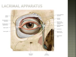
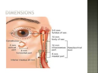
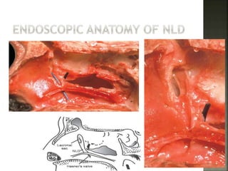
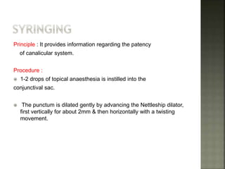
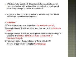
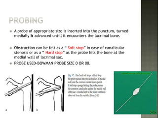
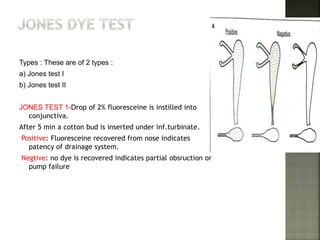
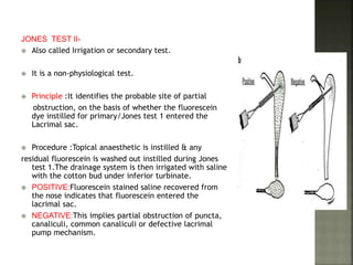
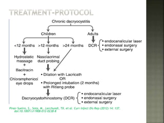
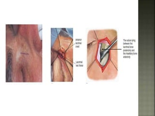
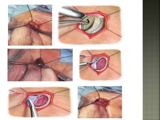
 Dacryocystitis is an infection of the lacrimal sac, secondary
to obstruction of the nasolacrimal duct at the junction of
lacrimal sac.
 The term derives from the Greek dákryon (tear), cysta (sac),
and –it is (inflammation).
 Although there are several causes, the main mechanism for
the occurrence of dacryocystitis is distal obstruction of the
nasolacrimal duct, which leads to the retention of tears
and detritus at the level of the lacrimal sac.
 Predisposing Factors.
 Factors causing stasis of tears.
 Source of infection.
 Causative organisms.
AGE:30-40 YEARS OF AGE.
GENDER:F>M.
LOW SOCIOECONOMIC STATUS.
POOR PERSONAL HYGEINE.
 Anatomical factors.
 Foreign bodies.
 Excessive lacrimation.
 Mild grade inflammation.
 Obstruction of lower end of NLD.
 GRAM POSITIVEStaphylococcus aureus and
Streptococcus pneumoniae being the most common
 Gram-negative bacteria Haemophilus influenzae,
Serratia marcescens and Pseudomonas aeruginosa
 Anaerobic microorganisms have been isolated in as many
as 15.7% of the positive cultures, in some studies, the most
common genus being Bacteroides (5.7%).
 As for fungi, they have been reported to be present in 4% to
7% of cases, the most commonly isolated genus being
Candida, although Aspergillus and Mucor may also be found
STAGE OF
CHRONIC
CATARRHAL
STAGE OF
LACRIMAL
MUCOCELE
STAGE OF
CHRONIC
SUPPORATIVE
DACROCYSTITIS
STAGE OF
CHRONIC
FIBROTIC SAC
 There is intermittent conjunctival hyperaemia and
epiphora, with mucoid discharge that is normally sterile.
 Lacrimal syringing –clear fluid or few fibrinous flakes
 Dacryocystography –block in the NLD, normal sized
lacrimal sac with healthy mucosa
 Stagnant tears collect and there is dilation of
the lacrimal sac, with mucoid content.
 Dacryocystography –distended sac with
blockage in the NLD.
 Epiphora and chronic conjunctivitis are
observed, with erythema of the lacrimal sac.
 There is reflux of purulent material with
pressure, and microorganisms are often isolated.
 •Dacryocystography – very small sac with
irregular mucosal folds.
 PHYSICAL EXAMINATION-EXCLUDE PUNCTAL
ANOMALIES,BLEPHARITIS
 ROPLAS TEST--ASSESS LACRIMAL DYSFUNCTION
 FLUORESCEIN DYE DISAPPEARANCE TEST ASSESS
LACRIMAL
DRAINAGE
 TEAR FILM BREAK UP TIME AND
PATENCY
 JONES DYE TEST
 SYRINGING AND PROBING--ASSESS LEVEL OF OBSTRUCTION
 Technique: The anterior lacrimal crest
is identified by tracing the inferior
orbital margin medially and superiorly.
 The index finger is then directed behind
the crest and used to apply
pressure on the sac area in an upward
and medial direction so as to express
the contents of the lacrimal sac into
the conjunctiva.
 Any reflux of fluid or purulent material
from the puncta is noted.
 INDICATES patent canalicular sysem
and obstruction distal to lower end of
lacrimal sac.
 Flourescein dye injected into both conjuctval
sacs & observed for 5 minutes.
 1 drop of 2% fluorescein is instilled into the
unanaesthesised conjunctival sac of both the
eyes.
 Prolonged retention indicates obstruction to
lacrimal apparatus.
Principle : It provides information regarding the patency
of canalicular system.
Procedure :
 1-2 drops of topical anaesthesia is instilled into the
conjunctival sac.
 The punctum is dilated gently by advancing the Nettleship dilator,
first vertically for about 2mm & then horizontally with a twisting
movement.
 With the eyelid stretched, dilator is withdrawn & the Lacrimal
cannula attached with syringe filled normal saline is advanced
horizontally through punctum & canaliculus.
 Irrigation is then done & the patient is asked to respond if fluid
passes into the oropharynx or nose.
 Inference :
If there is resistance to irrigation: obstruction is partial.
Regurgitation of fluid from same punctum indicates canalicular
block.
Regurgitation of fluid from upper punctum indicates blockage at
the level of common canalicular duct, lacrimal sac or
nasolacrimal duct.
 Relatively delayed regurgitation of fluid mixed with
mucous or pus usually indicates NLD blockage
 A probe of appropriate size is inserted into the punctum, turned
medially & advanced untill it encounters the lacrimal bone.
 Obstruction can be felt as a “ Soft stop” in case of canalicular
stenosis or as a “ Hard stop” as the probe hits the bone at the
medial wall of lacrimal sac.
 PROBE USED-BOWMAN PROBE SIZE 0 OR 00.
Types : These are of 2 types :
a) Jones test I
b) Jones test II
JONES TEST 1-Drop of 2% fluoresceine is instilled into
conjunctiva.
After 5 min a cotton bud is inserted under inf.turbinate.
Positive: Fluoresceine recovered from nose indicates
patency of drainage system.
Negtive: no dye is recovered indicates partial obsruction or
pump failure
JONES TEST II-
 Also called Irrigation or secondary test.
 It is a non-physiological test.
 Principle :It identifies the probable site of partial
obstruction, on the basis of whether the fluorescein
dye instilled for primary/Jones test 1 entered the
Lacrimal sac.
 Procedure :Topical anaesthetic is instilled & any
residual fluorescein is washed out instilled during Jones
test 1.The drainage system is then irrigated with saline
with the cotton bud under inferior turbinate.
 POSITIVE:Fluorescein stained saline recovered from
the nose indicates that fluorescein entered the
lacrimal sac.
 NEGATIVE:This implies partial obstruction of puncta,
canaliculi, common canaliculi or defective lacrimal
pump mechanism.
a ) Dacryocystography:
 DCG involves injection of radio-opaque dye into
the canaliculi & taking magnified images.
 Indications of DCG :
1. To confirm the site of
obstruction,especially prior to lacrimal
surgery.
2.To aid diagnosis of diverticula, fistulae &
filling defects caused by stones & tumours
 Technique:Inferior puncta are dilated with a
Nettleship punctum dilator.
 Contrast medium, usually 1-2ml Lipiodol,0.5-2ml of
water soluble iodinated contrast medium is injected
simultaneously on both the sides & anteroposterior
radiographs are taken.
b. Nuclear Lacrimal Scintigraphy:
 Nuclear lacrimal scintigraphy is a simple, noninvasive
physiological test that evaluates patency of the
lacrimal system.
 Scintigraphy uses a radiotracer (technetium-99m
pertechnetate), which is very easily detectable with
a gamma camera.
 The disadvantage as compared to DCG is that it
fails to show finer anatomical details.
 Apart from being a non-invasive procedure,radiation
exposure to lens is minimal compared to DCG.
 It is more sensitive in assessing incomplete blocks.
MRI:-ADVANTAGES--
MRI is reserved in cases
where tumors are
suspected.
a) Chemiluminiscene test : Cyalume, a chemiluminiscent material is
injected with a sialography catheter to demonstrate the patency of
outflow passages.
b) Dacryoscopy : Dacryoscope, a mini rigid endoscope allows the direct
visualisation of the interior & lining of lacrimal passages.
c) Standardised echography :Gross anatomical structural defects can be
evaluated.
d)Thermography : It is used in conjunction with routine lacrimal irrigation to
visualise the tear ducts in normal subjects and in patients with
obstructive epiphora.assess the degree of tissue inflammation.
Pinar-Sueiro, S., Sota, M., Lerchundi, TX. et al. Curr Infect Dis Rep (2012) 14: 137.
doi:10.1007/s11908-012-0238-8
 Balloons were first used by Becker and Berry in 1989.
 2 mm balloon is used for patients less than 30 months of
age and 3 mm for children more than 30 months of age.
Also available are 4mm and 5mm balloons.
INDICATIONS:
 Failed probing
 Failed intubation
 Older children (> 12 months of age)
 Down’s syndrome or any syndromic association with
CNLDO.
 Reduced operative trauma
 Less bleeding
 Faster and less time consuming
 No need for powered endoscopic instruments.
 Less postoperative morbidity.
 Early rehabilitation
 High success rates.
INDICATIONS:
1.Punctum and/or canalicular stenosis- Stent mechanically
dilates the punctum and vertical canaliculus, restores
normal tear outflow and prevents re-stenosis.
2.Traumatic canalicular laceration/tear.
3.Congenital nasolacrimal duct obstruction (CNLDO) – In case
primary probing fails, intubation with stents can be considered as
a good option.
4. To maintain the internal canalicular opening (ICO)
and ostium patency after DCR.
MONOCANALICULAR
MONOKA-TM STENT--The silicone
stent has an external diameter of
0.64mm
with 80 mm long and and have
metal bodkin
MONOKA-CRAWFORD STENT-
MODIFICATION OF MONOKA
BICANALICULAR
CRAWFORD STENTS-WIDER
DIAMETER OF 0.93MM
RITLENG INTUBATION DEVICE
MONAKA TM STENT MONOKA-CRAWFORD STENT
CRAWFORD BICANALICULAR RITLENG INTUBATION DEVICE
 Dacryocystorhinostomy (DCR) introduced by Toti in 1904 is a lacrimal
drainage operation in which a fistula is created between the lacrimal sac
and the nasal cavity in order to bypass an obstruction in the nasolacrimal
duct.
 Endonasal-DCR first described by caldwell 1893
 The first modern endoscopic endonasal DCR procedures were
described by McDonogh and Meiring in 1989.
Types: 1. EXTERNAL DCR
2. ENDONASAL/ENDOSCOPIC DCR
3. ENDOLASER DCR
 The main advantage of external DCR is visualization of the
anatomy that facilitates the precise removal of bone in the
lacrimal fossa and enables the exact anastomosis of the
nasal mucosa and lacrimal sac.
 Chronic epiphora due to a nasolacrimal duct
obstruction.
 Recurrent or chronic dacryocystitis.
 Failed probing and silicone intubations in a
child.
 Proposed intraocular surgery in the presence of
nasolacrimal duct obstruction.
Under GA;curved incision along medial to
medial canthus is givenMedial palpebral
ligament is exposed by blunt dissection to
expose anterior lacrimal crest.
Periosteum is seperated from anterior lacrimal
crest & lacrimal sac is reflected laterally with
blunt dissectorexpose nasal mucosa
Probe is introduced into sac through lower
canaliculus & sac is incised verticallyfashining of
nasal mucosa into H shaped
Suturing of flaps by 6-0 vicryl is done
Medial palpebral ligament is sutured to
periosteum;orbicularis muscle sutured
with 6-0 vicryl
 The reported success rate of this surgery ranges from 80 to 95 %, with the
major risks being wound complications (scar, infection, ectropion, or
disruption of the medial canthal ligament) and nosebleeds.
 Mean operative time is shorter in endonasal DCR than in external DCR
 Postoperative cutaneous scarring was unique to the external DCR
procedure.
 Post operative bleeding No significant difference
INDICATIONS:
 Patients with NLD obstruction and a previous history of
sinus surgery.
 Facial trauma
 Failed external DCR are good candidates for endoscopic
DCR.
 In cases of previously failed DCR, endoscopy
can help visualize previous scarring.
 Adolescents with anatomic variations from atypical forms of
congenital dacryostenosis may also benefit from endoscopic
DCR.
 Acute dacryocystitis.
 Patient is on anticoagulation medications and
is unable to stop perioperatively.
 Obstruction in the canaliculi or puncti.
 The success rates of endoscopic DCR reported in the literature
range from 79.4 to 96 %.
 Prevention of an external scar in the area of the medial
canthus.
 Preservation of the pump mechanism of the orbicularis
muscle.
 Decreased hospital stay.
 Less postoperative discomfort.
 Simultaneously can correct intranasal pathologic
conditions such as paranasal sinusitis and septal deviation
via endoscopic instrumentation.
 More rapid rehabilitation and a shorter duration of
surgery.
CREATE A POSTERIORLY BASED FLAP
IINCISIONSuperior icision made horizontally 10
mm above the axilla of middle turbinate—Extend
posteriorly to axilla to 10mm on to the frontal
process of maxila
Inferioly incision is made just superior to
inferior turbinate.
ELEVATION OF MUCOSAL FLAP—MADE BY
SUCTION FREER—IDENTIFY THE LACRIMAL BONE
REMOVAL OF LACRIMAL BONE.
REMOVAL OF FRONTAL PROCESS OF AXILLA –
KERISON PUNCH OR A DCR DRILL BURR—SAC IS
THEN EXPOSED AS A BULDGE.
EXPOSURE OF AGGER NASI CELL—
ALLOWING FOR WIDER
MARSUPIALIZATON OF LACRIMAL SAC
MARSUPALIZE OF SAC-PROBE IS THEN
INSERTED VIA INFERIOR CANALICULUS
AND TENT THE LACRIMAL SAC
INCISION AT SUPERIOR AND INFERIOR
SAC AND VERICALY INCISED TO CREATE
ANT AND POST FLAP—LYING FLAT ON
LATERAL WALL
REPOSITION OF THE MUCOSAL FLAP—
CUT THE MUCOSAL FLAP SO ONLY
SUPERIOR AND INFERIOR LIMB ARE
PRESENT
PASS OF CRAWFORD SILASTIC TUBES—
LACRIMAL TUBES ARE INSERTED INTO
THE SUP AND INFERIOR PUNCTAE AND
PASSED INTO NASAL CAVITY AND TIE
TOGETHER
 Patient hospitalization is 1 day, if no complications occur.
 A periodical wash cleaning of nasal fossae with saline
solution with local antibiotics, is recommended at home.
 The oral administration of antibiotics is
not usually recommended.
 Stents can be removed after four to six weeks.
ADVANTAGE
1.CAN BE PERFORMED
UNDER LA
2.CAN BE PERFORMED IN
ANTICOAGULATIVE
PATIENTS
3.SHORTER OPERATIVE
TIME
4.EFFECTIVE HEMOSTASIS
DISADVANTAGE
LOWER SUCCESS RATE 60-
80%
EXPENSIVE &THERMAL
DAMAGES
Weber, et al. Atlas of
Lacrimal
Surgery, Springer, 2009
 Lid hematoma and orbital fat prolapse (up to 28 % of cases).
 Epistaxis in 2% of patients.
 Less frequent adverse reactions include
Obstruction and subsequent rhinosinusitis—frontal and maxilla.
Orbital penetration with damage to EOM.
Retrobulbar hematoma leading to vision loss.
Canalicular stenosis.
Conjunctival fistula and subcutaneous emphysema.
CSF leak.
Ultrasonic or piezoelectric-assisted or powered
endoscopic DCR
A diamond cutting tip at a flow rate of 30 ml/min with D2 program
with power of 5 is used to begin the osteotomy from the inferior end
of the maxillary line
Endoilluminator and 23G Vitrectomy retinal light pipe
 The diode laser setting used is at an average of 10 W with continuous
laser delivery using the contact mode.
 600mico semirigid laser fiber optic is inserted in lower punctum into
the canaliculus up to the level of the lacrimal sac in a 45° fashion.
 0° nasal video endoscope attached to a TV monitor is inserted
through the nostril to visualize the transilluminated laser light (laser
glow )from sac
 Absence of a skin incision
 Preservation of the medical canthal structures
 Preservation of the lacrimal pump mechanism
 Less operative time
 Local anesthesia and outpatient surgery
 Minimal intraoperative and postoperative bleeding
 Decrease or no periorbital swelling postoperatively
 Low morbidity
 Shorter functional recovery
 Scott –brown.
 Metson R, Woog JJ, Puliafi to CA (1994) Endoscopic laser dacryocystorhinostomy. Laryngoscope.
 Sprekelsen MM, Barberan MT (1996) Endoscopic dacryocystorhinostomy: surgical technique and
results. Laryngoscope.
 Woog JJ, Kennedy RH, Custer PL, Kaltreider SA, Meyer DR, Camara JG (2001) Endonasal
dacryocystorhinostomy:a report by the American Academy of Ophthalmology. Ophthalmology 108:2369–2377
 Traquair H (1941) Chronic dacryocystitis: its causation and treatment. Arch Ophthalmol 26:165–180.
 Weidenbecher M, Hosemann W, Buhr W (1994) Endoscopic endonasal dacryocystorhinostomy: results in 56
patients. Ann Otol Rhinol Laryngol.
 Chandler PA (1936) Dacryocystorhinostomy. TransAm Ophthalmol Soc.
 Caldwell GW (1893) Two new operations for obstruction of the nasal duct, with preservation of the canaliculi
and an incidental description of a new lacrymalprobe. N Y Med J 57:581–582
 McDonogh M, Meiring JH (1989) Endoscopic transnasal dacryocystorhinostomy. J Laryngol Otol
 Parsons disease of eye.

CHRONIC DACROCYSTITIS AND ITS MANAGEMENT

  • 1.
  • 6.
     Dacryocystitis isan infection of the lacrimal sac, secondary to obstruction of the nasolacrimal duct at the junction of lacrimal sac.  The term derives from the Greek dákryon (tear), cysta (sac), and –it is (inflammation).  Although there are several causes, the main mechanism for the occurrence of dacryocystitis is distal obstruction of the nasolacrimal duct, which leads to the retention of tears and detritus at the level of the lacrimal sac.
  • 7.
     Predisposing Factors. Factors causing stasis of tears.  Source of infection.  Causative organisms.
  • 8.
    AGE:30-40 YEARS OFAGE. GENDER:F>M. LOW SOCIOECONOMIC STATUS. POOR PERSONAL HYGEINE.
  • 9.
     Anatomical factors. Foreign bodies.  Excessive lacrimation.  Mild grade inflammation.  Obstruction of lower end of NLD.
  • 10.
     GRAM POSITIVEStaphylococcusaureus and Streptococcus pneumoniae being the most common  Gram-negative bacteria Haemophilus influenzae, Serratia marcescens and Pseudomonas aeruginosa  Anaerobic microorganisms have been isolated in as many as 15.7% of the positive cultures, in some studies, the most common genus being Bacteroides (5.7%).  As for fungi, they have been reported to be present in 4% to 7% of cases, the most commonly isolated genus being Candida, although Aspergillus and Mucor may also be found
  • 12.
    STAGE OF CHRONIC CATARRHAL STAGE OF LACRIMAL MUCOCELE STAGEOF CHRONIC SUPPORATIVE DACROCYSTITIS STAGE OF CHRONIC FIBROTIC SAC
  • 13.
     There isintermittent conjunctival hyperaemia and epiphora, with mucoid discharge that is normally sterile.  Lacrimal syringing –clear fluid or few fibrinous flakes  Dacryocystography –block in the NLD, normal sized lacrimal sac with healthy mucosa
  • 14.
     Stagnant tearscollect and there is dilation of the lacrimal sac, with mucoid content.  Dacryocystography –distended sac with blockage in the NLD.
  • 15.
     Epiphora andchronic conjunctivitis are observed, with erythema of the lacrimal sac.  There is reflux of purulent material with pressure, and microorganisms are often isolated.
  • 16.
     •Dacryocystography –very small sac with irregular mucosal folds.
  • 17.
     PHYSICAL EXAMINATION-EXCLUDEPUNCTAL ANOMALIES,BLEPHARITIS  ROPLAS TEST--ASSESS LACRIMAL DYSFUNCTION  FLUORESCEIN DYE DISAPPEARANCE TEST ASSESS LACRIMAL DRAINAGE  TEAR FILM BREAK UP TIME AND PATENCY  JONES DYE TEST  SYRINGING AND PROBING--ASSESS LEVEL OF OBSTRUCTION
  • 18.
     Technique: Theanterior lacrimal crest is identified by tracing the inferior orbital margin medially and superiorly.  The index finger is then directed behind the crest and used to apply pressure on the sac area in an upward and medial direction so as to express the contents of the lacrimal sac into the conjunctiva.  Any reflux of fluid or purulent material from the puncta is noted.  INDICATES patent canalicular sysem and obstruction distal to lower end of lacrimal sac.
  • 19.
     Flourescein dyeinjected into both conjuctval sacs & observed for 5 minutes.  1 drop of 2% fluorescein is instilled into the unanaesthesised conjunctival sac of both the eyes.  Prolonged retention indicates obstruction to lacrimal apparatus.
  • 20.
    Principle : Itprovides information regarding the patency of canalicular system. Procedure :  1-2 drops of topical anaesthesia is instilled into the conjunctival sac.  The punctum is dilated gently by advancing the Nettleship dilator, first vertically for about 2mm & then horizontally with a twisting movement.
  • 21.
     With theeyelid stretched, dilator is withdrawn & the Lacrimal cannula attached with syringe filled normal saline is advanced horizontally through punctum & canaliculus.  Irrigation is then done & the patient is asked to respond if fluid passes into the oropharynx or nose.  Inference : If there is resistance to irrigation: obstruction is partial. Regurgitation of fluid from same punctum indicates canalicular block. Regurgitation of fluid from upper punctum indicates blockage at the level of common canalicular duct, lacrimal sac or nasolacrimal duct.  Relatively delayed regurgitation of fluid mixed with mucous or pus usually indicates NLD blockage
  • 22.
     A probeof appropriate size is inserted into the punctum, turned medially & advanced untill it encounters the lacrimal bone.  Obstruction can be felt as a “ Soft stop” in case of canalicular stenosis or as a “ Hard stop” as the probe hits the bone at the medial wall of lacrimal sac.  PROBE USED-BOWMAN PROBE SIZE 0 OR 00.
  • 23.
    Types : Theseare of 2 types : a) Jones test I b) Jones test II JONES TEST 1-Drop of 2% fluoresceine is instilled into conjunctiva. After 5 min a cotton bud is inserted under inf.turbinate. Positive: Fluoresceine recovered from nose indicates patency of drainage system. Negtive: no dye is recovered indicates partial obsruction or pump failure
  • 24.
    JONES TEST II- Also called Irrigation or secondary test.  It is a non-physiological test.  Principle :It identifies the probable site of partial obstruction, on the basis of whether the fluorescein dye instilled for primary/Jones test 1 entered the Lacrimal sac.  Procedure :Topical anaesthetic is instilled & any residual fluorescein is washed out instilled during Jones test 1.The drainage system is then irrigated with saline with the cotton bud under inferior turbinate.  POSITIVE:Fluorescein stained saline recovered from the nose indicates that fluorescein entered the lacrimal sac.  NEGATIVE:This implies partial obstruction of puncta, canaliculi, common canaliculi or defective lacrimal pump mechanism.
  • 25.
    a ) Dacryocystography: DCG involves injection of radio-opaque dye into the canaliculi & taking magnified images.  Indications of DCG : 1. To confirm the site of obstruction,especially prior to lacrimal surgery. 2.To aid diagnosis of diverticula, fistulae & filling defects caused by stones & tumours
  • 26.
     Technique:Inferior punctaare dilated with a Nettleship punctum dilator.  Contrast medium, usually 1-2ml Lipiodol,0.5-2ml of water soluble iodinated contrast medium is injected simultaneously on both the sides & anteroposterior radiographs are taken.
  • 27.
    b. Nuclear LacrimalScintigraphy:  Nuclear lacrimal scintigraphy is a simple, noninvasive physiological test that evaluates patency of the lacrimal system.  Scintigraphy uses a radiotracer (technetium-99m pertechnetate), which is very easily detectable with a gamma camera.  The disadvantage as compared to DCG is that it fails to show finer anatomical details.  Apart from being a non-invasive procedure,radiation exposure to lens is minimal compared to DCG.  It is more sensitive in assessing incomplete blocks.
  • 28.
    MRI:-ADVANTAGES-- MRI is reservedin cases where tumors are suspected.
  • 29.
    a) Chemiluminiscene test: Cyalume, a chemiluminiscent material is injected with a sialography catheter to demonstrate the patency of outflow passages. b) Dacryoscopy : Dacryoscope, a mini rigid endoscope allows the direct visualisation of the interior & lining of lacrimal passages. c) Standardised echography :Gross anatomical structural defects can be evaluated. d)Thermography : It is used in conjunction with routine lacrimal irrigation to visualise the tear ducts in normal subjects and in patients with obstructive epiphora.assess the degree of tissue inflammation.
  • 30.
    Pinar-Sueiro, S., Sota,M., Lerchundi, TX. et al. Curr Infect Dis Rep (2012) 14: 137. doi:10.1007/s11908-012-0238-8
  • 31.
     Balloons werefirst used by Becker and Berry in 1989.  2 mm balloon is used for patients less than 30 months of age and 3 mm for children more than 30 months of age. Also available are 4mm and 5mm balloons. INDICATIONS:  Failed probing  Failed intubation  Older children (> 12 months of age)  Down’s syndrome or any syndromic association with CNLDO.
  • 32.
     Reduced operativetrauma  Less bleeding  Faster and less time consuming  No need for powered endoscopic instruments.  Less postoperative morbidity.  Early rehabilitation  High success rates.
  • 33.
    INDICATIONS: 1.Punctum and/or canalicularstenosis- Stent mechanically dilates the punctum and vertical canaliculus, restores normal tear outflow and prevents re-stenosis. 2.Traumatic canalicular laceration/tear. 3.Congenital nasolacrimal duct obstruction (CNLDO) – In case primary probing fails, intubation with stents can be considered as a good option. 4. To maintain the internal canalicular opening (ICO) and ostium patency after DCR.
  • 34.
    MONOCANALICULAR MONOKA-TM STENT--The silicone stenthas an external diameter of 0.64mm with 80 mm long and and have metal bodkin MONOKA-CRAWFORD STENT- MODIFICATION OF MONOKA BICANALICULAR CRAWFORD STENTS-WIDER DIAMETER OF 0.93MM RITLENG INTUBATION DEVICE
  • 35.
    MONAKA TM STENTMONOKA-CRAWFORD STENT CRAWFORD BICANALICULAR RITLENG INTUBATION DEVICE
  • 36.
     Dacryocystorhinostomy (DCR)introduced by Toti in 1904 is a lacrimal drainage operation in which a fistula is created between the lacrimal sac and the nasal cavity in order to bypass an obstruction in the nasolacrimal duct.  Endonasal-DCR first described by caldwell 1893  The first modern endoscopic endonasal DCR procedures were described by McDonogh and Meiring in 1989. Types: 1. EXTERNAL DCR 2. ENDONASAL/ENDOSCOPIC DCR 3. ENDOLASER DCR
  • 37.
     The mainadvantage of external DCR is visualization of the anatomy that facilitates the precise removal of bone in the lacrimal fossa and enables the exact anastomosis of the nasal mucosa and lacrimal sac.
  • 38.
     Chronic epiphoradue to a nasolacrimal duct obstruction.  Recurrent or chronic dacryocystitis.  Failed probing and silicone intubations in a child.  Proposed intraocular surgery in the presence of nasolacrimal duct obstruction.
  • 39.
    Under GA;curved incisionalong medial to medial canthus is givenMedial palpebral ligament is exposed by blunt dissection to expose anterior lacrimal crest. Periosteum is seperated from anterior lacrimal crest & lacrimal sac is reflected laterally with blunt dissectorexpose nasal mucosa Probe is introduced into sac through lower canaliculus & sac is incised verticallyfashining of nasal mucosa into H shaped Suturing of flaps by 6-0 vicryl is done Medial palpebral ligament is sutured to periosteum;orbicularis muscle sutured with 6-0 vicryl
  • 42.
     The reportedsuccess rate of this surgery ranges from 80 to 95 %, with the major risks being wound complications (scar, infection, ectropion, or disruption of the medial canthal ligament) and nosebleeds.  Mean operative time is shorter in endonasal DCR than in external DCR  Postoperative cutaneous scarring was unique to the external DCR procedure.  Post operative bleeding No significant difference
  • 43.
    INDICATIONS:  Patients withNLD obstruction and a previous history of sinus surgery.  Facial trauma  Failed external DCR are good candidates for endoscopic DCR.  In cases of previously failed DCR, endoscopy can help visualize previous scarring.  Adolescents with anatomic variations from atypical forms of congenital dacryostenosis may also benefit from endoscopic DCR.
  • 44.
     Acute dacryocystitis. Patient is on anticoagulation medications and is unable to stop perioperatively.  Obstruction in the canaliculi or puncti.  The success rates of endoscopic DCR reported in the literature range from 79.4 to 96 %.
  • 45.
     Prevention ofan external scar in the area of the medial canthus.  Preservation of the pump mechanism of the orbicularis muscle.  Decreased hospital stay.  Less postoperative discomfort.  Simultaneously can correct intranasal pathologic conditions such as paranasal sinusitis and septal deviation via endoscopic instrumentation.  More rapid rehabilitation and a shorter duration of surgery.
  • 46.
    CREATE A POSTERIORLYBASED FLAP IINCISIONSuperior icision made horizontally 10 mm above the axilla of middle turbinate—Extend posteriorly to axilla to 10mm on to the frontal process of maxila Inferioly incision is made just superior to inferior turbinate. ELEVATION OF MUCOSAL FLAP—MADE BY SUCTION FREER—IDENTIFY THE LACRIMAL BONE REMOVAL OF LACRIMAL BONE. REMOVAL OF FRONTAL PROCESS OF AXILLA – KERISON PUNCH OR A DCR DRILL BURR—SAC IS THEN EXPOSED AS A BULDGE. EXPOSURE OF AGGER NASI CELL— ALLOWING FOR WIDER MARSUPIALIZATON OF LACRIMAL SAC
  • 47.
    MARSUPALIZE OF SAC-PROBEIS THEN INSERTED VIA INFERIOR CANALICULUS AND TENT THE LACRIMAL SAC INCISION AT SUPERIOR AND INFERIOR SAC AND VERICALY INCISED TO CREATE ANT AND POST FLAP—LYING FLAT ON LATERAL WALL REPOSITION OF THE MUCOSAL FLAP— CUT THE MUCOSAL FLAP SO ONLY SUPERIOR AND INFERIOR LIMB ARE PRESENT PASS OF CRAWFORD SILASTIC TUBES— LACRIMAL TUBES ARE INSERTED INTO THE SUP AND INFERIOR PUNCTAE AND PASSED INTO NASAL CAVITY AND TIE TOGETHER
  • 48.
     Patient hospitalizationis 1 day, if no complications occur.  A periodical wash cleaning of nasal fossae with saline solution with local antibiotics, is recommended at home.  The oral administration of antibiotics is not usually recommended.  Stents can be removed after four to six weeks.
  • 49.
    ADVANTAGE 1.CAN BE PERFORMED UNDERLA 2.CAN BE PERFORMED IN ANTICOAGULATIVE PATIENTS 3.SHORTER OPERATIVE TIME 4.EFFECTIVE HEMOSTASIS DISADVANTAGE LOWER SUCCESS RATE 60- 80% EXPENSIVE &THERMAL DAMAGES Weber, et al. Atlas of Lacrimal Surgery, Springer, 2009
  • 50.
     Lid hematomaand orbital fat prolapse (up to 28 % of cases).  Epistaxis in 2% of patients.  Less frequent adverse reactions include Obstruction and subsequent rhinosinusitis—frontal and maxilla. Orbital penetration with damage to EOM. Retrobulbar hematoma leading to vision loss. Canalicular stenosis. Conjunctival fistula and subcutaneous emphysema. CSF leak.
  • 51.
    Ultrasonic or piezoelectric-assistedor powered endoscopic DCR A diamond cutting tip at a flow rate of 30 ml/min with D2 program with power of 5 is used to begin the osteotomy from the inferior end of the maxillary line
  • 52.
    Endoilluminator and 23GVitrectomy retinal light pipe
  • 53.
     The diodelaser setting used is at an average of 10 W with continuous laser delivery using the contact mode.  600mico semirigid laser fiber optic is inserted in lower punctum into the canaliculus up to the level of the lacrimal sac in a 45° fashion.  0° nasal video endoscope attached to a TV monitor is inserted through the nostril to visualize the transilluminated laser light (laser glow )from sac
  • 54.
     Absence ofa skin incision  Preservation of the medical canthal structures  Preservation of the lacrimal pump mechanism  Less operative time  Local anesthesia and outpatient surgery  Minimal intraoperative and postoperative bleeding  Decrease or no periorbital swelling postoperatively  Low morbidity  Shorter functional recovery
  • 56.
     Scott –brown. Metson R, Woog JJ, Puliafi to CA (1994) Endoscopic laser dacryocystorhinostomy. Laryngoscope.  Sprekelsen MM, Barberan MT (1996) Endoscopic dacryocystorhinostomy: surgical technique and results. Laryngoscope.  Woog JJ, Kennedy RH, Custer PL, Kaltreider SA, Meyer DR, Camara JG (2001) Endonasal dacryocystorhinostomy:a report by the American Academy of Ophthalmology. Ophthalmology 108:2369–2377  Traquair H (1941) Chronic dacryocystitis: its causation and treatment. Arch Ophthalmol 26:165–180.  Weidenbecher M, Hosemann W, Buhr W (1994) Endoscopic endonasal dacryocystorhinostomy: results in 56 patients. Ann Otol Rhinol Laryngol.  Chandler PA (1936) Dacryocystorhinostomy. TransAm Ophthalmol Soc.  Caldwell GW (1893) Two new operations for obstruction of the nasal duct, with preservation of the canaliculi and an incidental description of a new lacrymalprobe. N Y Med J 57:581–582  McDonogh M, Meiring JH (1989) Endoscopic transnasal dacryocystorhinostomy. J Laryngol Otol  Parsons disease of eye.