call girls in Kamla Market (DELHI) 🔝 >༒9953330565🔝 genuine Escort Service 🔝✔️✔️
Extrapyramidal system.pdf very good details
1.
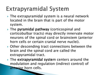
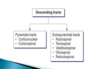
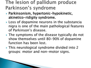
2. The extrapyramidal system is a neural network
located in the brain that is part of the motor
system.
The pyramidal pathway (corticospinal and
corticobulbar tracts) may directly innervate motor
neurons of the spinal cord or brainstem (anterior
horn cells or certain cranial nerve nuclei).
Other descending tract connections between the
brain and the spinal cord are called the
extrapyramidal tract.
The extrapyramidal system centers around the
modulation and regulation (indirect control) of
anterior horn cells.
3.
4. Responds for purposive movements
Regulates the muscle tone
Responds for nonvoluntary movements
5. The basal ganglia are a part of the motor system. The
principal nuclei of the basal ganglia are the caudate
nucleus, the putamen, and the globus pallidus, all of which
lie in the subcortical white matter of the telencephalon.
These nuclei are connected to each other, and to the
motor cortex, in complex regulatory circuits.
They exert both excitatory and inhibitory effects on the
motor cortex. They play an important role in the initiation
and modulation of movement and in the control of muscle
tone.
Lesions of the basal ganglia, and of other, functionally
related nuclei, such as the substantia nigra and the
subthalamic nucleus, can produce either an excess or a
deficiency of movement-related impulses, and/or
pathological alterations of muscle tone.
The most common disease of the basal ganglia is
Parkinson disease, which is characterized by the clinical
triad of rigidity, akinesia, and tremor.
8. The corpus striatum receives afferent input from extensive
areas of the cerebral cortex, particularly the motor areas
of the frontal lobe. These cortical afferents are derived
from projection neurons of the cerebral cortex (pyramidal
cells), are glutamatergic, run ipsilaterally. There are
probably no reciprocal fibers running from the corpus
striatum back to the cortex. A further point-to-point
afferent input to the corpus striatum is derived from the
centromedian nucleus of the thalamus, and is probably
excitatory.
This afferent pathway transmits impulses from the
cerebellum and the midbrain reticular formation to the
striatum.
The substantia nigra sends dopaminergic afferent fibers to
the striatum, whose loss is the cause of Parkinson disease.
The globus pallidus derives its major afferent input from
the corpus striatum and receives no direct afferent fibers
from the cerebral cortex.
9. The major efferent projections of the corpus
striatum go to the external and internal
segments of the globus pallidus. Further
efferent fibers travel to the substantia nigra.
The cells of origin of the striatal efferent fibers
are GABAergic neurons, the most common cell
type in the striatum.
Efferent pathways of the globus pallidus.
The major contingent of efferent fibers runs to
the thalamus, which, in turn, projects to the
cerebral cortex, completing a feedback loop.
11. Originated from
the red nucleus located
in the midbrain
Terminates in the
lateral column
of spinal cord
Functions-Facilities
flexor movements,
inhibits extensor muscles.
Motor function of skeletal
muscles of limbs, hands,
and feet.
12. A-pontine reticulospinal
tract (medial & excitatory)
Originated from pontine
reticular nuclei in pons
which terminate in the
medial anterior column.
B-medullary reticulospinal
tract(lateral & inhibitory)
Originated from medulla
and terminate in lateral
anterior column.
Function-it facilitates
extensor reflexes&
inhibits flexor reflexes.
13. Origin-vestibular nucleus
in medulla.
Terminate in the
anterior motor neuron
Function-Facilitates
extensors, inhibits
flexors.
Motor function of
muscle for maintaining
balance in response
to head movements.
14. Projects from the
midbrain to the spinal
cord and is important
for postural movements
that are driven by the
colliculus, which
is the tectum .
15. Romberg sign
Ability to maintain upright
position with feet together
and eyes open
Sway/fall when eyes closed
Indicates impaired
proprioception or
vestibular dysfunction
16. Control, precision, rhythm, synergy of
movement
Test at rest and with action in trunk and limbs:
Finger-nose-finger test
Rapid alternating movements
Finger or toe tapping test
17. Ask the patient to touch
your finger with his or
her index finger, then
to the tip of the nose.
You may move your
target finger in different
directions.
Do one arm at a time.
1.Patient accurately
performs the task: normal
2.Patient develops tremor
when approaching the
target (finger or his nose)-
intention tremor: cerebellar
disease or resting
tremor-lesion of pallidum.
3.Patient misses the target:
past-pointing or dysmetria.
18. Demonstrate to the
patient (finger tapping,
hand tapping, etc.),
first in slow motion,
and then faster.
If the patient is able
to do the task with
normal rate and
rhythm-normal.
If movements are
irregular, disorganized,
disrhythmic.
19. Posture of body and limbs
Length, speed, and rhythm of steps
Symmetry and base of gait
Steadiness
Arm swing
Turns
Test with normal gait, toe walking, heel
walking, tandem walking
20. Parkinsonism, hypertonic-hypokinetic,
akinetico-ridigity syndrome.
Loss of dopamine neurons in the substancia
nigra is one of the main pathological features
of Parkinson’s disease.
The symptoms of the disease typically do not
show themselves until 80-90% of dopamine
function has been lost.
This neurological syndrome divided into 2
groups: motor and non-motor signs.
21. Rigidity: stiffness; increased muscle tone in plastic
type. This produces a “cogwheel” rigidity when the
limb is passively moved. This resistance is present to
the same degree throughout the full range of
movement, affecting flexor and extensor muscle
groups equally and described as plastic rigidity.
Rigidity predominates in the flexor muscles of the
neck, trunk and limbs and result is Flexed-Bent
Posture.
The resting tremor, which is classically seen as a
“pill-rolling” action of the hands that may also affect
the chin, lips, legs, and trunk, can be markedly
increased by stressor emotions.
It occurs-at rest
Decrease-with movement
Disappear-during sleep.
Hypokinesia refers to slow or diminished movement
of body musculature.
Bradykinesia: this is a slowness of movement.
Postural instability: leads to impaired balance and
falls.
22.
23. Shuffing: gait is
characterized by short
steps, with feet barely
leaving the ground,
producing an audible
shuffling noise.
Acheirokinesis-
disappearance of
cooperation movements.
Stooped, forward-
flexed posture. In severe
forms, the head and
upper shoulders may be
bent at 90 degree relative
to the trunk.
Propultion-involuntary
jerky movements to ahead.
Lateropultion-to the sides
Retropultion-to the back
24. Festination: a combination of stooped
posture, imbalance, and short steps. It leads
to a gait that gets progressively faster and
faster, often ending in a fall.
Gait freezing: “freezing” is a manifestation of
akinesia (an inability to move). Gait freezing
is characterized by an inability to move the
feet which may worsen in tight, when
attempting to initiate gait.
25. Monotonic speech.
Bradilalia-is slowing of speech.
Drooling: (also known as sialorhea) is when saliva
flows outside the mouth. Most likely caused by a
weak, infrequent swallow and stooped posture.
Masked faces (a mask-like face also known as
hypomimia, with infrequent blinking.
Micrographia(small, cramped handwriting).
Impaired fine motor dexterity and motor
coordination.
Bradipsychizm- the slowing of mental processes.
Akayria means obtrusive (one asks the same
questions).
26. Hyperkines-is an involuntary, irregular jerking movements
affecting limb and axial muscle groups.
Disappear-during sleeping
Increase-with movements, emotions.
They are divided into 2 groups:
Rhythmic
Nonrhythmic
Rhythmic disorders are primarily tremors-regular
alternating or oscillatory movements, which can occur
mainly at rest, while maintaining a position, and/or during
attempted movement. However, in some cases, a tremor,
though rhythmic, is irregular, as occurs when tremor is
associated with dystonic disorders.
Nonrhythmic hyperkinetic disorders can be
-Slow (athetosis)
-Sustained (dystonia)
-Rapid(myoclonus, chorea, tics, hemiballismus)
Rapid nonrhytmic hyperkinetic disorders may be
-Suppressible (tics)
-Nonsuppressible(chorea)
27. Athetosis- irregular repetitive writhing
movements affecting face, distal part limbs.
Increase with emotions.
28. Chorea-irregular repetitive jerking movement
affecting limbs and axial muscles. It is like
dancing, clown around. Patient close eyes-
put out his tongue-make grimace-lift his
arm.
30. Torticollis(or wry neck)-unilateral deviation of
the head due to dystonic contraction of one
of the sternocleidomastoid muscles.
31. Torsion dystonia-slow sustained abnormal
movement, corkscrew movement of trunk
around his own axis.
32. Blepharospasm: a type of dystonia causes
involuntary contraction of the eyelids. The
main concern for this dystonia is that it can
cause the eyelids to close involuntary and for
indefinite periods of time.