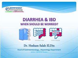
Diarrhea and IBD When to be woeeied ?
•Download as PPTX, PDF•
1 like•56 views

UCHID 2019

Recommended
More Related Content
What's hot
What's hot (20)
Similar to Diarrhea and IBD When to be woeeied ?
Similar to Diarrhea and IBD When to be woeeied ? (20)
More from Dr-Hesham Salah
Recently uploaded
Recently uploaded (20)
Diarrhea and IBD When to be woeeied ?
- 1. Head of Gastroenterology , Hepatology Department Qabbary Specialized Hospital
- 3. Diarrhea: An increase in the fluidity , frequency (3 motions /d) and the volume of daily stool output(>200 g/d) Acute Diarrhea : <14 days Persistent Diarrhea : 14-30 days Chronic Diarrhea : > 30 days
- 4. Pathophysiology of Diarrhea: 1- Osmotic Diarrhea . In which there is increased amounts of poorly absorbable osmoticaly active solutes in the gut lumen 2- Secretory Diarrhea in which there is increased Cl. And water secretion 3- Exudation of mucus , blood and proteins from sites of active inflammation in to the bowel lumen. 4- Abnormal intestinal motility
- 5. British Society of Gastroenterology Arasaradnam RP, et al. Gut 2018 British Society of Gastroenterology Arasaradnam RP, et al. Gut 2018
- 6. ETIOLOGY British Society of Gastroenterology Arasaradnam RP, et al. Gut 2018
- 10. INDICATIONS FOR REFERRAL TO A GASTROENTEROLOGIST GIT
- 11. Indications for referral include anyone of the following: • Alarm features • Severe diarrhea • Suspected inflammatory bowel disease • Inconclusive diagnosis after initial evaluation • Failure to respond to empiric therapy Referral to a gastroenterologist may also be appropriate in patients who require long-term management for the underlying cause (eg, inflammatory bowel disease, chronic pancreatitis)
- 12. INITIAL EVALUATION — The initial evaluation of patient with chronic diarrhea includes history, physical examination, and laboratory testing to determine whether the patient has any alarm findings which help to distinguish organic from functional diarrhea and characterizing the diarrhea in order to direct the need for additional evaluation. Evaluation for alarm features — Alarm features in patients with chronic diarrhea may be suggestive of an underlying organic etiology .These features include the following • Age of onset after age 50 years • Rectal bleeding or melena • Nocturnal pain or diarrhea • Progressive abdominal pain • Unexplained weight loss, fever, or other systemic symptoms • Laboratory abnormalities (iron deficiency anemia, elevated C-reactive protein or fecal calprotectin) • Family history of inflammatory bowel disease (IBD) or colorectal cancer
- 13. RECOMMENDATIONS 1. Stool and laboratory tests should be the initial step for the evaluation of clinical scenarios suggestive of infectious diarrhea. 2. In patients with chronic unexplained diarrhea, we suggest colonoscopy with random biopsies of the right and left side of the colon. Sigmoidoscopy is an alternative option, although this may miss right-sided organic disease.
- 14. 3. We recommend intubation of the terminal ileum during colonoscopy for patients undergoing evaluation of chronic diarrhea. There are insufficient data to determine whether biopsy of an endoscopically normal-appearing terminal ileum should be routinely performed, but the yield of this is likely low. 4. We recommend EGD with small-bowel biopsy in patients with chronic diarrhea or suspected malabsorption and inconclusive evaluation after colonoscopy with biopsy and in patients with positive celiac serology. 5. We recommend obtaining a minimum of 4 duodenal biopsy specimens for evaluation of suspected celiac disease. 6. Enteroscopy is not recommended for the routine evaluation of chronic diarrhea but may be useful for evaluation of small-bowel disease when other investigations are nondiagnostic. 7. VCE is not recommended for the routine evaluation of chronic diarrhea. 8. In patients with HIV and diarrhea, we suggest either flexible sigmoidoscopy or colonoscopy if laboratory evaluation is nondiagnostic.
- 16. Diarrhea continues to be a prevalent symptom in patients with inflammatory bowel disease (IBD) Mechanisms of Diarrhea in IBD Inflammation (Inflammation causes denudation of the epithelium, which results in leakage of plasma and blood and increased secretion of water and electrolytes. ) stimulation of anion secretion and impaired absorption. --- inhibition of fluid and electrolyte absorption Malabsorption Extensive ileal disease or ileal resection usually results in bile acid malabsorption inadequately compensated by hepatic synthesis.
- 17. Clinical presentation A. History Signs and symptoms Crohn’s diseaseUlcerative Colitis Fatigue,Wt loss, persistant abd. Pain. In More extensive involvement -> blood in stool bile acid malabsorbtion Cholesterol galstones Patients with Proctitis: tenesmus,urgency,mucus and bloody stool In More extensive involvement: - wieght loss ,fever,bloody diarrhea
- 18. Growth retardation and failure to develop sexual maturity Patients with CD may present with inflammatory,obstructive,fistulizing or preanal disease. Fistulas may be with • Another loop • The bladder • Vagina • Skin Extraintestinal manifestations Onset and course Both UC ,CD typically begin in childhood or early adulthood, although UC may develop in any age Asecond peak in the incidence of CD occure in elderly Most patient of UC experience intermittent exacerbation and remission
- 20. Physical examination General Patients with IBD often are thin and undernourished. Pallor due to Anemia of blood loss and chronic disease Tachycardia may result from dehydration and blood loss Low grade Fever may present Extraintestinal manifestations of IBD may be evident Clubbing of fingernails of chronic disease , prephral edema due to hypoalbuminamia Local Mild to moderate abdominal tenderness over the distended segment in UC A tender mass in the right lower quaderant in CD Abd. Distention, rebound tenderness absence of bowel sounds and high fever suggest Toxic megacolon or Abcess P/R bloody stool or frank blood in UC perianal scarring or fistula in CD
- 21. Diagnostic studies. - IBD is diagnosed based on integration of clinical ,endoscopic, histopathological and radiological data - Laboratory data are non specific and markers of chronic inflammatory may be normal in mild or moderate disease. - Infective causes of diarrhea should be excluded including C. difficile &CMV. - Initial investigations should include Laboratory Studies Anemia , leukocytosis , thrombocytosisCBC May be elevated in active diseaseESR & CRP ASCA+/pANCA –predicted Crohn’s dis. ASCA-/pANCA + predicted UC. Serologic examination of blood Stool Analysis Fecal calprotectin (relapse if above 200 ,remission if below 150 ) Examination of stool
- 22. Ilio-colonoscopy and upper GI Endoscopy Colonoscopy with intubation of the terminal ileum is useful in evaluation Colonoscopy is not recommended in severly active disease for fear of perforation Ulcerative colitis
- 24. Diagnosis – radiology (imaging) - Radiology and endoscopy are complementary techniques to define the site and extent of the disease - Radiology has a more evident role in CD than in UC Bowel lesions out of reach of the endoscope (10%). Penetrating lesions as fistulas, phlegmons or abscesses (15%). - Radiological tools include: Trans – abd US (high frequency, contrast enhanced, Doppler). Cross-sectional imaging (CT & MR enterography) represent the current standard (3) PET Barium studies, not preferred currently (low sensitivity). Plain film radiography (bowel obstruction).
- 25. Diagnosis – Endoscopy & histopathology UC - Endoscopy features: Endoscopic changes characteristically commence at the anal verge and extend proximally in a continuous confluent and concentric fashion with involvement of the rectum with clear demarcation between healthy & inflammed mucosa - Histopathological features: (3) A minimum of two biopsies from at least 5 sites around the colon (including the rectum and ileum). Characterized by: Mucosal atrophy Crypt architectural distortion Transmucosal inflammation with basal plasmacytosis (earliest diagnostic feature).
- 26. Diagnosis – Endoscopy & histopathology CD - Endoscopy features: Skip lesions Aphthus ulcers Cobble stone appearance Perianal lesions - Histopathological features: (same rule of biopsies as UC) Granuloma (epithelioid histocytes “monocytes/macrophages”) Focal crypt architectural abnormalities. Patchy chronic mucosal inflammation (plasma cells & lymphocytes).