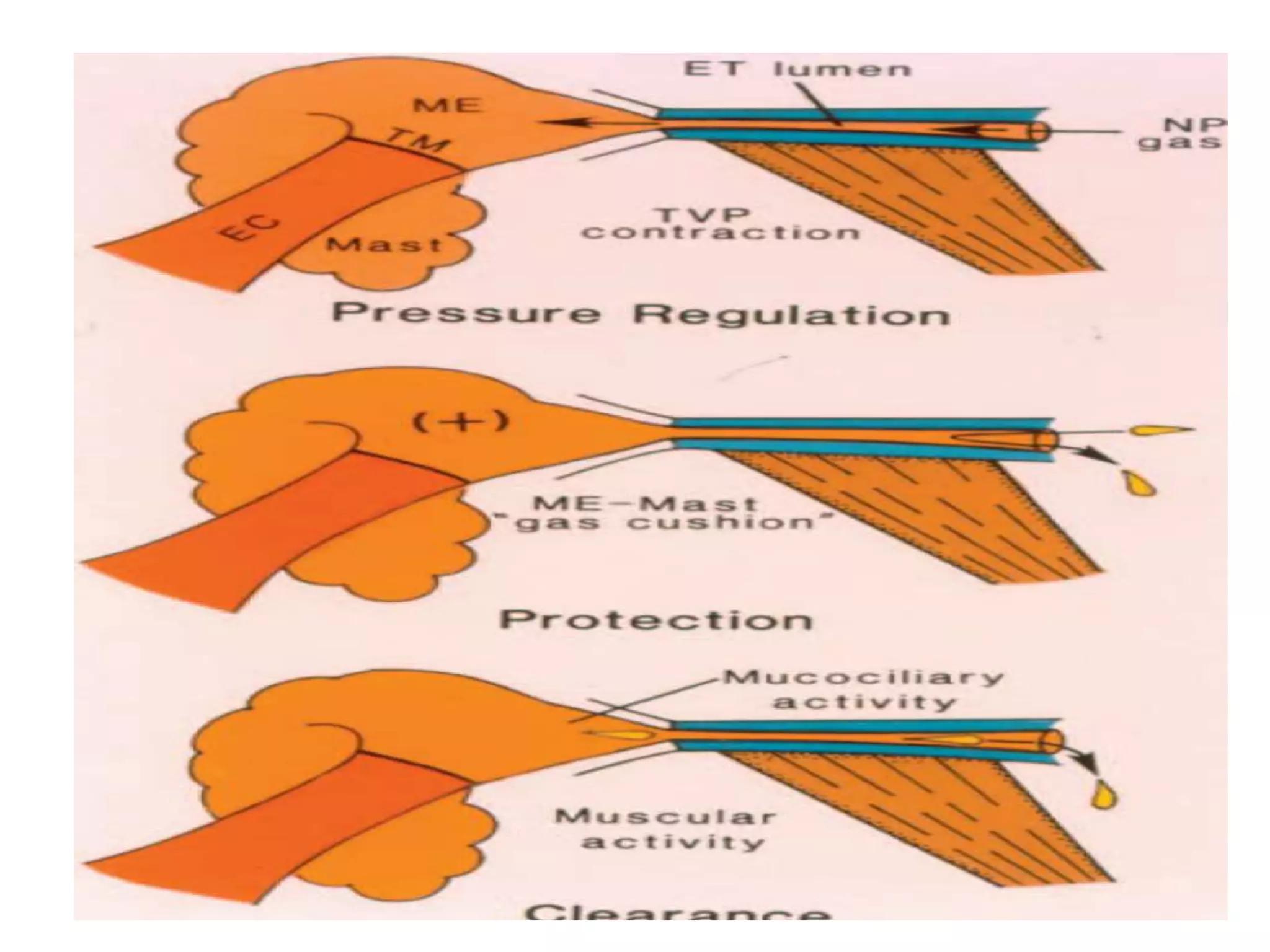
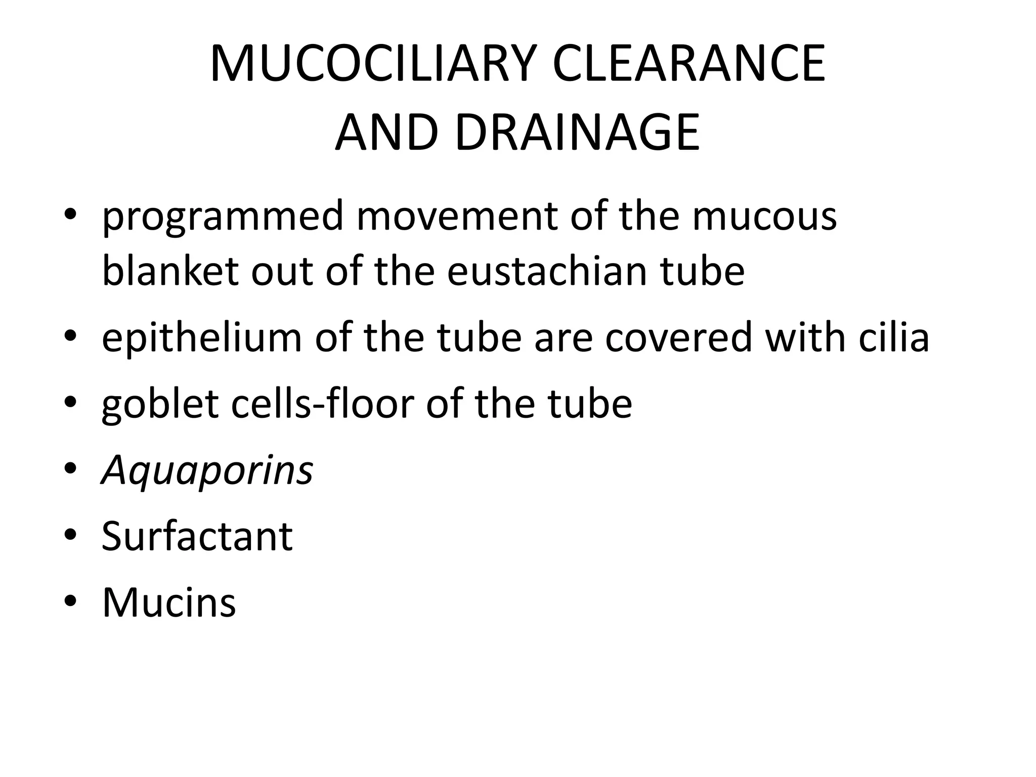
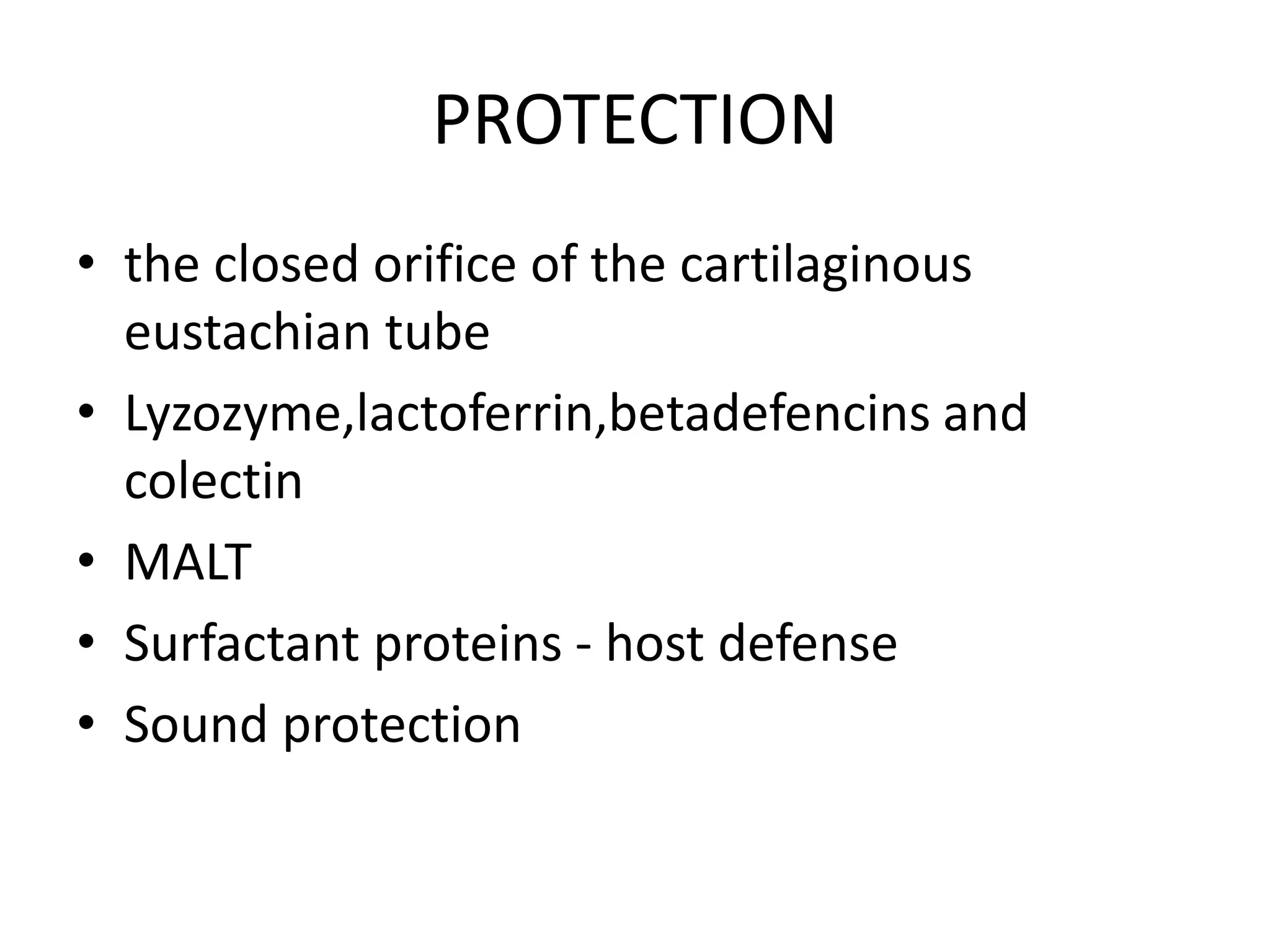
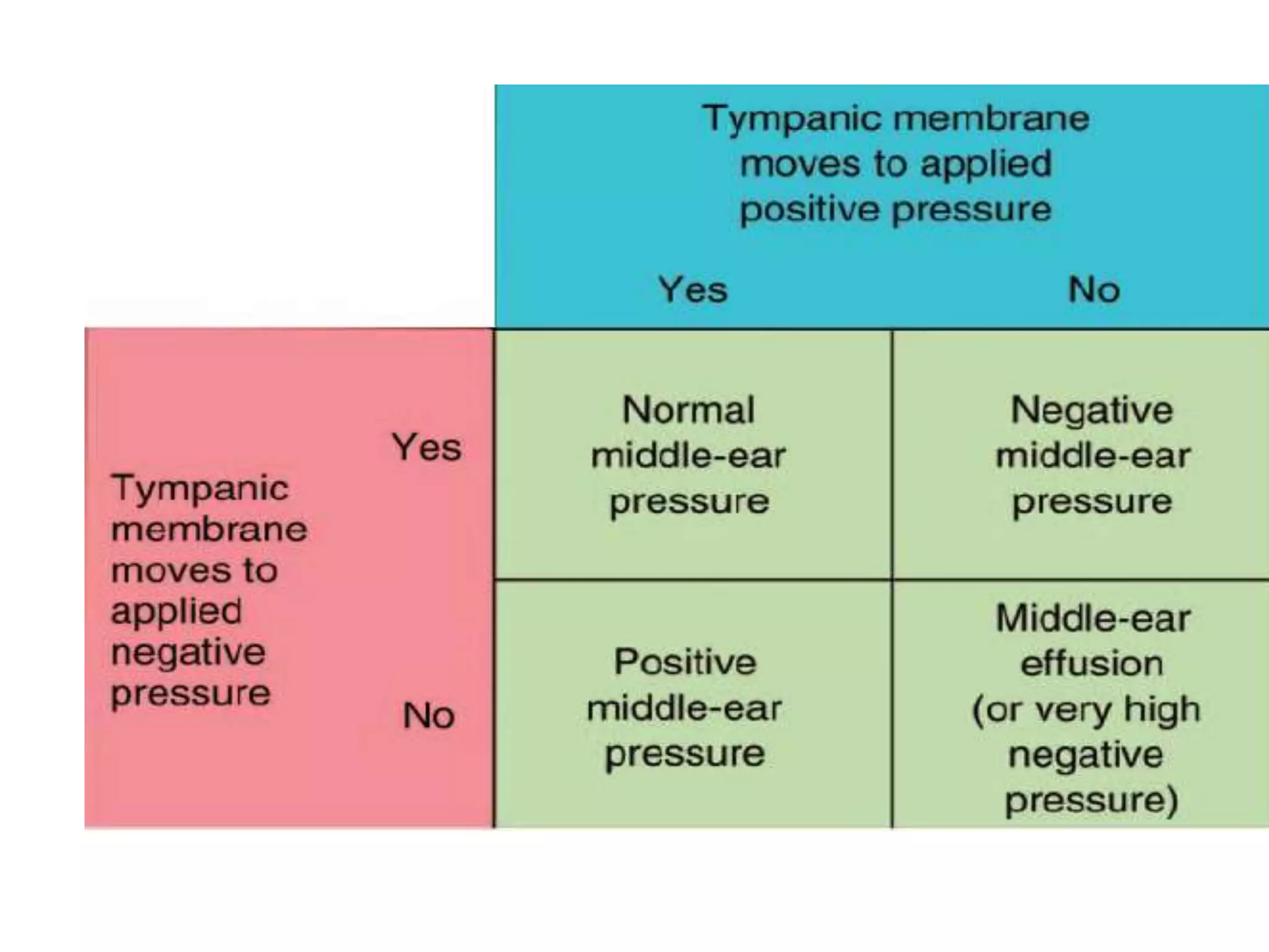
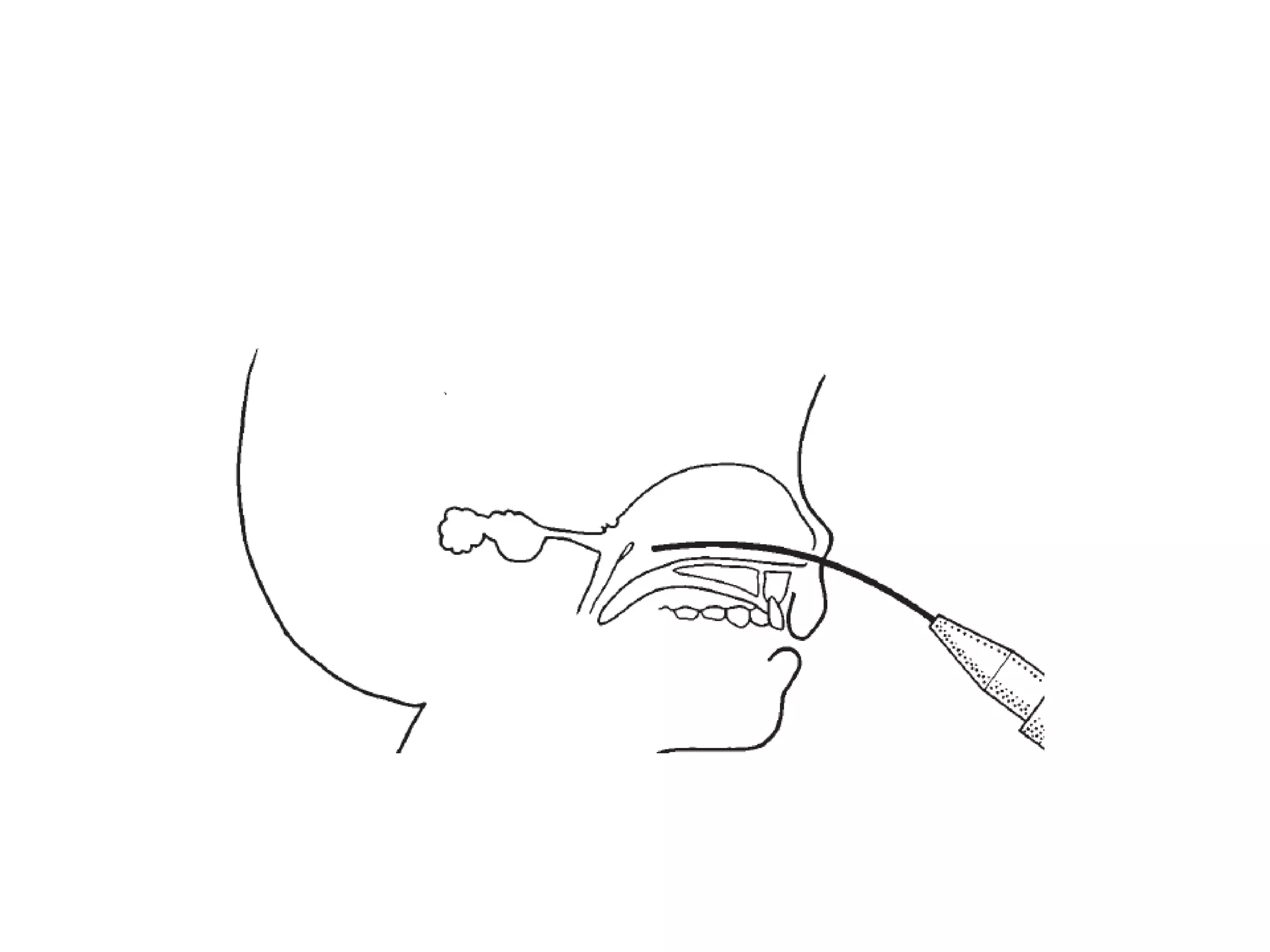
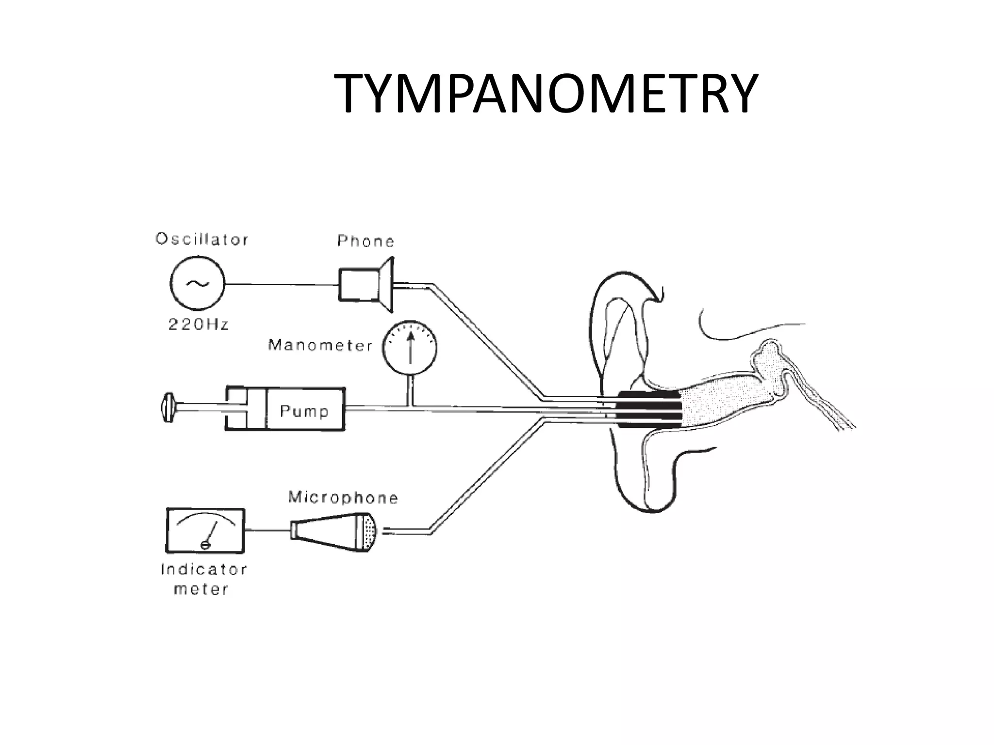
ETFUNCTION
GAJALAKSHMI
EMBRYOLOGY AND POSTNATAL
DEVELOPMENT
• The eustachian tube is derived from the first
pharyngeal pouch
ET Adults and Children
PHYSIOLOGY
• pressure equalization,
• mucociliary clearance and “drainage,”
• protection from both the influences of the
nasopharyngeal environment and loud
sounds.
• Role of surfactant
• Parasympathetic tone
• hydrops ex vacuo model-Politzer
• flask model-Bluestone
MUCOCILIARY CLEARANCE
AND DRAINAGE
• programmed movement of the mucous
blanket out of the eustachian tube
• epithelium of the tube are covered with cilia
• goblet cells-floor of the tube
• Aquaporins
• Surfactant
• Mucins
PROTECTION
• the closed orifice of the cartilaginous
eustachian tube
• Lyzozyme,lactoferrin,betadefencins and
colectin
• MALT
• Surfactant proteins - host defense
• Sound protection
EUSTACHIAN TUBE FUNCTION
ASSESSMENT
• History and Physical Examination
• Pneumatic Otoscopy
• Nasopharyngoscopy
• Imaging of the Eustachian Tube-MRI
Valsalva’s Test
POLITZER’S TEST
EUSTACHIAN TUBE CATHETERIZATION
ATTACHED TO A PRESSURE SYSTEM TO INSUFFLATE THE
MIDDLE EAR
TYMPANOMETRY
Holmquist’s method
1. A tympanogram is recorded to determine the initial
middle-ear pressure.
2. A negative pressure is created in the nasopharynx by a
pressure device connected to the nose, and the subject is
asked to swallow to establish a negative pressure of about
200 mm H2O in the middle ear.
3. A second tympanogram is recorded to evaluate the exact
negative middle-ear pressure achieved.
4. The patient is told to swallow repeatedly
5. A third tympanogram -the final middle-
ear pressure
PATULOUS EUSTACHIAN TUBE
Bluestone’s nine-step test
Sonotubometry
• An earphone: could generate a continuous
sound of 6, 7, or 8 kHz sound of 6, 7, or 8 kHz
Æ inserted into inserted into the nostril of the
test subject the nostril of the test subject
• „Sound source: „
• A microphone embedded in a circumaural ear
muff was placed in the ear muff was placed in
the ipsilateral external auditory canal external
auditory
• The microphone and insert earphone The
microphone and insert earphone Æ were
connected to a heterodyne
• A continuous tone generated by the oscillator
with filter set with filter set &the earphone
inserted into the nose „
• Test sound the external auditory canal - picked up
by a calibrated condenser microphone picked up
connected to a preamplifier connected to a
sound level recorder recorder
When TM is not intact
• Manometry
• Forced response test
• Clearance test
• Modified inflation and deflation test
My ref
• Cummings 3rd edition
• Bluestone CD, Shurin PA. Middle-ear diseases
in children: pathogenesis,diagnosis, and
management. Pediatr Clin North Am
1974;21:379–400.
• Sando I, Takahashi H, Matsune S, Aoki H.
Localization of function in the Eustachian
tube: a hypothesis. Ann Otol Rhinol Laryngol
1994;103

eustachian tube physiology ppt

  • 1.
  • 2.
    EMBRYOLOGY AND POSTNATAL DEVELOPMENT •The eustachian tube is derived from the first pharyngeal pouch
  • 4.
    ET Adults andChildren
  • 5.
    PHYSIOLOGY • pressure equalization, •mucociliary clearance and “drainage,” • protection from both the influences of the nasopharyngeal environment and loud sounds.
  • 10.
    • Role ofsurfactant • Parasympathetic tone
  • 11.
    • hydrops exvacuo model-Politzer • flask model-Bluestone
  • 14.
    MUCOCILIARY CLEARANCE AND DRAINAGE •programmed movement of the mucous blanket out of the eustachian tube • epithelium of the tube are covered with cilia • goblet cells-floor of the tube • Aquaporins • Surfactant • Mucins
  • 15.
    PROTECTION • the closedorifice of the cartilaginous eustachian tube • Lyzozyme,lactoferrin,betadefencins and colectin • MALT • Surfactant proteins - host defense • Sound protection
  • 16.
    EUSTACHIAN TUBE FUNCTION ASSESSMENT •History and Physical Examination • Pneumatic Otoscopy
  • 19.
    • Nasopharyngoscopy • Imagingof the Eustachian Tube-MRI
  • 21.
  • 22.
  • 23.
    EUSTACHIAN TUBE CATHETERIZATION ATTACHEDTO A PRESSURE SYSTEM TO INSUFFLATE THE MIDDLE EAR
  • 25.
  • 27.
    Holmquist’s method 1. Atympanogram is recorded to determine the initial middle-ear pressure. 2. A negative pressure is created in the nasopharynx by a pressure device connected to the nose, and the subject is asked to swallow to establish a negative pressure of about 200 mm H2O in the middle ear. 3. A second tympanogram is recorded to evaluate the exact negative middle-ear pressure achieved. 4. The patient is told to swallow repeatedly 5. A third tympanogram -the final middle- ear pressure
  • 28.
  • 29.
  • 30.
    Sonotubometry • An earphone:could generate a continuous sound of 6, 7, or 8 kHz sound of 6, 7, or 8 kHz Æ inserted into inserted into the nostril of the test subject the nostril of the test subject • „Sound source: „ • A microphone embedded in a circumaural ear muff was placed in the ear muff was placed in the ipsilateral external auditory canal external auditory
  • 31.
    • The microphoneand insert earphone The microphone and insert earphone Æ were connected to a heterodyne • A continuous tone generated by the oscillator with filter set with filter set &the earphone inserted into the nose „ • Test sound the external auditory canal - picked up by a calibrated condenser microphone picked up connected to a preamplifier connected to a sound level recorder recorder
  • 32.
    When TM isnot intact • Manometry • Forced response test • Clearance test • Modified inflation and deflation test
  • 33.
    My ref • Cummings3rd edition • Bluestone CD, Shurin PA. Middle-ear diseases in children: pathogenesis,diagnosis, and management. Pediatr Clin North Am 1974;21:379–400. • Sando I, Takahashi H, Matsune S, Aoki H. Localization of function in the Eustachian tube: a hypothesis. Ann Otol Rhinol Laryngol 1994;103

Editor's Notes

  • #4 The tensor veli palatini, dilator tubae, and tensor tympani are innervated by the mandibular branch of the trigeminal nerve, whereas the levator veli palatini is innervated by the vagus nerve.
  • #7 Functions of the Eustachian tube (ET)–middle ear (ME)–mastoid (Mast) gas cell system. Pressure regulation function is related to active dilation of the tube by contraction of the tensor veli palatini muscle (TVP) (upper figure). Protective function is dependent, in part, on an intact middle ear and mastoid gas cells to maintain a gas cushion (middle figure). Clearance function is enhanced by mucociliary activity and muscular activity during tubal closing (lower figure). EC = external canal; NP = nasopharynx; TM = tympanic membrane.
  • #8 Slow-motion videoendoscopy has revealed four steps in tubal opening: 1) palatal elevation with medial movement of the lateral pharyngeal wall and medial rotation of the medial lamina (initiation of opening of the distal cartilaginous tube, presumably by the levator veli palatini); 2) lateral movement of the lateral wall with dilation of the orifice laterally and vertically; 3) propagation of dilation of the tubal lumen from distal to proximal by the tensor veli palatini/dilator tubae; and 4) opening of the proximal cartilaginous tube adjacent to the junctional region with formation of a round to crescent-shaped lumen.58 During this event, the tube remains open for 0.3 to 0.5 seconds, but it is open much longer during yawning.56 In normal individuals, the eustachian tube fully opens once or twice hourly.59
  • #9 Illustration depicting the probable superior portion of the Eustachian tube lumen opening during active dilation (DL) by the tensor veli palatini muscle (TVPM/TVP), which is attached to the lateral lamina (LL) of the cartilage. Sando and colleagues suggested that the superior portion (R) is related to pressure regulation during active dilation and the inferior portion (F) of the tubal lumen is related to protection with its folds, glands (G), and goblet cells (GL).8 Elastin in the hinge portion (ie, junction between the medial and lateral laminae) aids in returning the medial lamina to the resting position.
  • #10 Illustration of the sequence of events during Eustachian tube (ET) dilation (active opening) owing to contraction of the tensor veli palatini muscle (TVP) during swallowing activity. A, The Eustachian tube at rest is closed. B, The proximal end of the cartilaginous lumen dilates first and is then followed by (C) dilation of the distal end and is open to the middle ear (ME). D, The Eustachian tube passively closes from the distal end to the proximal end to its resting, closed position.
  • #11 To be fully opened, the dilatory muscles must overcome the intraluminal surface tension generated by the apposition of the mucosal surfaces. Surfactant protein B, has been identified in the secretory granules of surface cells that line the eustachian tube Tubal surface tension is also influenced by the amount and composition of tubal secretion. This is under control of the autonomic nervous system, and increased parasympathetic tone has been shown to impair tubal opening.
  • #12 According to the classic theory of eustachian tube dysfunction suggested by Politzer67 in the nineteenth century, the gas contained in the middle ear/mastoid system is absorbed into the bloodstream by the capillaries of the middle ear/mastoid mucosa when the tube is hermetically sealed. This was termed the hydrops ex vacuo model. Bluestone and colleagues72 described a flask model, in which the volume supplied by the mastoid air cell system acts as a buffer to protect against abrupt pressure changes and reflux of nasopharyngeal secretions into the middle ear.
  • #14 Protective function of the Eustachian tube–middle ear–mastoid gas cell system can be visualized using the flask model. When liquid is instilled into the mouth of the flask (nasopharyngeal end of tube), the liquid stops in the narrow neck (isthmus of the cartilaginous portion of the tube) owing to the presence of positive (back) pressure built up in the bulbous portion and distal end of the narrow neck of the flask (middle-ear gas cushion). ET = Eustachian tube; Mast = mastoid gas cells; ME = middle ear. FIGURE
  • #15  Mucus of the tube is composed of a thick, more superficial gel phase and a thin sol phase. The cilia move in the sol phase, and their tips contact in the overlying gel phase, which propels the mucous blanket. and a group of integral membrane proteins called aquaporins that facilitate the passage of water through cell membranes. Surfactant potentiates the movement of the gel over the sol phase.61,78,79 Mucins are high-molecularweight glycoproteins and constitute the major component of the mucous secretions. They lubricate the epithelial surface and trap bacteria and viruses
  • #16 lysozyme, a muramidase that creates cell wall disruption; lactoferrin, an iron-binding glycoprotein that acts synergistically with immunoglobulins; β-defensins, antimicrobial peptides that increase permeability of cell walls; and colectins, oligomeric polypeptide chains that bind microbial carbohydrates and assist phagocytosis. Additionally, cellular proliferation in the lymphatic tissue associated with the eustachian tube (MALT) plays an important role in the local immune response. The primary role of SP-A seems to be mucosal defense through facilitation of phagocytosis, and the principal role of surfactants in antimicrobial defense may be as releasing agents and antiadhesives It has been shown experimentally that these sounds lead to a coordinated contraction of the stapedius, tensor tympani, tensor veli palatini, and dilator tubae muscles. This muscle contraction causes the middle ear cleft, mastoid, and nasopharynx to form one continuous cavity to assist in the dissipation of sound pressure
  • #17 a history of recent weight loss could indicate a patulous Eustachian tube (signs and symptoms of Eustachian tube dysfunction, such as fluctuating hearing loss, otalgia, vertigo, and tinnitus, including popping and snapping sounds in the ear or autophony). Otologic symptoms during pregnancy, puberty, flying in airplanes, swimming, and diving (especially scuba diving) can be helpful.
  • #18 When the middle- ear pressure is ambient, the normal tympanic membrane moves inward with slight positive pressure in the ear canal and outward with slight negative pressure. The motion observed is proportionate to the applied pressure and is best visualized in the posterosuperior quadrant of the tympanic membrane. If a two-layered membrane or an atrophic scar (owing to
  • #22 The tubal lumen is opened by a forced expiration with the nasal alae held between the thumb and forefinger with the mouth closed, which insufflates positive pressure into the middle ear through the Eustachian tube. Confirmation of tubal patency is by otoscopy (bulging tympanic membrane), by using a Toynbee tube (a rubber tube with one olive tip in the patient’s test ear and the other olive tip in the examiner’s ear; a pop can be heard in the test ear if the test is successful), or more modern and accurate with tympanometry. (Self-Valsalva is also a method to inflate the middle ear through the Eustachian tube when there is induced middle-ear negative pressure, such as during descent in an airplane or during scuba diving.)
  • #23 A nasal olive tip attached to a “Poltizer bag” (rubber tubing attached to a rubber bulb) is inserted into one naris while both nasal alae are compressed by finger pressure. The patient is asked to repeat the letter K or is asked to swallow, both of which close the velopharyngeal port, while the examiner compresses the rubber bulb. When normal tubal patency is present, positive pressure is insufflated into the middle ear through the Eustachian tube. Confirmation is by the same methods described in Figure 8–5.
  • #25 The Toynbee test of Eustachian tube function. Closed-nose swallowing results first in positive pressure in the nose and nasopharynx, followed by a negative pressure phase. When positive pressure is in the nasopharynx, air may enter the middle ear, creating positive pressure. During or after the negative pressure phase, negative pressure may develop in the middle ear, positive pressure may still be in the middle ear (no change in middle-ear pressure during negative phase), positive pressure may be followed by negative middle-ear pressure, or ambient pressure will be present if equilibration takes place before the tube closes. If the tube does not open during the positive or negative phase, no change in middle- ear pressure will occur.
  • #26 Block diagram of an immittance instrument in which a tympanogram can be obtained when the tympanic membrane is intact. The pump-manometer system can be employed to perform tests of Eustachian tube pressure regulation function when the tympanic membrane is not intact.
  • #27 one test represents the middleear pressure only at one moment. Serial determinations are more indicative of the dynamics of tubal function in a single patient First, a tympanogram is obtained to determine the resting middle-ear pressure. Then the subject is asked to perform a Toynbee maneuver, which normally leads to negative pressure in the middle ear. The establishment of this negative middle-ear pressure is verified by a second tympanogram. If the second tympanogram fails to record a change in middle-ear pressure, the subject is classified as Toynbee negative, indicating possible tubal dysfunction. If the maneuver is successful in inducing negative middle-ear pressure, then the subject is asked to swallow in an attempt to equilibrate the negative pressure. A third tympanogram is recorded to determine whether the equilibration was successful and, if so, to what degree. If the equilibration was not complete, the subject is asked to swallow repeatedly. A tympanogram is recorded between each swallow to monitor the progressive equilibration. The pressure remaining in the middle ear after several swallows is termed residual negative pressure. A similar approach is us
  • #28 measures the ability of the Eustachian tube to equilibrate induced negative middle-ear
  • #29 One tympanogram is obtained while the patient is breathing normally, and a second is obtained while the patient is holding his or her breath. Fluctuation of the tympanometric trace that coincides with breathing confirms the diagnosis of a patulous tube. Fluctuation can be exaggerated by asking the patient to occlude one nostril with the mouth closed during forced inspiration and expiration or by performing the Toynbee maneuver
  • #30 1. The tympanogram records resting middle-ear pressure. 2. Ear canal pressure is increased to +200 mm H2O with medial deflection of the tympanic membrane and a corresponding increase in middle-ear pressure. The subject swallows to equilibrate middle-ear overpressure. 3.While the subject refrains from swallowing, ear canal pressure is returned to normal, thus establishing a slight negative middle-ear pressure (as the tympanic membrane moves outward). The tympanogram documents the established middle-ear underpressure. 4. The subject swallows in an attempt to equilibrate negative middle-ear pressure. If equilibration is successful, airflow is from the nasopharynx to the middle ear. 5. The tympanogram records the extent of equilibration. 6. Ear canal pressure is decreased to 200 mm H2O, causing a lateral deflection of the tympanic membrane and a corresponding decrease in middle-ear pressure. The subject swallows to equilibrate negative middle-ear pressure; airflow is from the nasopharynx to the middle ear. 7. The subject refrains from swallowing while external ear canal pressure is returned to normal, thus establishing a slight positive pressure in the middle ear as the tympanic membrane moves medially. The tympanogram records the overpressure established. 8. The subject swallows to reduce overpressure. If equilibration is successful, airflow is from the middle ear to the nasopharynx. 9. The final tympanogram documents the extent of equilibration.
  • #31 Sound source: was fixed tightly within the Sound source: was fixed tightly within the nostril to minimize sound leakage nostril to minimize sound leakage
  • #32 connected to a heterodyne analyser analyser (consisted (consisted of an analyser analyser and a frequency oscillator) and a frequency oscillator) „ The test subject sat in a quiet room with his or her mouth closed and without moving the head He/she was asked to swallow water while the sound signal in the external ear was being recorded continuously recorded continuously „ Opening of the tube was reflected -sudden increase in signal in the external ear canal (5 dB)
  • #33 The active response is due to the contractions of the tensor veli palatini muscle, which displaces the lateral walls from the cartilage-supported medial wall of the tube. Thus, the clinician can determine whether tubal dysfunction is due to the material properties of the tube or to a defective active opening mechanism. During this test, the middle ear is inflated at a constant flow rate, forcing the Eustachian tube open. After the forced opening of the tube, the pump continues to deliver a constant airflow, maintaining a steady stream of air through the tube. Then the subject is instructed to swallow for assessment of the active dilatation of the tube. The method is unique in that it eliminates the “mucous forces” in the Eustachian tube lumen that may interfere with the results of the inflation-deflation test when an attempt is made to assess the active opening mechanisms and the compliance of the tube. In this test, the passive resistance is assessed, and the active resistance is determined during swallowing. Patients with