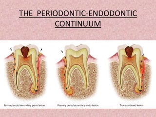
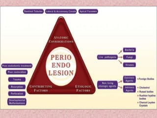
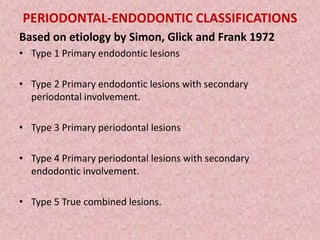
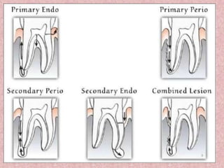
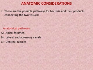
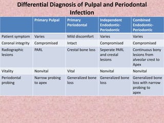
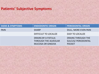
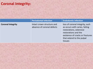
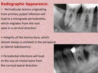
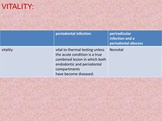
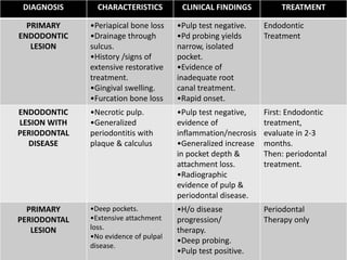
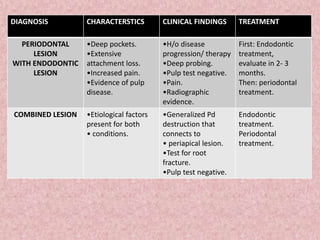
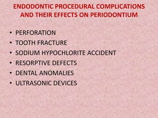
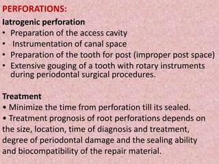
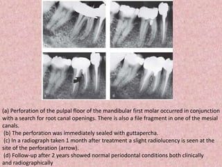
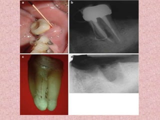
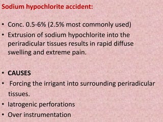
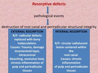
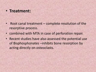
THE PERIODONTIC-ENDODONTIC
CONTINUUM
INTRODUCTION
• The endodontium and periodontium are closely related and
diseases of one tissue may lead to secondary diseases in
the other.
• Both pulpal and periodontal infections can coexist in the
same tooth, termed combined lesions.
• ENDODONTIC LESION is used to denote an
inflammatory process in the periodontal tissues
resulting from noxious agents present in the root
canal system of the tooth, usually a root canal
infection.
• PERIODONTAL LESION is used to denote an
inflammatory process in the periodontal tissues
resulting from accumulation of dental plaque on
the external tooth surfaces.
ETIOLOGICAL FACTORS
(1) instrumentation during periodontal, restorative, or prosthetic
dentistry;
(2) the progression of dental caries; and
(3) direct, local trauma such as tooth fracture
• Of these, dental caries is the most common cause of pulpal disease.
• Mutans streptococci and lactobacillus (aerobic) are the common
strains responsible for DC.
PERIODONTAL-ENDODONTIC CLASSIFICATIONS
Based on etiology by Simon, Glick and Frank 1972
• Type 1 Primary endodontic lesions
• Type 2 Primary endodontic lesions with secondary
periodontal involvement.
• Type 3 Primary periodontal lesions
• Type 4 Primary periodontal lesions with secondary
endodontic involvement.
• Type 5 True combined lesions.
ANATOMIC CONSIDERATIONS
• These are the possible pathways for bacteria and their products
connecting the two tissues:
Anatomical pathways
A) Apical foramen
B) Lateral and accessory canals
C) Dentinal tubules
• Persistent infection in the pulp tissue may lead to secondary
infection and breakdown of tissues in the periodontium.
• Conversely, severe periodontal disease may initiate or exacerbate
inflammatory changes in the pulp tissue.
• Advanced pulpitis will lead to pulp necrosis, which often is
accompanied by inflammatory bone resorption at the root apex, as
found in cases of chronic periradicular periodontitis (CPP) or chronic
radicular abscess (CPA)
• This is also known as retrograde periodontitis because it represents
the periodontal tissue breakdown from an apical to cervical
direction and is the opposite of orthograde periodontitis that results
from a sulcular infection.
• Alternatively, lateral or accessory canals may be the route of such
communications.
• Accessory canals are found along the length of the root canals, to
varying frequencies, depending on their location
• The third route is dentinal tubules.
• Dentinal tubules maintain a tapered structure along the length from
the pulpodentin complex (PDC) to the dentinoenamel junction (DEJ)
with the diameter of 2.5 μm at PDC and 0.9 μm at the DEJ.
• Permeability would increase when the carious lesion extends deeper
into the dentin.
Biologic Effects of Pulpal Infection on
Periodontal Tissues
• Early inflammatory changes in the pulp exert very little effect on the
periodontium. Even a pulp that is significantly inflamed may have
little or no effect on the surrounding periodontal tissues.
• When the pulp becomes necrotic, it produces an inflammatory
response, which traverse the apical foramen, the furcation, lateral
canals, dentinal tubules, or areas of trapped necrotic tissue along the
surface of the root, which extend past the periodontal ligament and
into the surrounding periradicular tissues
• Periodontal destruction that follows depends on
1. the virulence of the pathogens in the canal system,
2. the duration of the disease, and
3. the defense mechanisms of the host
• Extension of the infection through the periodontal ligament space
and surrounding bone occurs, and the patient begins to experience
a localized or diffuse swelling.
• Most often, the infection erupts through the labial, buccal, or
lingual mucosa and results in a draining sinus tract.
• In cases in which the path of least resistance for the infectious
process may be along the attached gingiva, the infection may dissect
the periodontal ligament space and result in a deep but narrow
periodontal pocket.
• This pocket will usually extend to the main site of the infection when
probed or traced with a gutta percha point.
Biologic Effects of Periodontal Infection on
Dental Pulp
Langeland et al 1974 indicated then when pathologic changes do occur in the pulp of
the tooth as a result of advanced periodontal disease
pulp does not usually undergo degenerative changes
as long as the main canal has not been involved.
If the vasculature of the pulp remains vital, no inflammatory reaction occurs and there
is no symptom of pulp pathosis
• Researchers and clinicians, however, have observed the spread of advanced
periodontal lesions that extend to the apical foramen and result in pulpal necrosis.
• This retrograde infection may proliferate through large accessory canals on the
periradicular surfaces of the tooth, canals positioned closer to the apical foramen,
and in the area in which the main canal exits the tooth apex
• The presence of an intact layer of cementum is important in protecting the pulp
from the plaque and other periodontal pathogens that migrate along the root
surface during the development of advanced periodontal disease.
• Excessive root planing and curettage that remove the cementum and dentin of the
root surface, encourages narrowing of the pulp canals
• Dentin thickness also contributes to the protection of the pulp
• Pulpal changes seen in periodontal disease were closely related to periodontal
treatment .
• Vigorous scaling and root planning removes cementum –
• - expose dentinal tubules
• - transport irritants
• - pulpal inflammation
• - necrosis of the dental pulp
• Weine summarized the precautions that can be taken during
the course of periodontal therapy as
(1) avoid using irritating chemicals on the root surface,
(2) minimize the use of ultrasonic scalers when there is less
than 2 mm of remaining dentin, and
(3) allow minor pulpal irritations to subside before completing
additional procedures
Differential Diagnosis of Pulpal and Periodontal
Infection
Primary Pulpal Primary
Periodontal
Independent
Endodontic-
Periodontic
Combined
Endodontic-
Periodontic
Patient symptom Varies Mild discomfort Varies Varies
Coronal integrity Compromised Intact Compromised Compromised
Radiographic
lesions
PARL Crestal bone loss Seperate PARL
and crestal
lesions
Continuous bony
lesions from
alveolar crest to
Apex
Vitality Nonvital Vital Nonvital Nonvital
Periodontal
probing
Narrow probing
to apex
Generalized bone
loss
Generalized bone
loss
Generalized bone
loss with narrow
probing to
apex
Patients’ Subjective Symptoms
SIGNS & SYMPTOMS ENDODONTIC ORIGIN PERIODONTAL ORIGIN
PAIN SHARP DULL, MORE EVEN PAIN
DIFFICULT TO LOCALIZE EASY TO LOCALIZE
DRAINS BY A FISTULA
THROUGH THE ALVEOLAR
MUCOSA OR GINGIVA
DRAINS THROUGH THE
SULCUS/ PERIODONTAL
POCKET
Coronal Integrity:
Periodontal infection Endodontic infection
Coronal Integrity Intact crown structure and
absence of coronal defects
loss of coronal integrity, such
as occurs with caries, failing
restorations, extensive
restorations and the
existence of cracks or fractures
that extend to the pulpal
tissues
Radiographic Appearance:
• Periradicular lesions originating
from primary pulpal infection will
lead to a retrograde periodontitis,
which migrates from the root
apex in a cervical direction.
• integrity of the lamina dura, which
almost always is violated in the periapical
or lateral radiolucency .
• Periodontal infections will lead
to the loss of crestal bone from
the cervical apical direction.
VITALITY:
periodontal infection periradicular
infection and a
periodontal abscess
vitality vital to thermal testing unless
the acute condition is a true
combined lesion in which both
endodontic and periodontal
compartments
have become diseased.
Nonvital
DIAGNOSIS CHARACTERISTICS CLINICAL FINDINGS TREATMENT
PRIMARY
ENDODONTIC
LESION
•Periapical bone loss
•Drainage through
sulcus.
•History /signs of
extensive restorative
treatment.
•Gingival swelling.
•Furcation bone loss
•Pulp test negative.
•Pd probing yields
narrow, isolated
pocket.
•Evidence of
inadequate root
canal treatment.
•Rapid onset.
Endodontic
Treatment
ENDODONTIC
LESION WITH
PERIODONTAL
DISEASE
•Necrotic pulp.
•Generalized
periodontitis with
plaque & calculus
•Pulp test negative,
evidence of
inflammation/necrosis
•Generalized increase
in pocket depth &
attachment loss.
•Radiographic
evidence of pulp &
periodontal disease.
First: Endodontic
treatment,
evaluate in 2-3
months.
Then: periodontal
treatment.
PRIMARY
PERIODONTAL
LESION
•Deep pockets.
•Extensive attachment
loss.
•No evidence of pulpal
disease.
•H/o disease
progression/
therapy.
•Deep probing.
•Pulp test positive.
Periodontal
Therapy only
DIAGNOSIS CHARACTERSTICS CLINICAL FINDINGS TREATMENT
PERIODONTAL
LESION
WITH ENDODONTIC
LESION
•Deep pockets.
•Extensive
attachment loss.
•Increased pain.
•Evidence of pulp
disease.
•H/o disease
progression/ therapy
•Deep probing.
•Pulp test negative.
•Pain.
•Radiographic
evidence.
First: Endodontic
treatment,
evaluate in 2- 3
months.
Then: periodontal
treatment.
COMBINED LESION •Etiological factors
present for both
• conditions.
•Generalized Pd
destruction that
connects to
• periapical lesion.
•Test for root
fracture.
•Pulp test negative.
Endodontic
treatment.
Periodontal
treatment.
ENDODONTIC PROCEDURAL COMPLICATIONS
AND THEIR EFFECTS ON PERIODONTIUM
• PERFORATION
• TOOTH FRACTURE
• SODIUM HYPOCHLORITE ACCIDENT
• RESORPTIVE DEFECTS
• DENTAL ANOMALIES
• ULTRASONIC DEVICES
PERFORATIONS:
Iatrogenic perforation
• Preparation of the access cavity
• Instrumentation of canal space
• Preparation of the tooth for post (improper post space)
• Extensive gouging of a tooth with rotary instruments
during periodontal surgical procedures.
Treatment
• Minimize the time from perforation till its sealed.
• Treatment prognosis of root perforations depends on
the size, location, time of diagnosis and treatment,
degree of periodontal damage and the sealing ability
and biocompatibility of the repair material.
(a) Perforation of the pulpal floor of the mandibular first molar occurred in conjunction
with a search for root canal openings. There is also a file fragment in one of the mesial
canals.
(b) The perforation was immediately sealed with guttapercha.
(c) In a radiograph taken 1 month after treatment a slight radiolucency is seen at the
site of the perforation (arrow).
(d) Follow-up after 2 years showed normal periodontal conditions both clinically
and radiographically
Tooth fracture:
• Crown-Root Fractures
• Horizontal Root Fractures in the Coronal Area
• Horizontal Fractures in the Midroot Area
• Horizontal Fractures in the Apical Area
• Vertical root fractures
Sodium hypochlorite accident:
• Conc. 0.5-6% (2.5% most commonly used)
• Extrusion of sodium hypochlorite into the
periradicular tissues results in rapid diffuse
swelling and extreme pain.
• CAUSES
• Forcing the irrigant into surrounding periradicular
tissues.
• Iatrogenic perforations
• Over instrumentation
Resorptive defects
pathological events
destruction of root canal and periradicular structural integrity
EXTERNAL RESORPTION
R/F- radicular defects
replaced with bony
trabeculation.
Causes: Trauma, damage in
cementoid layer, intracoronal
bleaching, excessive heat,
chronic inflammation of pulp
and periradicular tissues
EXTERNAL RESORPTION
R/F- radicular defects
replaced with bony
trabeculation.
Causes: Trauma, damage
incementoid layer,
intracoronal
bleaching, excessive heat,
chronic inflammation of
pulp and periradicular
tissues
INTERNAL RESORPTION
R/F- circular radiolucent
lesion centered within
the
root canal.
Causes: chronic
inflammation
of pulp and periradicular
tissues
• Treatment:
• Root canal treatment – complete resolution of the
resorptive process.
• combined with MTA in case of perforation repair.
• Recent studies have also assessed the potential use
of Bisphosphonates –inhibits bone resorption by
acting directly on osteoclasts.
Dental anomalies
Dens invaginatus Dens evaginatus Lingual groove
• Inner enamel epithelium
invaginates into the
dental papilla before
calcification.
• Increased labiolingual or
mesiodistal diameter
• Peg or conical
morphology.
• Talons cusp
• Orthograde
Endodontic or surgical
treatment
• Tubercle or protrusion of
enamel from the occlusal
tooth surface that has a
core of dentin usually
containing a thin extension
of pulpal tissue.
• Suspectible to pulpal
exposure caused by
fracture, wear, orthodontic
movement or occlusal
equilibrium.
• Prevention /prophylatic
treatment.
• Alloy or composite resin
restoration.
• Distolingual surface of
the maxillary lateral
incisor.
• Infolding of inner
enamel epithelium and
hertwig’s epithelial
root sheath.

The periodontic endodontic continuum.

  • 1.
  • 2.
    INTRODUCTION • The endodontiumand periodontium are closely related and diseases of one tissue may lead to secondary diseases in the other. • Both pulpal and periodontal infections can coexist in the same tooth, termed combined lesions.
  • 3.
    • ENDODONTIC LESIONis used to denote an inflammatory process in the periodontal tissues resulting from noxious agents present in the root canal system of the tooth, usually a root canal infection. • PERIODONTAL LESION is used to denote an inflammatory process in the periodontal tissues resulting from accumulation of dental plaque on the external tooth surfaces.
  • 4.
    ETIOLOGICAL FACTORS (1) instrumentationduring periodontal, restorative, or prosthetic dentistry; (2) the progression of dental caries; and (3) direct, local trauma such as tooth fracture • Of these, dental caries is the most common cause of pulpal disease. • Mutans streptococci and lactobacillus (aerobic) are the common strains responsible for DC.
  • 6.
    PERIODONTAL-ENDODONTIC CLASSIFICATIONS Based onetiology by Simon, Glick and Frank 1972 • Type 1 Primary endodontic lesions • Type 2 Primary endodontic lesions with secondary periodontal involvement. • Type 3 Primary periodontal lesions • Type 4 Primary periodontal lesions with secondary endodontic involvement. • Type 5 True combined lesions.
  • 8.
    ANATOMIC CONSIDERATIONS • Theseare the possible pathways for bacteria and their products connecting the two tissues: Anatomical pathways A) Apical foramen B) Lateral and accessory canals C) Dentinal tubules
  • 9.
    • Persistent infectionin the pulp tissue may lead to secondary infection and breakdown of tissues in the periodontium. • Conversely, severe periodontal disease may initiate or exacerbate inflammatory changes in the pulp tissue. • Advanced pulpitis will lead to pulp necrosis, which often is accompanied by inflammatory bone resorption at the root apex, as found in cases of chronic periradicular periodontitis (CPP) or chronic radicular abscess (CPA) • This is also known as retrograde periodontitis because it represents the periodontal tissue breakdown from an apical to cervical direction and is the opposite of orthograde periodontitis that results from a sulcular infection.
  • 10.
    • Alternatively, lateralor accessory canals may be the route of such communications. • Accessory canals are found along the length of the root canals, to varying frequencies, depending on their location • The third route is dentinal tubules. • Dentinal tubules maintain a tapered structure along the length from the pulpodentin complex (PDC) to the dentinoenamel junction (DEJ) with the diameter of 2.5 μm at PDC and 0.9 μm at the DEJ. • Permeability would increase when the carious lesion extends deeper into the dentin.
  • 11.
    Biologic Effects ofPulpal Infection on Periodontal Tissues • Early inflammatory changes in the pulp exert very little effect on the periodontium. Even a pulp that is significantly inflamed may have little or no effect on the surrounding periodontal tissues. • When the pulp becomes necrotic, it produces an inflammatory response, which traverse the apical foramen, the furcation, lateral canals, dentinal tubules, or areas of trapped necrotic tissue along the surface of the root, which extend past the periodontal ligament and into the surrounding periradicular tissues • Periodontal destruction that follows depends on 1. the virulence of the pathogens in the canal system, 2. the duration of the disease, and 3. the defense mechanisms of the host
  • 12.
    • Extension ofthe infection through the periodontal ligament space and surrounding bone occurs, and the patient begins to experience a localized or diffuse swelling. • Most often, the infection erupts through the labial, buccal, or lingual mucosa and results in a draining sinus tract. • In cases in which the path of least resistance for the infectious process may be along the attached gingiva, the infection may dissect the periodontal ligament space and result in a deep but narrow periodontal pocket. • This pocket will usually extend to the main site of the infection when probed or traced with a gutta percha point.
  • 14.
    Biologic Effects ofPeriodontal Infection on Dental Pulp Langeland et al 1974 indicated then when pathologic changes do occur in the pulp of the tooth as a result of advanced periodontal disease pulp does not usually undergo degenerative changes as long as the main canal has not been involved. If the vasculature of the pulp remains vital, no inflammatory reaction occurs and there is no symptom of pulp pathosis • Researchers and clinicians, however, have observed the spread of advanced periodontal lesions that extend to the apical foramen and result in pulpal necrosis. • This retrograde infection may proliferate through large accessory canals on the periradicular surfaces of the tooth, canals positioned closer to the apical foramen, and in the area in which the main canal exits the tooth apex
  • 15.
    • The presenceof an intact layer of cementum is important in protecting the pulp from the plaque and other periodontal pathogens that migrate along the root surface during the development of advanced periodontal disease. • Excessive root planing and curettage that remove the cementum and dentin of the root surface, encourages narrowing of the pulp canals • Dentin thickness also contributes to the protection of the pulp • Pulpal changes seen in periodontal disease were closely related to periodontal treatment . • Vigorous scaling and root planning removes cementum – • - expose dentinal tubules • - transport irritants • - pulpal inflammation • - necrosis of the dental pulp
  • 16.
    • Weine summarizedthe precautions that can be taken during the course of periodontal therapy as (1) avoid using irritating chemicals on the root surface, (2) minimize the use of ultrasonic scalers when there is less than 2 mm of remaining dentin, and (3) allow minor pulpal irritations to subside before completing additional procedures
  • 17.
    Differential Diagnosis ofPulpal and Periodontal Infection Primary Pulpal Primary Periodontal Independent Endodontic- Periodontic Combined Endodontic- Periodontic Patient symptom Varies Mild discomfort Varies Varies Coronal integrity Compromised Intact Compromised Compromised Radiographic lesions PARL Crestal bone loss Seperate PARL and crestal lesions Continuous bony lesions from alveolar crest to Apex Vitality Nonvital Vital Nonvital Nonvital Periodontal probing Narrow probing to apex Generalized bone loss Generalized bone loss Generalized bone loss with narrow probing to apex
  • 18.
    Patients’ Subjective Symptoms SIGNS& SYMPTOMS ENDODONTIC ORIGIN PERIODONTAL ORIGIN PAIN SHARP DULL, MORE EVEN PAIN DIFFICULT TO LOCALIZE EASY TO LOCALIZE DRAINS BY A FISTULA THROUGH THE ALVEOLAR MUCOSA OR GINGIVA DRAINS THROUGH THE SULCUS/ PERIODONTAL POCKET
  • 19.
    Coronal Integrity: Periodontal infectionEndodontic infection Coronal Integrity Intact crown structure and absence of coronal defects loss of coronal integrity, such as occurs with caries, failing restorations, extensive restorations and the existence of cracks or fractures that extend to the pulpal tissues
  • 20.
    Radiographic Appearance: • Periradicularlesions originating from primary pulpal infection will lead to a retrograde periodontitis, which migrates from the root apex in a cervical direction. • integrity of the lamina dura, which almost always is violated in the periapical or lateral radiolucency . • Periodontal infections will lead to the loss of crestal bone from the cervical apical direction.
  • 21.
    VITALITY: periodontal infection periradicular infectionand a periodontal abscess vitality vital to thermal testing unless the acute condition is a true combined lesion in which both endodontic and periodontal compartments have become diseased. Nonvital
  • 22.
    DIAGNOSIS CHARACTERISTICS CLINICALFINDINGS TREATMENT PRIMARY ENDODONTIC LESION •Periapical bone loss •Drainage through sulcus. •History /signs of extensive restorative treatment. •Gingival swelling. •Furcation bone loss •Pulp test negative. •Pd probing yields narrow, isolated pocket. •Evidence of inadequate root canal treatment. •Rapid onset. Endodontic Treatment ENDODONTIC LESION WITH PERIODONTAL DISEASE •Necrotic pulp. •Generalized periodontitis with plaque & calculus •Pulp test negative, evidence of inflammation/necrosis •Generalized increase in pocket depth & attachment loss. •Radiographic evidence of pulp & periodontal disease. First: Endodontic treatment, evaluate in 2-3 months. Then: periodontal treatment. PRIMARY PERIODONTAL LESION •Deep pockets. •Extensive attachment loss. •No evidence of pulpal disease. •H/o disease progression/ therapy. •Deep probing. •Pulp test positive. Periodontal Therapy only
  • 23.
    DIAGNOSIS CHARACTERSTICS CLINICALFINDINGS TREATMENT PERIODONTAL LESION WITH ENDODONTIC LESION •Deep pockets. •Extensive attachment loss. •Increased pain. •Evidence of pulp disease. •H/o disease progression/ therapy •Deep probing. •Pulp test negative. •Pain. •Radiographic evidence. First: Endodontic treatment, evaluate in 2- 3 months. Then: periodontal treatment. COMBINED LESION •Etiological factors present for both • conditions. •Generalized Pd destruction that connects to • periapical lesion. •Test for root fracture. •Pulp test negative. Endodontic treatment. Periodontal treatment.
  • 24.
    ENDODONTIC PROCEDURAL COMPLICATIONS ANDTHEIR EFFECTS ON PERIODONTIUM • PERFORATION • TOOTH FRACTURE • SODIUM HYPOCHLORITE ACCIDENT • RESORPTIVE DEFECTS • DENTAL ANOMALIES • ULTRASONIC DEVICES
  • 25.
    PERFORATIONS: Iatrogenic perforation • Preparationof the access cavity • Instrumentation of canal space • Preparation of the tooth for post (improper post space) • Extensive gouging of a tooth with rotary instruments during periodontal surgical procedures. Treatment • Minimize the time from perforation till its sealed. • Treatment prognosis of root perforations depends on the size, location, time of diagnosis and treatment, degree of periodontal damage and the sealing ability and biocompatibility of the repair material.
  • 26.
    (a) Perforation ofthe pulpal floor of the mandibular first molar occurred in conjunction with a search for root canal openings. There is also a file fragment in one of the mesial canals. (b) The perforation was immediately sealed with guttapercha. (c) In a radiograph taken 1 month after treatment a slight radiolucency is seen at the site of the perforation (arrow). (d) Follow-up after 2 years showed normal periodontal conditions both clinically and radiographically
  • 27.
    Tooth fracture: • Crown-RootFractures • Horizontal Root Fractures in the Coronal Area • Horizontal Fractures in the Midroot Area • Horizontal Fractures in the Apical Area • Vertical root fractures
  • 30.
    Sodium hypochlorite accident: •Conc. 0.5-6% (2.5% most commonly used) • Extrusion of sodium hypochlorite into the periradicular tissues results in rapid diffuse swelling and extreme pain. • CAUSES • Forcing the irrigant into surrounding periradicular tissues. • Iatrogenic perforations • Over instrumentation
  • 31.
    Resorptive defects pathological events destructionof root canal and periradicular structural integrity EXTERNAL RESORPTION R/F- radicular defects replaced with bony trabeculation. Causes: Trauma, damage in cementoid layer, intracoronal bleaching, excessive heat, chronic inflammation of pulp and periradicular tissues EXTERNAL RESORPTION R/F- radicular defects replaced with bony trabeculation. Causes: Trauma, damage incementoid layer, intracoronal bleaching, excessive heat, chronic inflammation of pulp and periradicular tissues INTERNAL RESORPTION R/F- circular radiolucent lesion centered within the root canal. Causes: chronic inflammation of pulp and periradicular tissues
  • 32.
    • Treatment: • Rootcanal treatment – complete resolution of the resorptive process. • combined with MTA in case of perforation repair. • Recent studies have also assessed the potential use of Bisphosphonates –inhibits bone resorption by acting directly on osteoclasts.
  • 33.
    Dental anomalies Dens invaginatusDens evaginatus Lingual groove • Inner enamel epithelium invaginates into the dental papilla before calcification. • Increased labiolingual or mesiodistal diameter • Peg or conical morphology. • Talons cusp • Orthograde Endodontic or surgical treatment • Tubercle or protrusion of enamel from the occlusal tooth surface that has a core of dentin usually containing a thin extension of pulpal tissue. • Suspectible to pulpal exposure caused by fracture, wear, orthodontic movement or occlusal equilibrium. • Prevention /prophylatic treatment. • Alloy or composite resin restoration. • Distolingual surface of the maxillary lateral incisor. • Infolding of inner enamel epithelium and hertwig’s epithelial root sheath.