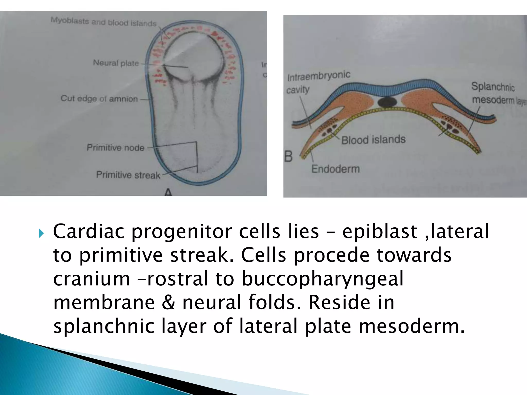
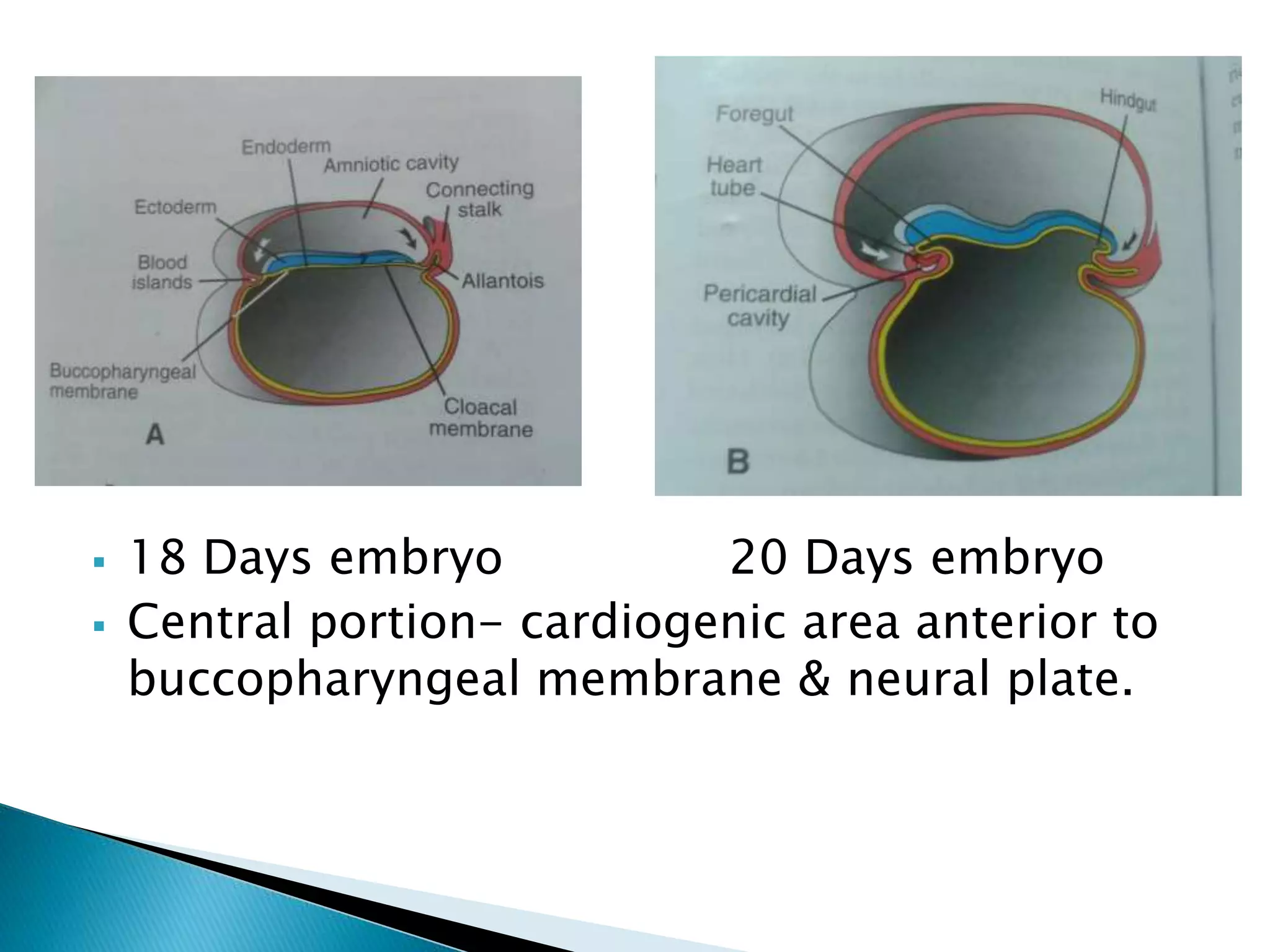
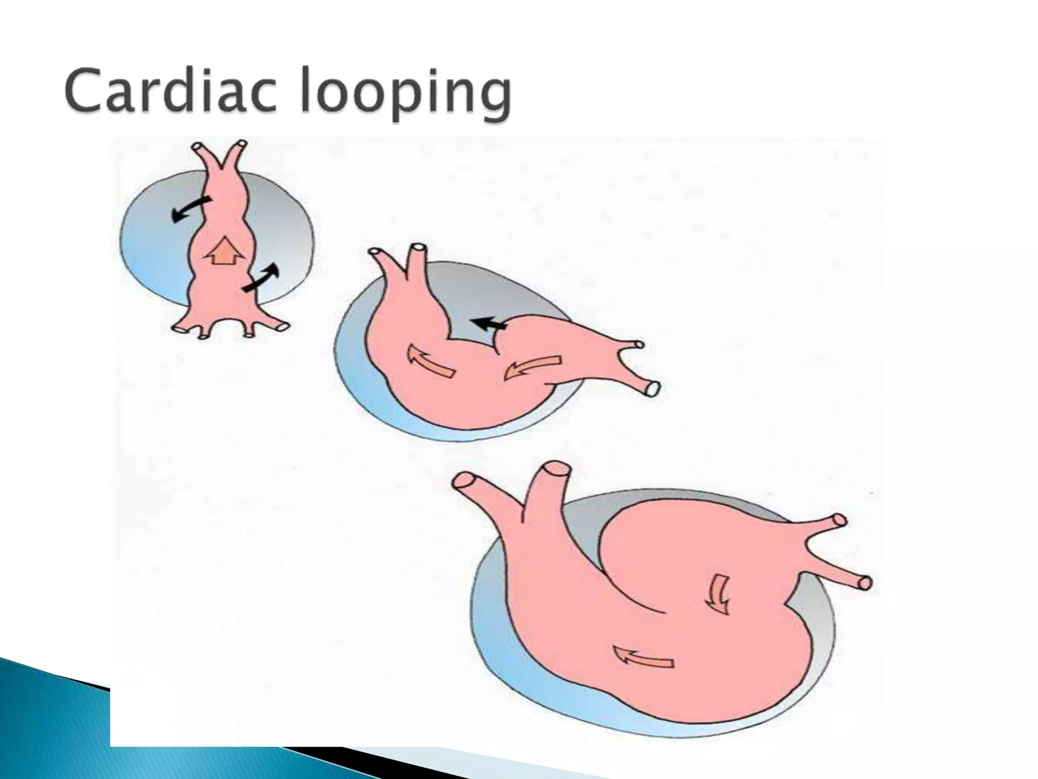
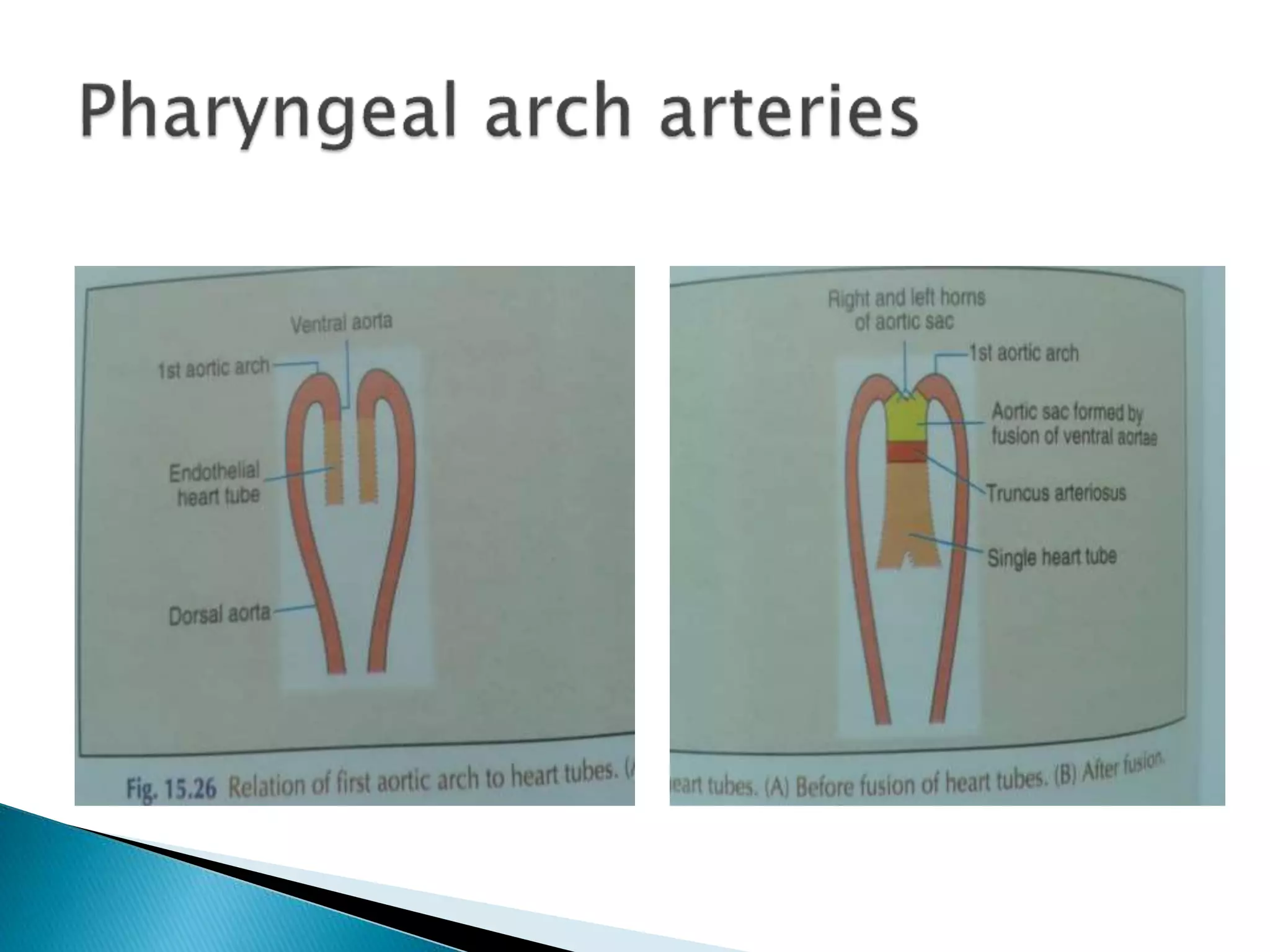
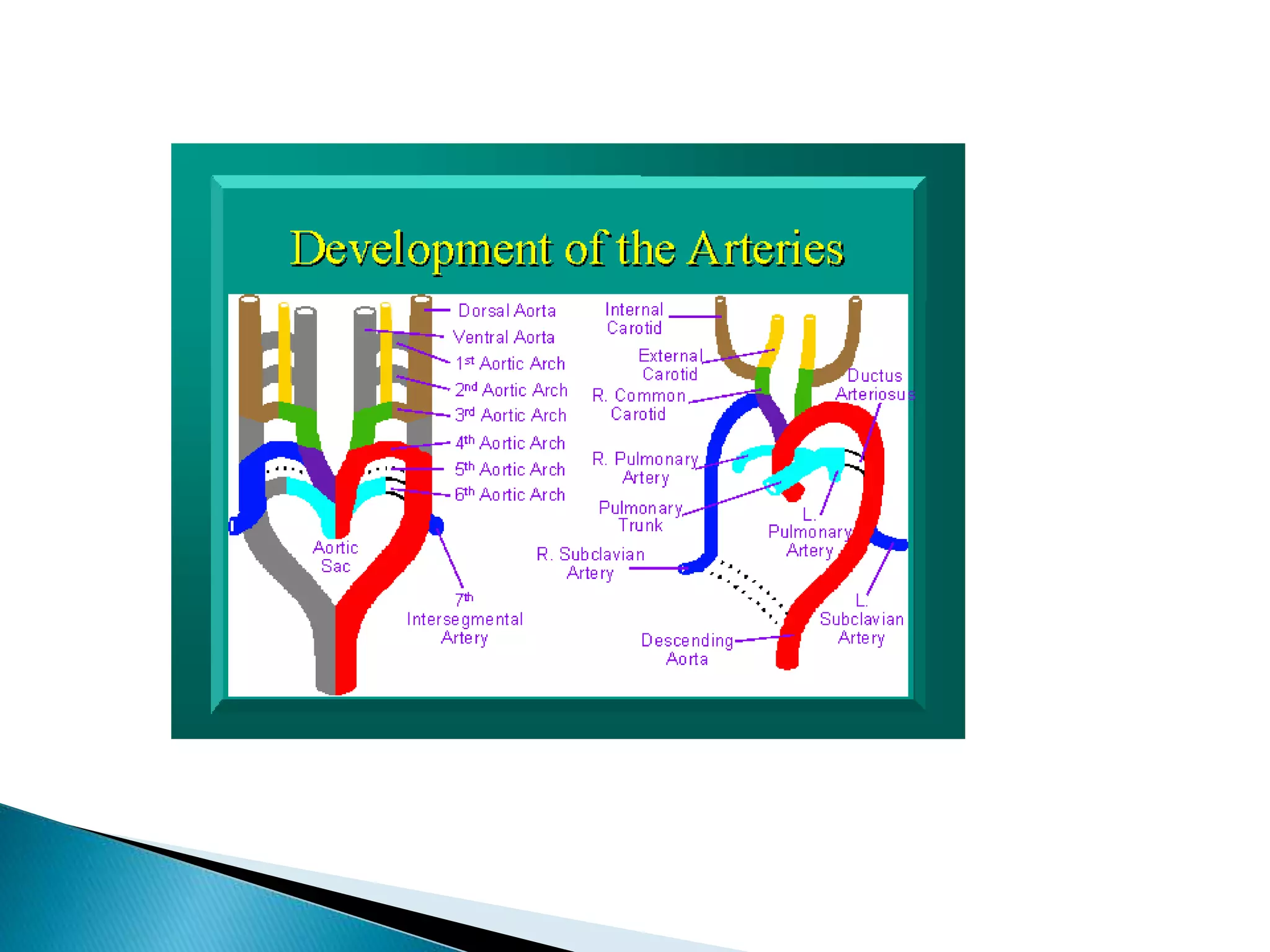
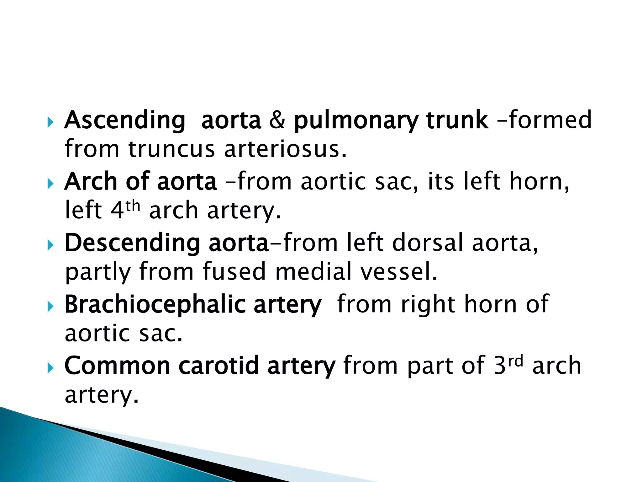
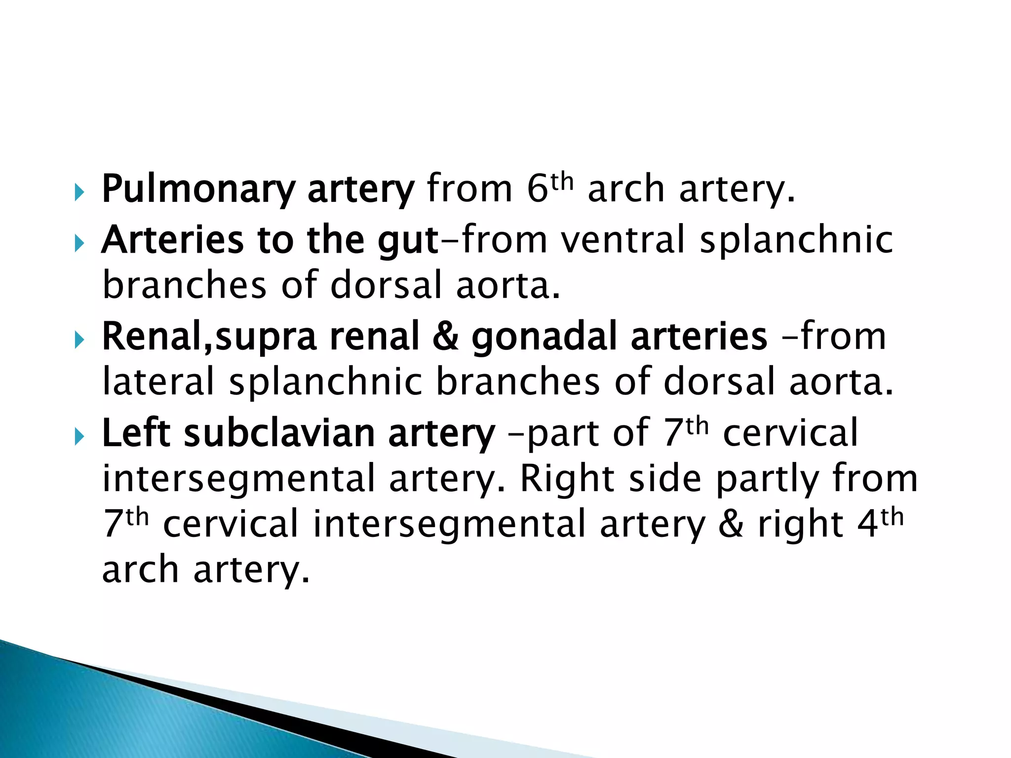
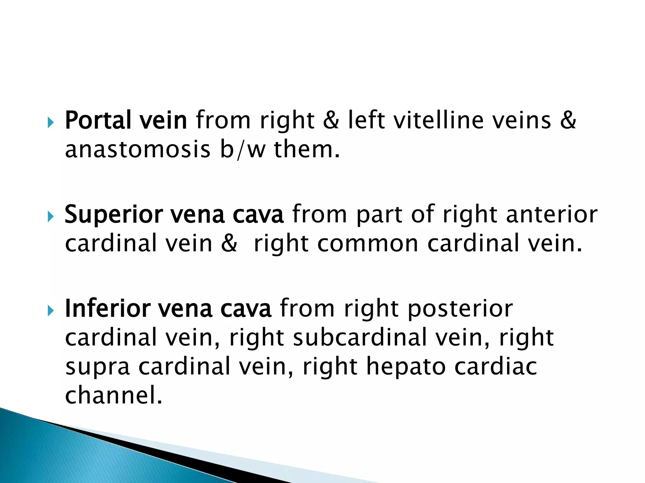
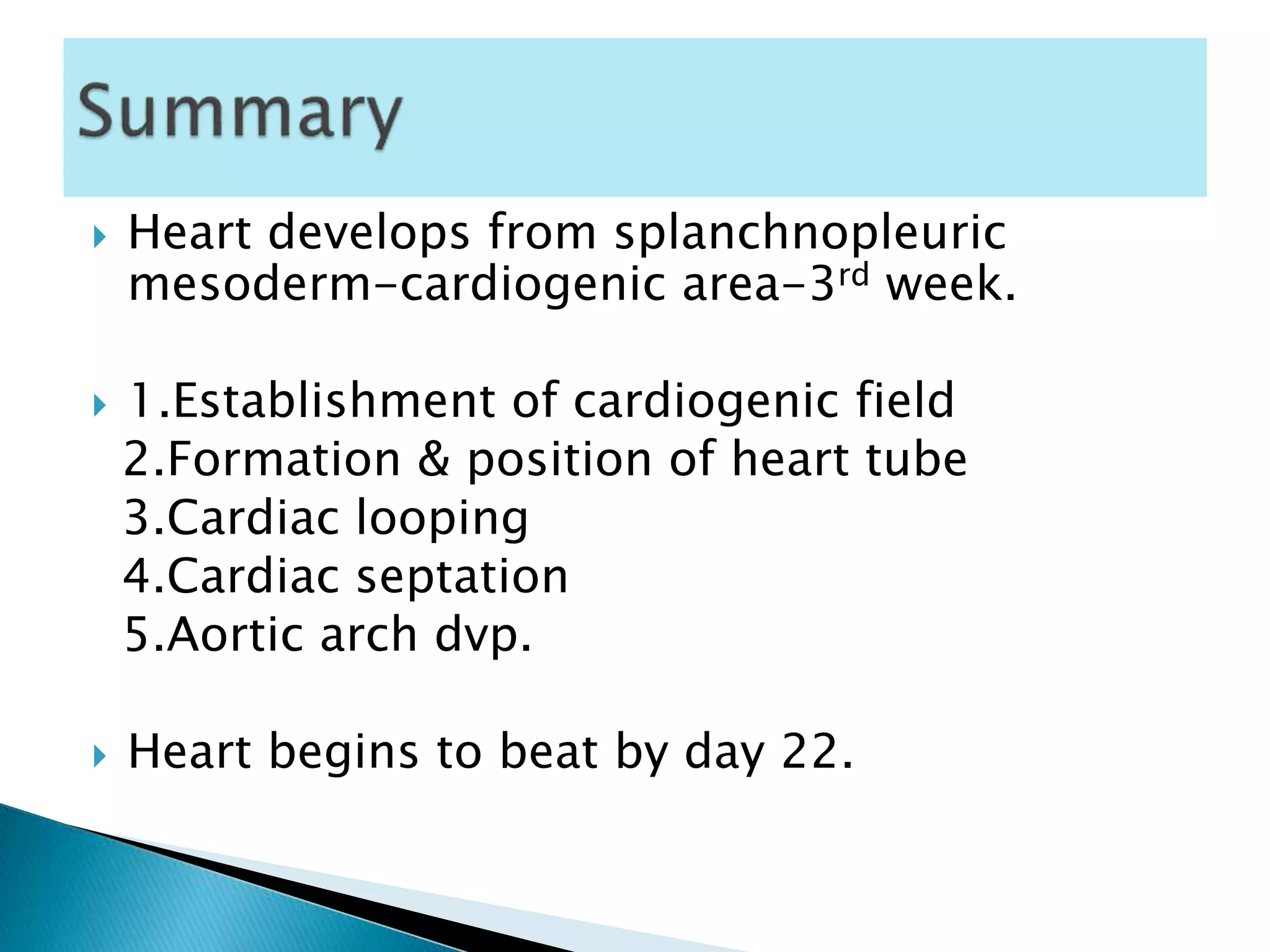
By Dr Robin Thomas
By Dr Robin Thomas
 The vascular system appears in the middle of
3rd week , when embryo is no longer able to
satisfy its nutritional requirement .
 The Heart develops from splanchnopleuric
mesoderm related - intraembryonic coelom
that forms pericardial cavity. This mesoderm
called Cardiogenic area.
Cardiac development involves :
 1.Establishment of cardiogenic field
 2.Formation & position of heart tube
 3.Cardiac looping
 4.Cardiac septation
 Cardiac progenitor cells lie in the epiblast ,
lateral to primitive streak.
 Migrate towards primitive streak
 Cells destined to form cranial segments of heart ,
outflow tract, migrate first & cells forming more
caudal portions , right ventricle,
left ventricle & sinus venosus migrate in
sequential order.
 Cells procede towards cranium & position
themselves rostral to buccopharyngeal
membrane & neural folds- reside in splanchnic
layer of lateral plate mesoderm.
 Cardiac progenitor cells lies – epiblast ,lateral
to primitive streak. Cells procede towards
cranium –rostral to buccopharyngeal
membrane & neural folds. Reside in
splanchnic layer of lateral plate mesoderm.
 Blood islands appear in this mesoderm –form
bloodcells & vessels –vasculogenesis.
 Blood islands unite & form horseshoe shaped
endothelial lined tube surrounded by
myoblasts- cardiogenic field – intraembryonic
cavity over it develops into pericardial cavity.
 Initially central portion of cardiogenic area is
anterior to buccopharyngeal membrane & neural
plate.
 Closure of neural tube & formation of brain
vesicles – central nervous system grows cephalad
 Buccopharyngeal membrane is pulled forward –
heart & pericardial cavity move first to cervical
region & finally to thorax.
 Embryo fold cephalocaudally & laterally- caudal
region of paired cardiac primordia merge.
 18 Days embryo 20 Days embryo
 Central portion- cardiogenic area anterior to
buccopharyngeal membrane & neural plate.
 21Days embryo 22 Days embryo
growth of brain & buccopharyngeal memb
cephalic folding of pulled forward –heart &
embryo. pericardial cavity moves
first to cervical region-
thorax.
 22 day embryo
 Embryo folds cephalocaudally & laterally-caudal
region of paired cardiac primordia merge-
 Heart –continuous expanded tube with inner
endothelial lining & outer myocardial layer. Pump
blood –first aortic arch –dorsal aorta.
 Heart - continuous expanded tube – inner
endothelial lining & outer myocardial layer.
 Receives venous drainage at caudal pole - pump
blood - first aortic arch into dorsal aorta .
 Heart tube - attached to pericardial cavity by
dorsal mesocardium.
 Dorsal mesocardium disappears-creates
transverse pericardial sinus-connects both sides
of pericardial cavity.
 Myocardium thickens & secrete thick layer of
extracellular matrix – rich in hyaluronic acid –
separates from endothelium.
 Mesothelial cells on surface of septum
transversum form proepicardium-later form
epicardium.
 Heart tube consists of 3 layers
Endocardium-internal endothelial lining
Myocardium-muscular wall
Epicardium-outside of heart tube
 Right & left endothelial
heart tubes.
 Fusion of heart tubes
from cranial to caudal
ends.
 Arterial end of heart tube Venous end of
heart tube
 Single endothelial heart tube shows series of
dilatations.
 1.Bulbus cordis
 2.ventricle
 3.atrium
 4.sinus venosus
 Ventricle & atrium are connected by
atrioventricular canal.
 Sinus venosus has prolongations - right & left
horns.
 Bulbus cordis lies at arterial end of heart tube
–divisible into proximal part - conus &
distal part - truncus arteriosus.
 Truncus arteriosus continuous distally with
aortic sac-continuous with right & left
pharyngeal arch arteries-continuous with
right & left dorsal aorta.
 Sinus venosus lies at venous end of heart-has
right & left horns.
 One vitelline vein (yolk sac), umbilical vein
(placenta), common cardinal vein (bodywall)
join each horn of sinus venosus.
 Heart tube elongate & bend on day 21.
 Cephalic portion bend ventrally,caudally & to
right.
 Atrial portion shift dorsocranially & to left.
 Bending creates cardiac loop-complete by
day 28.
 Cephalic portion heart tube bend ventrally,
caudally & to right.
 Atrial portion shift dorso-cranially & to left.
Creates Cardiac loop-complete by day 28.
 Dextrocardia-heart loops to left instead of
right.
 Heart lies on right side of thorax instead of
left.
 May coincide with situs inversus-complete
reversal of asymmetry in all organs.
 Truncus arteriosus form ascending aorta &
pulmonary trunk.
 Conus absorbed into ventricle & form
chamber –partitioned into right & left
ventricles.
 Primitive atrium partitioned into right & left
atria.
 Sinus venosus absorbed into right atrium.
 Right common cardinal vein forms superior
vena cava.
 Umbilical vein disappears.
 Right vitelline vein forms part of inferior vena
cava.
 Left horn of sinus venosus retrogresses-form
part of coronary sinus.
 Sinus venosus & primitive atrial chamber are
connected by a wide opening
 Opening becomes narrow-shifts to right-
narrow slit-right & left venous valves.
 Cranially 2 valves fuse to form septum
spurium.
 Atrioventricular canal divides into right & left
halves.
 2 thickenings-atrioventricular cushions
appear on dorsal & ventral walls.
 Atrio ventricular cushions grow towards each
other & fuse –form septum intermedium.
 Begins at day 26-ingrowth of large tissue
masses.
 Endocardial cushions at atrio ventricular &
conotruncal junctions.
 Endocardial cells de differentiate & form part of
atrio- ventricular valves.
 Complete septation of atrioventricular canal –
fusion of endocardial cushions.
 Most of atrioventricular valve tissue –derived
from ventricular myocardium –undermining of
ventricular walls.
 Physical separation of 2 valves (mitral &
tricuspid valve) produce atrioventricular
septum.
 Absence causes-Atrio ventricular canal
defects. (Endocardial cushion defects)
 1.Complete endocardial cushion defects
Ostium primum ASD, VSD in interventricular
septum, clefts in anterior mitral valve & septal
leaflet of tricuspid valve.
 2.Partial endocardial cushion defects
Ostium primum ASD, clefts in mitral &
tricuspid valve.
 Septation of atria begins at day 30 day .
 Atrial chamber undergoes division into right
& left halves-formation of 2 septa.
 Septum primum arises from roof of atrium ,
to right of septum spurium.
 Septum primum grows downwards & fuses
with atrioventricular cushions.
 Upper part of septum primum degenerates-
form foramen secundum.
 Septum secundum formed to right of septum
primum.
 Septum secundum grows downwards from
roof of atrial chamber-overlapps free edge of
septum primum.
 Blood now flows from left to right through
oblique cleft b/w 2 septa-foramen ovale.
 During fetal life oxygenated blood reaches
right atrium from placenta.
 Blood reaches left atrium tru foramen ovale.
 After birth left atrium receives oxygenated
blood from lungs- foramen ovale obliterated
by fusion of septum primum & septum
secundum.
 Annulus ovalis –septum secundum.
 Fossa ovalis-septum primum.
Inter atrial septal defects
 1.Septum primum defect
 2.Septum secundum defect
 3.Patent foramen ovale
 4.Premature closure of foramen ovale
 Right atrium derived from :
1.Right half of primitive atrium-main part.
2.Sinus venosus.
3.Right half of Atrio ventricular canal.
 SINUS VENOSUS
 Left horn of sinus venosus –part of coronary
sinus.
 Right common cardinal vein becomes part of
superior vena cava.
 Right vitelline vein forms terminal part of
inferior vena cava.
 Right venous valve (right margin of sinu atrial
orifice) forms crista terminalis, valve of IVC,
& valve of coronary sinus.
 Left atrium is derived from :
1.Primitive atrial chamber-left half
2.Atrioventricular canal-left half
3.Pulmonary veins -absorbed proximal parts
 Single pulmonary vein opens into left half of
atrium –when septum primum just beginning to
form.
 Pulmonary vein bifurcates-right & left branches-
bifurcates-drain corresp. lung bud.
 Parts of pulmonary vein nearest to left atrium –
absorbed -4 pulmonary veins
 Fate of Bulbus Cordis
 Bulbus Cordis –divisible into conus (proximal
part) & truncus arteriosus (distal part).
 Spiral septum –union of right superior & left
inferior truncus swelling or cushions.
 Spiral septum appears within truncus
arteriosus & subdivides into ascending aorta
& pulmonary trunk.
 Conus (bulbus cordis proper) merges with
cavity of primitive ventricle .
 Right & left ventricle formed –partition of this
chamber.
 Bulboventricular cavity consists of :
Dilated lower part –communicates with atria.
Conical upper part-communicates with
truncus arteriosus.
 Septation of ventricles begins at day 25.
 Interventricular septum grows upwards from
floor of bulboventricular cavity-divides lower
dilated part into right & left halves.
 Interventricular septum fuses with fused
atrioventricular cushions.
 Right & left bulbar ridges arise from conical part
of bulboventricular cavity & fuse to form bulbar
septum.
 Bulbar septum grows towards interventricular
septum , doesnot reach it –gap left b/w two.
 Gap b/w upper edge of interventricular
septum & lower edge of bulbar septum –filled
by proliferation of tissue from atrioventricular
cushions.
 Inter ventricular septal defects
 1.VSD
 2.Tetralogy of Fallot
 3.Persistant truncus arteriosus
 4.Transposition of Great arteries
 Mitral & tricuspid valves formed –proliferation
of connective tissue under endocardium of
right & left atrioventricular canals.
 Pulmonary & aortic valves –endocardial
cushions formed at junction of truncus -
arteriosus & conus.
 3rd week of life ( 15-21 days)
 1.Splanchnopleuric mesoderm-18 days.
 2.Cavitation of mesoderm-intraembryonic
coelom-pericardial, pleural, peritoneal
cavities-18 days.
 3.Endocardial heart tube-20 days.
 4.Cardiac looping-21days.
 4 th week of life ( 22-28 days )
 1.Dvp. of left ventricle & right ventricle.
 2.Circulation commences.
 3.Cardiovascular septation.
 4.Aortic arches begins.
 5 th week of life ( 29-35 days )
 1.LV,RV,ventricular septum continue to grow.
 2.Approximation of aorta to interventricular
foramen, mitral valve,LV.
 3.Separation of ascending aorta,main
pulmonary artery.
 4.Separation of mitral & tricuspid valve.
 5.Tricuspid valve opens into right ventricle.
 6.Ostium primum closed by endocardial
cushions.
 6th & 7th week of life ( 36-49 days )
 1.Closure of conal septum
 2.Closure of membraneous part of ventricular
septum 38-45 days
 Blood vessel development by 2 mechanisms.
 1.Vasculogenesis-vessels arise by coalesence
of angioblasts.
 2.Angiogenesis-vessels sprout from existing
vessels.
 Dorsal aorta & cardinal veins –vasculogenesis.
 VEGF-vascular endothelial growth factor.
 Pharyngeal arches develop during 4 th & 5th
weeks of development.
 First arteries to appear in embryo-right & left
primitive aortae.
 Continuous with 2 endocardial heart tubes.
 Each primitive aorta-ventral aorta (lying
ventral to foregut), arched portion & dorsal
aorta (lying dorsal to gut).
 Fusion of 2 endocardial heart tubes-2 ventral
aorta partially fuse to form aortic sac.
 Unfused parts remain as right & left horns of
sac.
 Successive arterial arches appear in 2-6
pharyngeal arches, each connected ventrally
to right or left horn of aortic sac & dorsally to
dorsal aorta.
 Greater part of 1st & 2nd arch arteries
disappear ( 29 day embryo )
 Small portion of 1st arch artery persists to
form maxillary artery.
 Second arch artery form hyoid & stapedial
artery.
 Fifth arch artery also disappears.
 Aortic sac now connected with arteries of 3,4
& 6th arches.

 3rd & 4th arteries open into ventral part & 6th
arch arteries into dorsal part of aortic sac.
 Spiral septum formed in truncus arteriosus
,extends into aortic sac –fuses with posterior
wall-blood from pulmonary trunk passes into
6th arch artery –blood from ascending aorta
passes into 3rd & 4th arch arteries.
 Dorsal aorta b/w 3rd & 4th arches disappear
on both sides.
 Each 6th arch artery gives off artery to
developing lung bud.
 Right side-portion of 6th arch artery b/w lung
bud & dorsal aorta disappears.
 Left side-remains patent & forms ductus
arteriosus.
 Each 3rd arch artery gives off a bud –grows
cranially & form external carotid artery.
 Dorsal aorta gives off series of lateral
intersegmental branches to body wall.
 7th cervical intersegmental artery forms left
subclavian artery.
Arch Arterial derivative
1 arch Maxillary artery
2 arch Hyoid & stapedial artery
3 arch Common carotid & 1st
part of internal carotid
artery.
Left side of 4th arch Arch of aorta ( from left
common carotid to left
subclavian arteries ).
Right side of 4th arch Right subclavian artery
(proximal portion)
Left side of 6th arch Left pulmonary artery &
ductus arteriosus
Right side of 6th arch Right pulmonary artery
 Ascending aorta & pulmonary trunk –formed
from truncus arteriosus.
 Arch of aorta –from aortic sac, its left horn,
left 4th arch artery.
 Descending aorta-from left dorsal aorta,
partly from fused medial vessel.
 Brachiocephalic artery from right horn of
aortic sac.
 Common carotid artery from part of 3rd arch
artery.
 Pulmonary artery from 6th arch artery.
 Arteries to the gut-from ventral splanchnic
branches of dorsal aorta.
 Renal,supra renal & gonadal arteries –from
lateral splanchnic branches of dorsal aorta.
 Left subclavian artery –part of 7th cervical
intersegmental artery. Right side partly from
7th cervical intersegmental artery & right 4th
arch artery.
 Portal vein from right & left vitelline veins &
anastomosis b/w them.
 Superior vena cava from part of right anterior
cardinal vein & right common cardinal vein.
 Inferior vena cava from right posterior
cardinal vein, right subcardinal vein, right
supra cardinal vein, right hepato cardiac
channel.
 Heart develops from splanchnopleuric
mesoderm-cardiogenic area-3rd week.
 1.Establishment of cardiogenic field
2.Formation & position of heart tube
3.Cardiac looping
4.Cardiac septation
5.Aortic arch dvp.
 Heart begins to beat by day 22.
 Cardiogenesis begins on 18th day of life &
normally completed by 45th day of life with
formation of membraneous part of
interventricular septum.
 1.Kliegman, Stanton, Geme ST, Schor, Behrman. Nelson
Textbook of Pediatrics 19th edition 2012 :1527-1529.
 2.Sadler WT. Langmans Medical Embryology 10th edition
2012 :159-189.
 3.John FK, James EL, Donald CF Nadas Pediatric
Cardiology 2nd edition :14-25
 4.Myung K Park .Pediatric cardiology for Practitioners 2012
5th edition 120-180
 5.Inderbir singh, Pal PK. Human Embryology 8th edition
2012 :191-231
Development of heart finale   copy

Development of heart finale copy

  • 1.
  • 2.
  • 3.
     The vascularsystem appears in the middle of 3rd week , when embryo is no longer able to satisfy its nutritional requirement .  The Heart develops from splanchnopleuric mesoderm related - intraembryonic coelom that forms pericardial cavity. This mesoderm called Cardiogenic area.
  • 4.
    Cardiac development involves:  1.Establishment of cardiogenic field  2.Formation & position of heart tube  3.Cardiac looping  4.Cardiac septation
  • 5.
     Cardiac progenitorcells lie in the epiblast , lateral to primitive streak.  Migrate towards primitive streak  Cells destined to form cranial segments of heart , outflow tract, migrate first & cells forming more caudal portions , right ventricle, left ventricle & sinus venosus migrate in sequential order.  Cells procede towards cranium & position themselves rostral to buccopharyngeal membrane & neural folds- reside in splanchnic layer of lateral plate mesoderm.
  • 6.
     Cardiac progenitorcells lies – epiblast ,lateral to primitive streak. Cells procede towards cranium –rostral to buccopharyngeal membrane & neural folds. Reside in splanchnic layer of lateral plate mesoderm.
  • 7.
     Blood islandsappear in this mesoderm –form bloodcells & vessels –vasculogenesis.  Blood islands unite & form horseshoe shaped endothelial lined tube surrounded by myoblasts- cardiogenic field – intraembryonic cavity over it develops into pericardial cavity.
  • 8.
     Initially centralportion of cardiogenic area is anterior to buccopharyngeal membrane & neural plate.  Closure of neural tube & formation of brain vesicles – central nervous system grows cephalad  Buccopharyngeal membrane is pulled forward – heart & pericardial cavity move first to cervical region & finally to thorax.  Embryo fold cephalocaudally & laterally- caudal region of paired cardiac primordia merge.
  • 9.
     18 Daysembryo 20 Days embryo  Central portion- cardiogenic area anterior to buccopharyngeal membrane & neural plate.
  • 10.
     21Days embryo22 Days embryo growth of brain & buccopharyngeal memb cephalic folding of pulled forward –heart & embryo. pericardial cavity moves first to cervical region- thorax.
  • 11.
     22 dayembryo  Embryo folds cephalocaudally & laterally-caudal region of paired cardiac primordia merge-  Heart –continuous expanded tube with inner endothelial lining & outer myocardial layer. Pump blood –first aortic arch –dorsal aorta.
  • 12.
     Heart -continuous expanded tube – inner endothelial lining & outer myocardial layer.  Receives venous drainage at caudal pole - pump blood - first aortic arch into dorsal aorta .  Heart tube - attached to pericardial cavity by dorsal mesocardium.  Dorsal mesocardium disappears-creates transverse pericardial sinus-connects both sides of pericardial cavity.  Myocardium thickens & secrete thick layer of extracellular matrix – rich in hyaluronic acid – separates from endothelium.
  • 13.
     Mesothelial cellson surface of septum transversum form proepicardium-later form epicardium.  Heart tube consists of 3 layers Endocardium-internal endothelial lining Myocardium-muscular wall Epicardium-outside of heart tube
  • 14.
     Right &left endothelial heart tubes.  Fusion of heart tubes from cranial to caudal ends.
  • 16.
     Arterial endof heart tube Venous end of heart tube
  • 17.
     Single endothelialheart tube shows series of dilatations.  1.Bulbus cordis  2.ventricle  3.atrium  4.sinus venosus
  • 18.
     Ventricle &atrium are connected by atrioventricular canal.  Sinus venosus has prolongations - right & left horns.  Bulbus cordis lies at arterial end of heart tube –divisible into proximal part - conus & distal part - truncus arteriosus.  Truncus arteriosus continuous distally with aortic sac-continuous with right & left pharyngeal arch arteries-continuous with right & left dorsal aorta.
  • 19.
     Sinus venosuslies at venous end of heart-has right & left horns.  One vitelline vein (yolk sac), umbilical vein (placenta), common cardinal vein (bodywall) join each horn of sinus venosus.
  • 20.
     Heart tubeelongate & bend on day 21.  Cephalic portion bend ventrally,caudally & to right.  Atrial portion shift dorsocranially & to left.  Bending creates cardiac loop-complete by day 28.
  • 21.
     Cephalic portionheart tube bend ventrally, caudally & to right.  Atrial portion shift dorso-cranially & to left. Creates Cardiac loop-complete by day 28.
  • 23.
     Dextrocardia-heart loopsto left instead of right.  Heart lies on right side of thorax instead of left.  May coincide with situs inversus-complete reversal of asymmetry in all organs.
  • 25.
     Truncus arteriosusform ascending aorta & pulmonary trunk.  Conus absorbed into ventricle & form chamber –partitioned into right & left ventricles.  Primitive atrium partitioned into right & left atria.  Sinus venosus absorbed into right atrium.  Right common cardinal vein forms superior vena cava.
  • 26.
     Umbilical veindisappears.  Right vitelline vein forms part of inferior vena cava.  Left horn of sinus venosus retrogresses-form part of coronary sinus.
  • 29.
     Sinus venosus& primitive atrial chamber are connected by a wide opening  Opening becomes narrow-shifts to right- narrow slit-right & left venous valves.  Cranially 2 valves fuse to form septum spurium.  Atrioventricular canal divides into right & left halves.  2 thickenings-atrioventricular cushions appear on dorsal & ventral walls.
  • 30.
     Atrio ventricularcushions grow towards each other & fuse –form septum intermedium.
  • 32.
     Begins atday 26-ingrowth of large tissue masses.  Endocardial cushions at atrio ventricular & conotruncal junctions.  Endocardial cells de differentiate & form part of atrio- ventricular valves.  Complete septation of atrioventricular canal – fusion of endocardial cushions.  Most of atrioventricular valve tissue –derived from ventricular myocardium –undermining of ventricular walls.
  • 33.
     Physical separationof 2 valves (mitral & tricuspid valve) produce atrioventricular septum.  Absence causes-Atrio ventricular canal defects. (Endocardial cushion defects)
  • 34.
     1.Complete endocardialcushion defects Ostium primum ASD, VSD in interventricular septum, clefts in anterior mitral valve & septal leaflet of tricuspid valve.  2.Partial endocardial cushion defects Ostium primum ASD, clefts in mitral & tricuspid valve.
  • 39.
     Septation ofatria begins at day 30 day .  Atrial chamber undergoes division into right & left halves-formation of 2 septa.  Septum primum arises from roof of atrium , to right of septum spurium.  Septum primum grows downwards & fuses with atrioventricular cushions.  Upper part of septum primum degenerates- form foramen secundum.
  • 40.
     Septum secundumformed to right of septum primum.  Septum secundum grows downwards from roof of atrial chamber-overlapps free edge of septum primum.  Blood now flows from left to right through oblique cleft b/w 2 septa-foramen ovale.  During fetal life oxygenated blood reaches right atrium from placenta.  Blood reaches left atrium tru foramen ovale.
  • 41.
     After birthleft atrium receives oxygenated blood from lungs- foramen ovale obliterated by fusion of septum primum & septum secundum.  Annulus ovalis –septum secundum.  Fossa ovalis-septum primum.
  • 42.
    Inter atrial septaldefects  1.Septum primum defect  2.Septum secundum defect  3.Patent foramen ovale  4.Premature closure of foramen ovale
  • 44.
     Right atriumderived from : 1.Right half of primitive atrium-main part. 2.Sinus venosus. 3.Right half of Atrio ventricular canal.
  • 45.
     SINUS VENOSUS Left horn of sinus venosus –part of coronary sinus.  Right common cardinal vein becomes part of superior vena cava.  Right vitelline vein forms terminal part of inferior vena cava.  Right venous valve (right margin of sinu atrial orifice) forms crista terminalis, valve of IVC, & valve of coronary sinus.
  • 47.
     Left atriumis derived from : 1.Primitive atrial chamber-left half 2.Atrioventricular canal-left half 3.Pulmonary veins -absorbed proximal parts  Single pulmonary vein opens into left half of atrium –when septum primum just beginning to form.  Pulmonary vein bifurcates-right & left branches- bifurcates-drain corresp. lung bud.  Parts of pulmonary vein nearest to left atrium – absorbed -4 pulmonary veins
  • 49.
     Fate ofBulbus Cordis  Bulbus Cordis –divisible into conus (proximal part) & truncus arteriosus (distal part).  Spiral septum –union of right superior & left inferior truncus swelling or cushions.  Spiral septum appears within truncus arteriosus & subdivides into ascending aorta & pulmonary trunk.
  • 50.
     Conus (bulbuscordis proper) merges with cavity of primitive ventricle .  Right & left ventricle formed –partition of this chamber.  Bulboventricular cavity consists of : Dilated lower part –communicates with atria. Conical upper part-communicates with truncus arteriosus.
  • 54.
     Septation ofventricles begins at day 25.  Interventricular septum grows upwards from floor of bulboventricular cavity-divides lower dilated part into right & left halves.  Interventricular septum fuses with fused atrioventricular cushions.  Right & left bulbar ridges arise from conical part of bulboventricular cavity & fuse to form bulbar septum.
  • 55.
     Bulbar septumgrows towards interventricular septum , doesnot reach it –gap left b/w two.  Gap b/w upper edge of interventricular septum & lower edge of bulbar septum –filled by proliferation of tissue from atrioventricular cushions.
  • 56.
     Inter ventricularseptal defects  1.VSD  2.Tetralogy of Fallot  3.Persistant truncus arteriosus  4.Transposition of Great arteries
  • 61.
     Mitral &tricuspid valves formed –proliferation of connective tissue under endocardium of right & left atrioventricular canals.  Pulmonary & aortic valves –endocardial cushions formed at junction of truncus - arteriosus & conus.
  • 63.
     3rd weekof life ( 15-21 days)  1.Splanchnopleuric mesoderm-18 days.  2.Cavitation of mesoderm-intraembryonic coelom-pericardial, pleural, peritoneal cavities-18 days.  3.Endocardial heart tube-20 days.  4.Cardiac looping-21days.
  • 64.
     4 thweek of life ( 22-28 days )  1.Dvp. of left ventricle & right ventricle.  2.Circulation commences.  3.Cardiovascular septation.  4.Aortic arches begins.
  • 65.
     5 thweek of life ( 29-35 days )  1.LV,RV,ventricular septum continue to grow.  2.Approximation of aorta to interventricular foramen, mitral valve,LV.  3.Separation of ascending aorta,main pulmonary artery.  4.Separation of mitral & tricuspid valve.  5.Tricuspid valve opens into right ventricle.  6.Ostium primum closed by endocardial cushions.
  • 66.
     6th &7th week of life ( 36-49 days )  1.Closure of conal septum  2.Closure of membraneous part of ventricular septum 38-45 days
  • 67.
     Blood vesseldevelopment by 2 mechanisms.  1.Vasculogenesis-vessels arise by coalesence of angioblasts.  2.Angiogenesis-vessels sprout from existing vessels.  Dorsal aorta & cardinal veins –vasculogenesis.  VEGF-vascular endothelial growth factor.
  • 73.
     Pharyngeal archesdevelop during 4 th & 5th weeks of development.  First arteries to appear in embryo-right & left primitive aortae.  Continuous with 2 endocardial heart tubes.  Each primitive aorta-ventral aorta (lying ventral to foregut), arched portion & dorsal aorta (lying dorsal to gut).
  • 74.
     Fusion of2 endocardial heart tubes-2 ventral aorta partially fuse to form aortic sac.  Unfused parts remain as right & left horns of sac.  Successive arterial arches appear in 2-6 pharyngeal arches, each connected ventrally to right or left horn of aortic sac & dorsally to dorsal aorta.
  • 75.
     Greater partof 1st & 2nd arch arteries disappear ( 29 day embryo )  Small portion of 1st arch artery persists to form maxillary artery.  Second arch artery form hyoid & stapedial artery.  Fifth arch artery also disappears.  Aortic sac now connected with arteries of 3,4 & 6th arches. 
  • 76.
     3rd &4th arteries open into ventral part & 6th arch arteries into dorsal part of aortic sac.  Spiral septum formed in truncus arteriosus ,extends into aortic sac –fuses with posterior wall-blood from pulmonary trunk passes into 6th arch artery –blood from ascending aorta passes into 3rd & 4th arch arteries.  Dorsal aorta b/w 3rd & 4th arches disappear on both sides.
  • 77.
     Each 6tharch artery gives off artery to developing lung bud.  Right side-portion of 6th arch artery b/w lung bud & dorsal aorta disappears.  Left side-remains patent & forms ductus arteriosus.  Each 3rd arch artery gives off a bud –grows cranially & form external carotid artery.
  • 78.
     Dorsal aortagives off series of lateral intersegmental branches to body wall.  7th cervical intersegmental artery forms left subclavian artery.
  • 79.
    Arch Arterial derivative 1arch Maxillary artery 2 arch Hyoid & stapedial artery 3 arch Common carotid & 1st part of internal carotid artery. Left side of 4th arch Arch of aorta ( from left common carotid to left subclavian arteries ). Right side of 4th arch Right subclavian artery (proximal portion) Left side of 6th arch Left pulmonary artery & ductus arteriosus Right side of 6th arch Right pulmonary artery
  • 80.
     Ascending aorta& pulmonary trunk –formed from truncus arteriosus.  Arch of aorta –from aortic sac, its left horn, left 4th arch artery.  Descending aorta-from left dorsal aorta, partly from fused medial vessel.  Brachiocephalic artery from right horn of aortic sac.  Common carotid artery from part of 3rd arch artery.
  • 81.
     Pulmonary arteryfrom 6th arch artery.  Arteries to the gut-from ventral splanchnic branches of dorsal aorta.  Renal,supra renal & gonadal arteries –from lateral splanchnic branches of dorsal aorta.  Left subclavian artery –part of 7th cervical intersegmental artery. Right side partly from 7th cervical intersegmental artery & right 4th arch artery.
  • 82.
     Portal veinfrom right & left vitelline veins & anastomosis b/w them.  Superior vena cava from part of right anterior cardinal vein & right common cardinal vein.  Inferior vena cava from right posterior cardinal vein, right subcardinal vein, right supra cardinal vein, right hepato cardiac channel.
  • 83.
     Heart developsfrom splanchnopleuric mesoderm-cardiogenic area-3rd week.  1.Establishment of cardiogenic field 2.Formation & position of heart tube 3.Cardiac looping 4.Cardiac septation 5.Aortic arch dvp.  Heart begins to beat by day 22.
  • 84.
     Cardiogenesis beginson 18th day of life & normally completed by 45th day of life with formation of membraneous part of interventricular septum.
  • 85.
     1.Kliegman, Stanton,Geme ST, Schor, Behrman. Nelson Textbook of Pediatrics 19th edition 2012 :1527-1529.  2.Sadler WT. Langmans Medical Embryology 10th edition 2012 :159-189.  3.John FK, James EL, Donald CF Nadas Pediatric Cardiology 2nd edition :14-25  4.Myung K Park .Pediatric cardiology for Practitioners 2012 5th edition 120-180  5.Inderbir singh, Pal PK. Human Embryology 8th edition 2012 :191-231