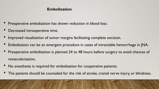
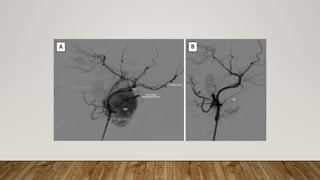
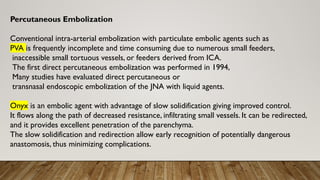
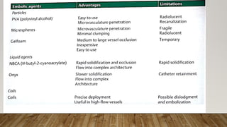
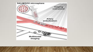
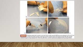
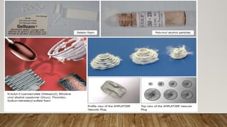
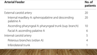
EMBOLIZATION IN
JNA SURGERY
MODERATOR
DR .T. SUMAN KRISHNA
ASSISTANT PROFESSOR ENT
NMCH
PRESENTOR
DR. V . SANKAR NAIK
JUNIOR RESIDENT 3rd
year ENT
NMCH
Embolization
 Preoperative embolization has shown reduction in blood loss.
 Decreased intraoperative time.
 Improved visualization of tumor margins facilitating complete excision.
 Embolization can be an emergent procedure in cases of intractable hemorrhage in JNA.
 Preoperative embolization is planned 24 to 48 hours before surgery to avoid chances of
revascularization.
 No anesthesia is required for embolization for cooperative patients.
 The patients should be counseled for the risk of stroke, cranial nerve injury, or blindness.
Embolization Procedures
Transarterial Embolization
• The catheter is positioned in external carotid artery (ECA) and selective angiogram is done
to visualize the tumor blush.
• Microcatheterization of the ECA system is done at the level of internal maxillary artery.
• If spasm is encountered, injections of vasodilators such as nicardipine, verapamil, or
nitroglycerin can be given. Catheter size depends on the size of the vessel that has to be
embolized.
• Digital subtraction angiography is done.The angiographic pattern is analyzed both in arterial
phase and venous phase.
• The arterial phase shows reticulated pattern in early phase and dense homogenous blush
persists in the venous phase.
• Postembolization, the tumor blush disappears .A systematic approach of embolization
described by Davis et al (1987) states embolization of maxillary artery offers the best chance
of devascularizing the tumor, followed by selective embolization of residual feeding vessels.
• Detailed angiographic assessment of the feeders and anastomosis should be done prior to
embolization.
• Contralateral ECA and ipsilateral ICA angiograms are always done to assess feeders from
them, if any.
Percutaneous Embolization
Conventional intra-arterial embolization with particulate embolic agents such as
PVA is frequently incomplete and time consuming due to numerous small feeders,
inaccessible small tortuous vessels, or feeders derived from ICA.
The first direct percutaneous embolization was performed in 1994,
Many studies have evaluated direct percutaneous or
transnasal endoscopic embolization of the JNA with liquid agents.
Onyx is an embolic agent with advantage of slow solidification giving improved control.
It flows along the path of decreased resistance, infiltrating small vessels. It can be redirected,
and it provides excellent penetration of the parenchyma.
The slow solidification and redirection allow early recognition of potentially dangerous
anastomosis, thus minimizing complications.
Complications
• Immediate complications can be pain and hypersensitivity reactions.
• Accidental introduction of embolic material from ECA to ICA can result in stroke,
• in case of ophthalmic artery can cause blindness,
• the superficial branches of face can cause facial pain.
• Embolization by percutaneous technique is associated with triggering
trigeminocardiac reflex causing bradycardia.
Multidisciplinary management of juvenile nasopharyngeal angiofibroma
by Abouzeid et al.
Year 2021
Egypt
Introduction
• The study focuses on juvenile nasopharyngeal angiofibroma (JNA), a rare benign but
locally aggressive vascular tumor that predominantly affects adolescent males.
JNA constitutes only about 0.5% of head and neck tumors, it is the most common
benign nasopharyngeal tumor.
• The tumor typically presents with symptoms like recurrent epistaxis, nasal
obstruction, and chronic otomastoiditis.
Objective
• The study aims to evaluate the efficiency and safety of using an
endovascular modality, specifically pre-operative trans-arterial
embolization followed by endoscopic surgical resection, in managing JNA.
Methods
Study Period :The study was conducted between January 2012 and December 2017.
It involved 20 male patients treated at three university hospitals in Egypt.
Patient Selection: Male patients aged between 6 and 20 years with a diagnosis of JNA
were included.All patients underwent a thorough clinical and radiological evaluation
before treatment.
Procedure:
Imaging: CT and MRI were used to assess the extent of the tumor, especially regarding
bony involvement and intracranial extension.
Embolization
• Preoperative super-selective trans-arterial embolization was performed using polyvinyl
alcohol (PVA) particles to achieve tumor devascularization.
• Surgery: Endoscopic surgical resection was performed within 48 hours after embolization.
The degree of tumor resection, intraoperative blood loss, and operative time were
evaluated.
• Follow-Up: Clinical and radiological follow-ups were conducted for at least one year to
monitor for recurrence or complications.
Results
• The average age of the patients was 13 years.Tumor Grading :The tumors were graded
according to the Radkowski grading system, with patients having varying grades from IA to
IIIB.
• Embolization Outcomes:All patients successfully underwent embolization, with total
obliteration of the vascular supply in 15 patients and near-total obliteration in 5 patients.
• Surgical Outcomes:The average intraoperative blood loss was 225 ml, and the average surgery
duration was 2.5 hours.
• There were no significant complications, and no transfusions were required in 70% of the
cases.
• Only one patient experienced tumor recurrence after 1.5 years, which was managed with re-
embolization and surgery.
Discussion
• Efficacy of Embolization :The study highlighted the effectiveness of pre-operative
embolization in reducing intraoperative blood loss, shortening surgery duration, and
minimizing the risk of complications.
• Comparison with Other Studies:The study results were consistent with other research,
showing a decrease in intraoperative bleeding and surgery time following embolization.
• Challenges: Some challenges noted included the technical difficulties of embolizing small
feeders and the potential risks associated with embolization near vital structures.
Conclusion
• Recommendation:The study strongly recommends trans-arterial embolization as a valuable
pre-operative step in the management of JNA.
• It assists surgeons by reducing blood loss, improving the extent of tumor resection,
decreasing operative time, and lowering the incidence of recurrence.
• The multidisciplinary approach combining embolization and surgery was found to be both
safe and effective for managing juvenile nasopharyngeal angiofibroma.
Preoperative embolization of nasopharyngeal angiofibromas.
The study specifically explores the role of direct percutaneous injection of
cyanoacrylate glue in conjunction with a particulate endovascular approach to
minimize blood loss during surgical resection of these tumors
Introduction
• Nasopharyngeal Angiofibromas (JNAs): Highly vascular benign
tumors occurring in adolescent males.
• Typically cause recurrent epistaxis and can extend into neighboring
structures.
• Importance of managing blood supply to the tumor to reduce
intraoperative risks.
Objective of the Study
• Assess the clinical application, extent of tumor devascularization,
and surgical outcomes after preoperative embolization using
cyanoacrylate glue and particulate materials
Study Design
• Retrospective study of 29 patients.
• Preoperative embolization followed by surgical resection within
24-72 hours.
Procedure Details
• Combination of endovascular embolization and direct
percutaneous injection of cyanoacrylate glue.
• Imaging techniques used (CT, MRI, angiography).
• Assessment of embolization success and intraoperative
blood loss
Results
• Efficacy of the Procedure -Near-complete radiographic devascularization achieved
in most cases.
• Average blood transfused during surgery was significantly reduced (mean 150 mL).
• Safety - No major complications reported from the embolization procedure.
• Outcome - The technique was effective in reducing intraoperative bleeding,
making surgery safer
Discussion
• Comparison with Other Techniques: Benefits of combining
percutaneous glue injection with endovascular particulate
embolization.
• Potential complications and how they were managed in this study.
• Clinical Implications: Applicability of the method in clinical practice.
• Long-term outcomes and follow-up considerations
Conclusion
• The combination technique proved to be a safe and effective
approach to managing JNAs preoperatively.
• Suggestions for further research or improvements in the
technique
References
1. Boghani Z, Husain Q, KanumuriVV, Khan MN, Sangvhi S, Liu JK, et al.
Juvenile Nasopharyngeal Angiofbroma: a Systematic review and
comparison of endoscopic, endoscopic-assisted, and open resection in
1047 cases. Laryngoscope. 2013;123(4):859–69.
2. ParikhV, Hennemeyer C. Microspheres embolization of juvenile
nasopharyngeal angiofbroma in an adult. Int J Surg Case Rep.
2014;5(12):1203–6.
3. Moorthy PN, Ranganatha Reddy B, Qaiyum HA, Madhira S, Kolloju S. Management of juvenile nasopharyngeal
angiofbroma: a
fve-year retrospective study. Indian J Otolaryngol Head Neck Surg.
2010;62(4):390–4.
4. Mishra S, Praveena NM, Panigrahi RG, GuptaYM. Imaging in the
diagnosis of juvenile nasopharyngeal angiofbroma. J Clin Imaging Sci.
2013;3(Suppl 1):1
EMBOLIZATION IN JNA SURGERY. Before surgery

EMBOLIZATION IN JNA SURGERY. Before surgery

  • 1.
    EMBOLIZATION IN JNA SURGERY MODERATOR DR.T. SUMAN KRISHNA ASSISTANT PROFESSOR ENT NMCH PRESENTOR DR. V . SANKAR NAIK JUNIOR RESIDENT 3rd year ENT NMCH
  • 2.
    Embolization  Preoperative embolizationhas shown reduction in blood loss.  Decreased intraoperative time.  Improved visualization of tumor margins facilitating complete excision.  Embolization can be an emergent procedure in cases of intractable hemorrhage in JNA.  Preoperative embolization is planned 24 to 48 hours before surgery to avoid chances of revascularization.  No anesthesia is required for embolization for cooperative patients.  The patients should be counseled for the risk of stroke, cranial nerve injury, or blindness.
  • 3.
    Embolization Procedures Transarterial Embolization •The catheter is positioned in external carotid artery (ECA) and selective angiogram is done to visualize the tumor blush. • Microcatheterization of the ECA system is done at the level of internal maxillary artery. • If spasm is encountered, injections of vasodilators such as nicardipine, verapamil, or nitroglycerin can be given. Catheter size depends on the size of the vessel that has to be embolized. • Digital subtraction angiography is done.The angiographic pattern is analyzed both in arterial phase and venous phase.
  • 4.
    • The arterialphase shows reticulated pattern in early phase and dense homogenous blush persists in the venous phase. • Postembolization, the tumor blush disappears .A systematic approach of embolization described by Davis et al (1987) states embolization of maxillary artery offers the best chance of devascularizing the tumor, followed by selective embolization of residual feeding vessels. • Detailed angiographic assessment of the feeders and anastomosis should be done prior to embolization. • Contralateral ECA and ipsilateral ICA angiograms are always done to assess feeders from them, if any.
  • 6.
    Percutaneous Embolization Conventional intra-arterialembolization with particulate embolic agents such as PVA is frequently incomplete and time consuming due to numerous small feeders, inaccessible small tortuous vessels, or feeders derived from ICA. The first direct percutaneous embolization was performed in 1994, Many studies have evaluated direct percutaneous or transnasal endoscopic embolization of the JNA with liquid agents. Onyx is an embolic agent with advantage of slow solidification giving improved control. It flows along the path of decreased resistance, infiltrating small vessels. It can be redirected, and it provides excellent penetration of the parenchyma. The slow solidification and redirection allow early recognition of potentially dangerous anastomosis, thus minimizing complications.
  • 12.
    Complications • Immediate complicationscan be pain and hypersensitivity reactions. • Accidental introduction of embolic material from ECA to ICA can result in stroke, • in case of ophthalmic artery can cause blindness, • the superficial branches of face can cause facial pain. • Embolization by percutaneous technique is associated with triggering trigeminocardiac reflex causing bradycardia.
  • 14.
    Multidisciplinary management ofjuvenile nasopharyngeal angiofibroma by Abouzeid et al. Year 2021 Egypt
  • 15.
    Introduction • The studyfocuses on juvenile nasopharyngeal angiofibroma (JNA), a rare benign but locally aggressive vascular tumor that predominantly affects adolescent males. JNA constitutes only about 0.5% of head and neck tumors, it is the most common benign nasopharyngeal tumor. • The tumor typically presents with symptoms like recurrent epistaxis, nasal obstruction, and chronic otomastoiditis.
  • 16.
    Objective • The studyaims to evaluate the efficiency and safety of using an endovascular modality, specifically pre-operative trans-arterial embolization followed by endoscopic surgical resection, in managing JNA.
  • 17.
    Methods Study Period :Thestudy was conducted between January 2012 and December 2017. It involved 20 male patients treated at three university hospitals in Egypt. Patient Selection: Male patients aged between 6 and 20 years with a diagnosis of JNA were included.All patients underwent a thorough clinical and radiological evaluation before treatment. Procedure: Imaging: CT and MRI were used to assess the extent of the tumor, especially regarding bony involvement and intracranial extension.
  • 18.
    Embolization • Preoperative super-selectivetrans-arterial embolization was performed using polyvinyl alcohol (PVA) particles to achieve tumor devascularization. • Surgery: Endoscopic surgical resection was performed within 48 hours after embolization. The degree of tumor resection, intraoperative blood loss, and operative time were evaluated. • Follow-Up: Clinical and radiological follow-ups were conducted for at least one year to monitor for recurrence or complications.
  • 19.
    Results • The averageage of the patients was 13 years.Tumor Grading :The tumors were graded according to the Radkowski grading system, with patients having varying grades from IA to IIIB. • Embolization Outcomes:All patients successfully underwent embolization, with total obliteration of the vascular supply in 15 patients and near-total obliteration in 5 patients. • Surgical Outcomes:The average intraoperative blood loss was 225 ml, and the average surgery duration was 2.5 hours. • There were no significant complications, and no transfusions were required in 70% of the cases. • Only one patient experienced tumor recurrence after 1.5 years, which was managed with re- embolization and surgery.
  • 22.
    Discussion • Efficacy ofEmbolization :The study highlighted the effectiveness of pre-operative embolization in reducing intraoperative blood loss, shortening surgery duration, and minimizing the risk of complications. • Comparison with Other Studies:The study results were consistent with other research, showing a decrease in intraoperative bleeding and surgery time following embolization. • Challenges: Some challenges noted included the technical difficulties of embolizing small feeders and the potential risks associated with embolization near vital structures.
  • 23.
    Conclusion • Recommendation:The studystrongly recommends trans-arterial embolization as a valuable pre-operative step in the management of JNA. • It assists surgeons by reducing blood loss, improving the extent of tumor resection, decreasing operative time, and lowering the incidence of recurrence. • The multidisciplinary approach combining embolization and surgery was found to be both safe and effective for managing juvenile nasopharyngeal angiofibroma.
  • 24.
    Preoperative embolization ofnasopharyngeal angiofibromas. The study specifically explores the role of direct percutaneous injection of cyanoacrylate glue in conjunction with a particulate endovascular approach to minimize blood loss during surgical resection of these tumors
  • 25.
    Introduction • Nasopharyngeal Angiofibromas(JNAs): Highly vascular benign tumors occurring in adolescent males. • Typically cause recurrent epistaxis and can extend into neighboring structures. • Importance of managing blood supply to the tumor to reduce intraoperative risks.
  • 26.
    Objective of theStudy • Assess the clinical application, extent of tumor devascularization, and surgical outcomes after preoperative embolization using cyanoacrylate glue and particulate materials
  • 27.
    Study Design • Retrospectivestudy of 29 patients. • Preoperative embolization followed by surgical resection within 24-72 hours.
  • 28.
    Procedure Details • Combinationof endovascular embolization and direct percutaneous injection of cyanoacrylate glue. • Imaging techniques used (CT, MRI, angiography). • Assessment of embolization success and intraoperative blood loss
  • 29.
    Results • Efficacy ofthe Procedure -Near-complete radiographic devascularization achieved in most cases. • Average blood transfused during surgery was significantly reduced (mean 150 mL). • Safety - No major complications reported from the embolization procedure. • Outcome - The technique was effective in reducing intraoperative bleeding, making surgery safer
  • 30.
    Discussion • Comparison withOther Techniques: Benefits of combining percutaneous glue injection with endovascular particulate embolization. • Potential complications and how they were managed in this study. • Clinical Implications: Applicability of the method in clinical practice. • Long-term outcomes and follow-up considerations
  • 31.
    Conclusion • The combinationtechnique proved to be a safe and effective approach to managing JNAs preoperatively. • Suggestions for further research or improvements in the technique
  • 32.
    References 1. Boghani Z,Husain Q, KanumuriVV, Khan MN, Sangvhi S, Liu JK, et al. Juvenile Nasopharyngeal Angiofbroma: a Systematic review and comparison of endoscopic, endoscopic-assisted, and open resection in 1047 cases. Laryngoscope. 2013;123(4):859–69. 2. ParikhV, Hennemeyer C. Microspheres embolization of juvenile nasopharyngeal angiofbroma in an adult. Int J Surg Case Rep. 2014;5(12):1203–6. 3. Moorthy PN, Ranganatha Reddy B, Qaiyum HA, Madhira S, Kolloju S. Management of juvenile nasopharyngeal angiofbroma: a fve-year retrospective study. Indian J Otolaryngol Head Neck Surg. 2010;62(4):390–4. 4. Mishra S, Praveena NM, Panigrahi RG, GuptaYM. Imaging in the diagnosis of juvenile nasopharyngeal angiofbroma. J Clin Imaging Sci. 2013;3(Suppl 1):1