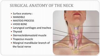
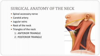
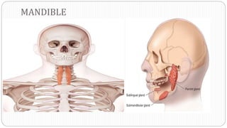
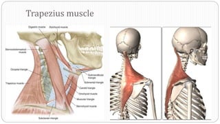
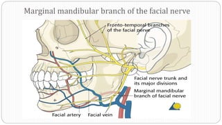
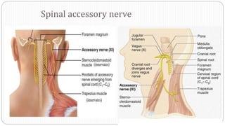
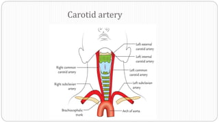
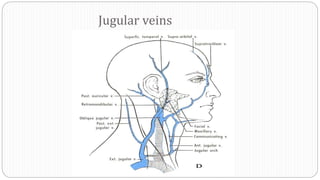
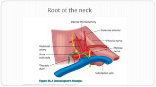
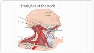
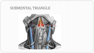
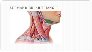
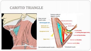
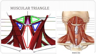
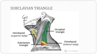
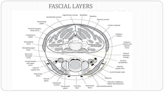
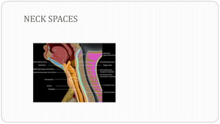
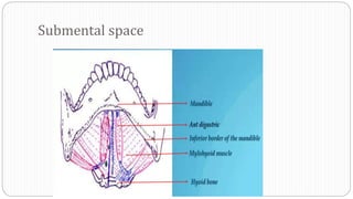
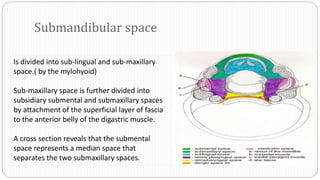
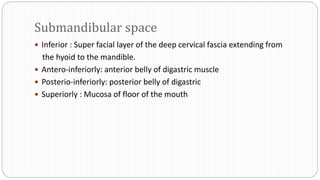
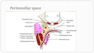
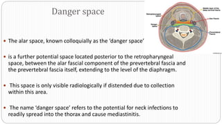
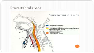
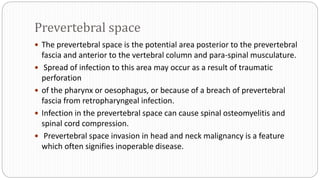
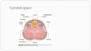
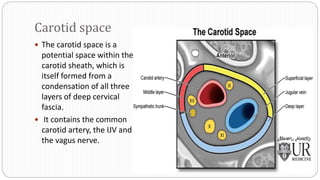
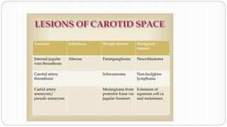
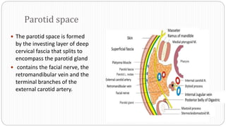
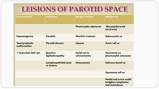
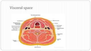
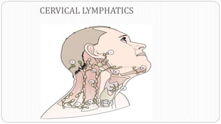
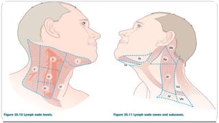
NECK SPACE AND LYMPHATIC SYSTEM
MODERATER
DR SHAMSHEER SIR
ASSOCIATE PROFESSOR
NMCH
BY
DR . V SANKAR NAIK
M.S ENT JUNIOR RESIDENT
NMCH
SURGICAL ANATOMY OF THE NECK
 Surface anatomy :
 MANDIBLE
 MASTOID PROCESS
 HYOID BONE
 Laryngeal cartilages and trachea
 Thyroid
 Sternocleidomastoid muscle
 Trapezius muscle
 Marginal mandibular branch of
the facial nerve
SURGICAL ANATOMY OF THE NECK
 Spinal accessory nerve
 Carotid artery
 Jugular veins
 Root of the neck
 Triangles of the neck
1 : ANTERIOR TRIANGLE
2 : POSTERIOR TRIANGLE
MANDIBLE
MASTOID PROCESS
HYOID BONE
Laryngeal cartilages and trachea
Sternocleidomastoid muscle
Trapezius muscle
Marginal mandibular branch of the facial nerve
Spinal accessory nerve
Carotid artery
Jugular veins
Root of the neck
Triangles of the neck
SUBMENTAL TRIANGLE
SUBMANDIBULAR TRIANGLE
CAROTID TRIANGLE
MUSCULAR TRIANGLE
SUBCLAVIAN TRIANGLE
FASCIAL LAYERS
NECK SPACES
Submental space
Submandibular space
Is divided into sub-lingual and sub-maxillary
space.( by the mylohyoid)
Sub-maxillary space is further divided into
subsidiary submental and submaxillary spaces
by attachment of the superficial layer of fascia
to the anterior belly of the digastric muscle.
A cross section reveals that the submental
space represents a median space that
separates the two submaxillary spaces.
Submandibular space
 Inferior : Super facial layer of the deep cervical fascia extending from
the hyoid to the mandible.
 Antero-inferiorly: anterior belly of digastric muscle
 Posterio-inferiorly: posterior belly of digastric
 Superiorly : Mucosa of floor of the mouth
 Ludwig’s angina is a life-threatening airway
 complication of submandibular space infection, characterized by rapid
onset of cellulitis of the floor of mouth tissues, causing swelling,
induration and tongue elevation resulting in a compromised airway.
 Submandibular space infections are most commonly odontogenic in
origin but can occur secondary to submandibular sialadenitis,
lymphadenitis, trauma or after surgery.
Peritonsillar space
 The potential space between the palatine tonsil and the superior
constrictor muscle is known as the peritonsillar space.
The condensation of the pharyngobasillar which forms the tonsillar capsule
may be breached by bacterial infections of the tonsil, resulting in abscess
formation in the peritonsillar space, also known as a quinsy.
 Untreated peritonsillar infections may spread to involve the
parapharyngeal space.
Parapharyngeal space
 The parapharyngeal space is an inverted
pyramid-shaped space, extending from
the petrous temporal bone superiorly to
the level of the hyoid.
 The space is bounded by the superior
constrictor muscle medially and the
pterygoid muscles, the parotid salivary
gland and the mandible laterally.
 The styloid process and its attachments
pass through the space, dividing it into
pre-styloid and post-styloid
compartments.
containts
 The pre-styloid compartment contains fat, connective tissue, the
maxillary artery, the inferior alveolar nerve, the lingual nerve and the
auriculotemporal nerve.
 The post-styloid compartment contains the carotid sheath and its
contents, the glossopharyngeal and hypoglossal nerves, the sympathetic
chain and lymph nodes.
 Pre-styloid lesions are most commonly associated with the deep lobe of
the parotid gland and will deflect the carotid sheath and parapharyngeal
fat posteromedially.
 Lesions in the post-styloid compartment are frequently of
neuroendocrine origin, arising from the carotid sheath as carotid body
tumours or vagal schwannomas, or neuromas of the sympathetic chain.
 Post-styloid lesions displace the parapharyngeal fat pad anteriorly.
Surgical access to the parapharyngeal space can be challenging and may
require mobilization of the parotid gland or lip split mandibulotomy.
 Abscesses of the parapharyngeal space may occur because of spread from the
submandibular space secondary to oro-dental causes, or from tperitonsiller space.
 Parapharyngeal space abscesses readily result in airway compromise if untreated.
 Medial displacement of the tonsil and lateral oropharyngeal wall will be seen but neck
swelling or asymmetry may or may not be present.
 Trismus may also occur secondary to inflammation of the pterygoid muscles.
 The two parapharyngeal spaces communicate with the retropharyngeal space posteriorly
and infection may readily spread into this compartment.
Retropharyngeal space
 Between the two parapharyngeal spaces is the retropharyngeal space which is
continuous with both.
 The superior limit of the retropharyngeal space is the skull base
 inferiorly the posterior mediastinum, down to the level of the carina.
 The anterior boundary in the neck is the buccopharyngeal fascia which encases
the pharyngeal constrictors.
 the posterior limit is the alar fascial component of the prevertebral fascia.
 The only contents of this space are the retropharyngeal lymph nodes, which
typically regress by adulthood but can represent a route of metastatic nodal
spread from midline or posterior tumours of the pharynx
 Retropharyngeal abscesses are more commonly seen in children than in
adults, secondary to bacterial infection of the upper aerodigestive tract
and may result in airway obstruction due to anterior displacement of the
airway.
Danger space
 The alar space, known colloquially as the ‘danger space’
 is a further potential space located posterior to the retropharyngeal
space, between the alar fascial component of the prevertebral fascia and
the prevertebral fascia itself, extending to the level of the diaphragm.
 This space is only visible radiologically if distended due to collection
within this area.
 The name ‘danger space’ refers to the potential for neck infections to
readily spread into the thorax and cause mediastinitis.
Prevertebral space
Prevertebral space
 The prevertebral space is the potential area posterior to the prevertebral
fascia and anterior to the vertebral column and para-spinal musculature.
 Spread of infection to this area may occur as a result of traumatic
perforation
 of the pharynx or oesophagus, or because of a breach of prevertebral
fascia from retropharyngeal infection.
 Infection in the prevertebral space can cause spinal osteomyelitis and
spinal cord compression.
 Prevertebral space invasion in head and neck malignancy is a feature
which often signifies inoperable disease.
Carotid space
Carotid space
 The carotid space is a
potential space within the
carotid sheath, which is
itself formed from a
condensation of all three
layers of deep cervical
fascia.
 It contains the common
carotid artery, the IJV and
the vagus nerve.
Parotid space
 The parotid space is formed
by the investing layer of deep
cervical fascia that splits to
encompass the parotid gland
 contains the facial nerve, the
retromandibular vein and the
terminal branches of the
external carotid artery.
Visceral space
 The visceral space is bounded by the middle layer of deep cervical fascia
that envelops the thyroid and the trachea anteriorly and posteriorly by
the pretracheal fascia.
 It contains the larynx, hypopharynx, cervical oesophagus, proximal
trachea, thyroid and parathyroid glands and lymphatics of level VI.
CERVICAL LYMPHATICS
NECK SPACE AND LYMPHATIC SYSTEM.pptx

NECK SPACE AND LYMPHATIC SYSTEM.pptx

  • 1.
    NECK SPACE ANDLYMPHATIC SYSTEM MODERATER DR SHAMSHEER SIR ASSOCIATE PROFESSOR NMCH BY DR . V SANKAR NAIK M.S ENT JUNIOR RESIDENT NMCH
  • 2.
    SURGICAL ANATOMY OFTHE NECK  Surface anatomy :  MANDIBLE  MASTOID PROCESS  HYOID BONE  Laryngeal cartilages and trachea  Thyroid  Sternocleidomastoid muscle  Trapezius muscle  Marginal mandibular branch of the facial nerve
  • 3.
    SURGICAL ANATOMY OFTHE NECK  Spinal accessory nerve  Carotid artery  Jugular veins  Root of the neck  Triangles of the neck 1 : ANTERIOR TRIANGLE 2 : POSTERIOR TRIANGLE
  • 4.
  • 5.
  • 6.
  • 7.
  • 8.
  • 9.
  • 10.
    Marginal mandibular branchof the facial nerve
  • 11.
  • 12.
  • 13.
  • 14.
  • 15.
  • 16.
  • 17.
  • 18.
  • 19.
  • 20.
  • 21.
  • 23.
  • 24.
  • 25.
    Submandibular space Is dividedinto sub-lingual and sub-maxillary space.( by the mylohyoid) Sub-maxillary space is further divided into subsidiary submental and submaxillary spaces by attachment of the superficial layer of fascia to the anterior belly of the digastric muscle. A cross section reveals that the submental space represents a median space that separates the two submaxillary spaces.
  • 26.
    Submandibular space  Inferior: Super facial layer of the deep cervical fascia extending from the hyoid to the mandible.  Antero-inferiorly: anterior belly of digastric muscle  Posterio-inferiorly: posterior belly of digastric  Superiorly : Mucosa of floor of the mouth
  • 27.
     Ludwig’s anginais a life-threatening airway  complication of submandibular space infection, characterized by rapid onset of cellulitis of the floor of mouth tissues, causing swelling, induration and tongue elevation resulting in a compromised airway.  Submandibular space infections are most commonly odontogenic in origin but can occur secondary to submandibular sialadenitis, lymphadenitis, trauma or after surgery.
  • 28.
  • 29.
     The potentialspace between the palatine tonsil and the superior constrictor muscle is known as the peritonsillar space. The condensation of the pharyngobasillar which forms the tonsillar capsule may be breached by bacterial infections of the tonsil, resulting in abscess formation in the peritonsillar space, also known as a quinsy.  Untreated peritonsillar infections may spread to involve the parapharyngeal space.
  • 30.
    Parapharyngeal space  Theparapharyngeal space is an inverted pyramid-shaped space, extending from the petrous temporal bone superiorly to the level of the hyoid.  The space is bounded by the superior constrictor muscle medially and the pterygoid muscles, the parotid salivary gland and the mandible laterally.  The styloid process and its attachments pass through the space, dividing it into pre-styloid and post-styloid compartments.
  • 31.
    containts  The pre-styloidcompartment contains fat, connective tissue, the maxillary artery, the inferior alveolar nerve, the lingual nerve and the auriculotemporal nerve.  The post-styloid compartment contains the carotid sheath and its contents, the glossopharyngeal and hypoglossal nerves, the sympathetic chain and lymph nodes.
  • 32.
     Pre-styloid lesionsare most commonly associated with the deep lobe of the parotid gland and will deflect the carotid sheath and parapharyngeal fat posteromedially.  Lesions in the post-styloid compartment are frequently of neuroendocrine origin, arising from the carotid sheath as carotid body tumours or vagal schwannomas, or neuromas of the sympathetic chain.  Post-styloid lesions displace the parapharyngeal fat pad anteriorly. Surgical access to the parapharyngeal space can be challenging and may require mobilization of the parotid gland or lip split mandibulotomy.
  • 33.
     Abscesses ofthe parapharyngeal space may occur because of spread from the submandibular space secondary to oro-dental causes, or from tperitonsiller space.  Parapharyngeal space abscesses readily result in airway compromise if untreated.  Medial displacement of the tonsil and lateral oropharyngeal wall will be seen but neck swelling or asymmetry may or may not be present.  Trismus may also occur secondary to inflammation of the pterygoid muscles.  The two parapharyngeal spaces communicate with the retropharyngeal space posteriorly and infection may readily spread into this compartment.
  • 34.
  • 35.
     Between thetwo parapharyngeal spaces is the retropharyngeal space which is continuous with both.  The superior limit of the retropharyngeal space is the skull base  inferiorly the posterior mediastinum, down to the level of the carina.  The anterior boundary in the neck is the buccopharyngeal fascia which encases the pharyngeal constrictors.  the posterior limit is the alar fascial component of the prevertebral fascia.  The only contents of this space are the retropharyngeal lymph nodes, which typically regress by adulthood but can represent a route of metastatic nodal spread from midline or posterior tumours of the pharynx
  • 36.
     Retropharyngeal abscessesare more commonly seen in children than in adults, secondary to bacterial infection of the upper aerodigestive tract and may result in airway obstruction due to anterior displacement of the airway.
  • 37.
    Danger space  Thealar space, known colloquially as the ‘danger space’  is a further potential space located posterior to the retropharyngeal space, between the alar fascial component of the prevertebral fascia and the prevertebral fascia itself, extending to the level of the diaphragm.  This space is only visible radiologically if distended due to collection within this area.  The name ‘danger space’ refers to the potential for neck infections to readily spread into the thorax and cause mediastinitis.
  • 38.
  • 39.
    Prevertebral space  Theprevertebral space is the potential area posterior to the prevertebral fascia and anterior to the vertebral column and para-spinal musculature.  Spread of infection to this area may occur as a result of traumatic perforation  of the pharynx or oesophagus, or because of a breach of prevertebral fascia from retropharyngeal infection.  Infection in the prevertebral space can cause spinal osteomyelitis and spinal cord compression.  Prevertebral space invasion in head and neck malignancy is a feature which often signifies inoperable disease.
  • 40.
  • 41.
    Carotid space  Thecarotid space is a potential space within the carotid sheath, which is itself formed from a condensation of all three layers of deep cervical fascia.  It contains the common carotid artery, the IJV and the vagus nerve.
  • 43.
    Parotid space  Theparotid space is formed by the investing layer of deep cervical fascia that splits to encompass the parotid gland  contains the facial nerve, the retromandibular vein and the terminal branches of the external carotid artery.
  • 45.
  • 46.
     The visceralspace is bounded by the middle layer of deep cervical fascia that envelops the thyroid and the trachea anteriorly and posteriorly by the pretracheal fascia.  It contains the larynx, hypopharynx, cervical oesophagus, proximal trachea, thyroid and parathyroid glands and lymphatics of level VI.
  • 47.