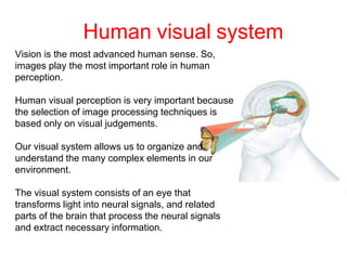
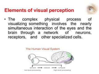
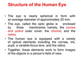
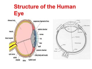
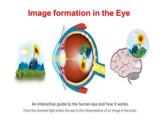
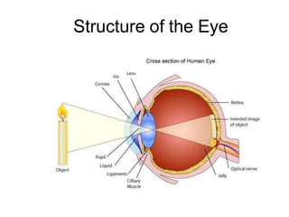
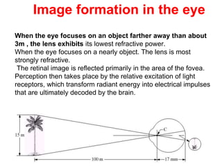
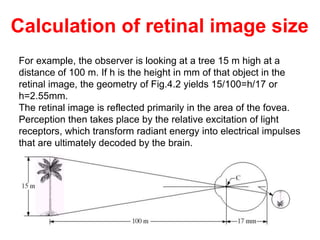
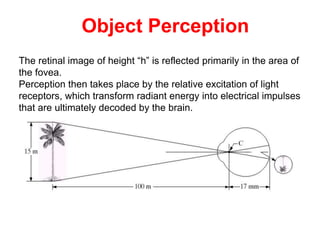
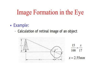
SlideShare a Scribd company logo
1 of 46
Elements of visual
perception
Visual Perception
Although the digital image processing field is built on a foundation of
mathematical and probabilistic formulations,
DIP depends human intuition and analysis play a central role in the choice of
one technique versus another, and this choice often is made based on
subjective, visual judgments.
It is important to study human visual perception.
Visual Perception
Human visual system
Vision is the most advanced human sense. So,
images play the most important role in human
perception.
Human visual perception is very important because
the selection of image processing techniques is
based only on visual judgements.
Our visual system allows us to organize and
understand the many complex elements in our
environment.
The visual system consists of an eye that
transforms light into neural signals, and related
parts of the brain that process the neural signals
and extract necessary information.
Elements of visual perception
• The complex physical process of
visualizing something involves the nearly
simultaneous interaction of the eyes and the
brain through a network of neurons,
receptors, and other specialized cells.
Structure of the Human Eye
• The eye is nearly spherical in form with
an average diameter of approximately 20 mm.
• The eye, called the optic globe is enclosed
by three membranes namely, the cornea
and sclera outer cover, the choroid, and the
retina.
• The human eye is equipped with a variety
of optical elements including the cornea, iris,
pupil, a variable-focus lens, and the retina.
• Together, these elements work to form images
of the objects in a person's field of view.
Structure of the Human
Eye
The Cornea and Sclera outer cover
• The cornea is a tough, transparent strong bulge located
at the front surface of the eye.
• Adult cornea has a radius of approximately 8mm.
• The cornea (convex outer layer )contributes to the image
forming process by refracting light entering the eye.
• The sclera , tough white sheath around the outside of the
eye-ball, is continuous with the cornea.
• It is the opaque membrane that encloses the
remainder of the eye.
• Sclera- White of the Eye
Choroid
The choroid layer is located behind the retina directly below the
sclera and absorbs unused radiation.
•This membrane contains a network of blood vessels that serve as
the major source of nutrition to the eye.
•The choroid coat is heavily pigmented and hence helps to reduce
the amount of extraneous light entering the eye and the
backscatter within the optical globe.
At its anterior extreme , the choroid is divided into the ciliary body
and the iris diaphragm.
Ciliary muscle:
The ciliary muscle is a ring shaped muscle attached to the Iris.
It is important because contraction and relaxation of the ciliary
muscle controls the shape of the lens.
The choroid and Iris Diaphragm
Iris Diaphragm:
It contracts or expands to control the amount of light that enters
the eye.
The central opening of the iris (the pupil) varies in diameter from
approximately 2 to 8 mm.
The front of the iris contains the visible pigment of the eye,
whereas the back contains a black pigment.
Lens
•The lens is made up of concentric layers of fibrous cells
• It is suspended by fibers that attach to the ciliary body.
• It contains 60 to 70%water, about 6%fat, and more protein than
any other tissue in the eye.
Cataracts:
•The lens is colored by a slightly yellow pigmentation that
increases with age.
• In extreme cases, excessive clouding of the lens, caused by the
affliction commonly referred to as cataracts, can lead to poor
color discrimination and loss of clear vision.
•The lens absorbs approximately 8% of the visible light spectrum,
with relatively higher absorption at shorter wavelengths.
•Both infrared and ultraviolet light are absorbed appreciably by
proteins within the lens structure and, in excessive amounts, can
damage the eye.
Retina
The retina may be described as the “screen” on which an image
is formed by light that has passed into the eye via the cornea,
aqueous humour, pupil , lens , then the hyaloid canal and finally
the vitreous humour before reaching the retina.
The retina contains photosensitive elements (called rods and
cones) that convert the light they detect into nerve impulses that
are sent onto the brain along the optic nerve.
Optic Nerve:
The optic nerve is the second cranial nerve and is responsible for
vision.
Each nerve contains approx. one million fibres transmitting
information from the rod and cone cells of the retina.
.
The Retina
When the eye is properly focused, light from an object outside
the eye is imaged on the retina.
Pattern vision is afforded by the distribution of discrete light
receptors over the surface of the retina.
Light receptors : Rods & Cones
There are two classes of receptors: cones and rods. Rods and
cones operate differently. Rods are more sensitive to light than
cones. They photo receptors are located primarily in the central
portion of the retina, called the fovea, and are highly sensitive
to color.
Visual Axis:
A straight line that passes through both the centre of the pupil
and the Centre of the fovea.
photoreceptors
Retina has two kinds of photoreceptors: cones and rods.
Cones
•The cones are highly sensitive to color.
•Their number is 6-7 million and they are mainly located at the
central part of the retina.
•Each cone is connected to one nerve end.
•Cone vision is the photopic or bright light vision.
Rods
• Rods serve to view the general picture of the vision field.
•They are sensitive to low levels of illumination and cannot
discriminate colors. This is the scotopic or dim-light vision.
•Their number is 75 to 150 million and they are distributed over the
retinal surface.
•Several rods are connected to a single nerve end. This fact and
their large spatial distribution explain their low resolution.
The larger area of distribution and the fact that several rods are
connected to a single nerve end reduce the amount of detail
discernible by these receptors.
Rods serve to give a general, overall picture of the field of view.
Rods are not involved in color vision and are sensitive to low levels
of illumination.
Humans can resolve fine details with these cones largely because
each one is connected to its own nerve end.
Muscles controlling the eye rotate the eyeball until the image of an
object of interest falls on the fovea.
Cones respond to higher levels of illumination
The number of rods is much larger: Some 75 to 150 million are
distributed over the retinal surface.
Rods and cones
Cones and Rods
Distribution of Rods & Cones
Figure shows the density of rods and cones for a cross section of
the right eye passing through the region of emergence of the
optic nerve from the eye.
Types of Vision
Scotopic or dim-light vision
For example, objects that appear brightly colored in daylight
when seen by moonlight appear as colorless forms because only
the rods are stimulated. This phenomenon is known as scotopic
or dim-light vision.
photopic or bright-light vision
Cone vision is called photopic or bright-light vision. Cones
respond to higher levels of illumination; their response is called
photopic vision.
Blind spot / papilla :
The area in which there is no response or presence of light
receptors
The absent of reciprocators is called blind spot.
Role of lens in Image formation in the eye
•The major difference between the lens of the eye and an
ordinary optical lens in that the former is flexible.
•The radius of curvature of the anterior surface of the lens >
radius of curvature of its posterior surface.
•The shape of the lens of the eye is controlled by tension in the
fiber of the ciliary body.
To focus on distant objects, the controlling muscles cause
the lens to be relatively flattened and lens will have lowest
refractive power.
•Similarly,these muscles allow the lens to become thicker in
order to focus on objects near the eye and most strongly
refractive.
•The distance between the optical center of the lens and the
retina is called the focal length
•Focal length varies from 17mm to 14mm as the refractive power
of the lens increases from its minimum to its maximum.
Image formation in the Eye
Structure of the Eye
Image formation in the eye
When the eye focuses on an object farther away than about
3m , the lens exhibits its lowest refractive power.
When the eye focuses on a nearly object. The lens is most
strongly refractive.
The retinal image is reflected primarily in the area of the fovea.
Perception then takes place by the relative excitation of light
receptors, which transform radiant energy into electrical impulses
that are ultimately decoded by the brain.
For example, the observer is looking at a tree 15 m high at a
distance of 100 m. If h is the height in mm of that object in the
retinal image, the geometry of Fig.4.2 yields 15/100=h/17 or
h=2.55mm.
The retinal image is reflected primarily in the area of the fovea.
Perception then takes place by the relative excitation of light
receptors, which transform radiant energy into electrical impulses
that are ultimately decoded by the brain.
Calculation of retinal image size
The retinal image of height “h” is reflected primarily in the area of
the fovea.
Perception then takes place by the relative excitation of light
receptors, which transform radiant energy into electrical impulses
that are ultimately decoded by the brain.
Object Perception
Brightness Adaptation
Human visual system cannot operate over a wide range of light
levels simultaneously.
Rather, it accomplishes this large variation by changes in its
overall sensitivity. This phenomenon is known as brightness
adaptation.
For a given set of conditions the current sensitivity level of the
visual system is called the brightness adaptation level.
Brightness Discrimination
The eye’s ability to discriminate i.e differentiate various intensity
levels since digital Images are displayed as a discrete set of
intensity.
The total range of distinct intensity levels it can discriminate
simultaneously is rather small when compared with the total
adaptation range.
Brightness Adaptation and
Discrimination
Brightness Adaptation and
Discrimination
For any given set of conditions, the current
sensitivity level of the visual system is called the
brightness adaptation level, which may
correspond, for example, to brightness Ba .
The short intersecting curve represents the
range of subjective brightness that the eye can
perceive when adapted to this level.
This range is rather restricted, having a level Bb
at and below which all stimuli are perceived as
indistinguishable blacks.
The upper (dashed) portion of the curve is not
actually restricted but, if extended too far, loses
its meaning because much higher intensities
would simply raise the adaptation level higher
than Ba.
Weber’s Law
Weber’s law states that the ratio of the
incremental threshold to the background
intensity is a constant.
It predicts a linear relation between
incremental threshold and the background
intensity
Weber Ratio:
Weber ratio = ΔIc /I
– I: the background illumination
– ΔIc : the increment of illumination which is
distinguishable 50% of the time with
background illumination.
– Small Weber ratio indicates good
discrimination
– Larger Weber ratio indicates poor
discrimination
Psychovisual effects
The perceived brightness is not a simple function of intensity
•Mach bands- Human Visual system tends to undershoot or
overshoot around the boundaries of regions with different
intensities.
•Mach bands or the Mach effect refers to an optical phenomenon
from edge enhancement due to lateral inhibition of the retina .
This is an inbuilt edge enhancement mechanism of the retina,
where the edges of darker objects next to lighter objects will
appear darker and vice versa, creating a false shadow.
Mach Bands
Mach Bands
• The Mach bands effect is due to the
spatial high-boost filtering performed by
the human visual system on the
luminance channel of the image
captured by the retina.
• Along the boundary between
adjacent shades of grey in the
Mach bands illusion, lateral inhibition
makes the darker area falsely appear
even darker and the lighter area
falsely appear even lighter.
Mach Bands
• In the case of visual information, such
(spatial) differentiation causes gradual changes
in the contrast between an object and its
background to be enhanced, i.e., to become
more visible.
• In human perception, this contrast
enhancement produces what is known as Mach
Bands: between two regions of different intensity a
thin bright band appears at the lighter side and a
thin dark band appears on the darker size.
• These bands are not physically present are
"overshoot" and "undershoot" caused by our
neural circuits in processing a step discontinuity
in illumination.
Psychovisual effects
Simultaneous Contrast
Two colors, side by side, interact with one another and
change our perception accordingly. The effect of this interaction
is called simultaneous contrast.
Simultaneous Contrast phenomenon a region is
perceived brightness does not depend simply on its
intensity.
Since we rarely see colors in isolation, simultaneous
contrast affects our sense of the color that we see.
Psychovisual effects
For example,
Consider an intense beam of blue light, surrounded by white
light, striking our retinas.
Where the blue light strikes, the blue cones will be stimulated,
overloaded and fatigued. The horizontal cells that link the blue
cones will cause blue cones, outside of but close to the blue
beam, to also become fatigued.
In the surround of the blue beam where the white light falls, the
blue receptors will be fatigued and the white light will appear to
our brain as yellow. (Recall that blue light plus yellow light equals
white light.)
Simultaneous contrast causes the white around the blue to seem
yellow. Similarly, white light around a yellow beam will seem blue
Psychovisual effects
Simultaneous contrast affects every pair of adjacent colors.
For our example of the red and blue flowerbeds, the red bed
makes the blue bed seem green because it induces its
complementary color, green, in the blue bed.
The blue bed makes the red bed seem orange because it
induces its complementary color, yellow, in the red bed.
The real colors are not altered; only our perception of them
changes.
https://www.webexhibits.org/colorart/contrast.html#:~:text=For%2
0example%2C%20red%20and%20blue,explanation%20that%20
we%20will%20uncover.
Optical illusion
• An optical illusion (also called a visual
illusion is an illusion caused by the
visual system and characterized by a
visual percept that appears to differ from
reality.
• The eye fills in non-existing
information or wrongly perceives
geometrical properties of objects
Optical Illusions
Optical Illusions can use color, light and patterns to create
images that can be deceptive or misleading to our brains.
The information gathered by the eye is processed by the brain,
creating a perception that in reality, does not match the true
image.
Perception refers to the interpretation of what we take in through
our eyes. Optical illusions occur because our brain is trying to
interpret what we see and make sense of the world around us.
Optical illusions simply trick our brains into seeing things which
may or may not be real.
Optical Illusions
Are the pinwheels moving?
Answer: No, the wheels are
not turning. The Moiré effect
can produce interesting and
beautiful geometric patterns.
Optical Illusion
Optical illusion

More Related Content

Similar to Elements of visual perception Eye vision .pptx

The human eye 10 13-2011
The human eye 10 13-2011The human eye 10 13-2011
The human eye 10 13-2011
Varunitya
 
Opticsandphotoactivation
OpticsandphotoactivationOpticsandphotoactivation
Opticsandphotoactivation
starfish57
 
Mechanism of Human Behavior
Mechanism of Human BehaviorMechanism of Human Behavior
Mechanism of Human Behavior
Agnetha Monje
 
VISION IN ANIMALS VETERINARY PHYSIOLOGY.pdf
VISION IN ANIMALS VETERINARY PHYSIOLOGY.pdfVISION IN ANIMALS VETERINARY PHYSIOLOGY.pdf
VISION IN ANIMALS VETERINARY PHYSIOLOGY.pdf
TatendaMageja
 

Similar to Elements of visual perception Eye vision .pptx (20)

HUMAN EYE AND COLOURFUL WORLD.pdf
HUMAN EYE AND COLOURFUL WORLD.pdfHUMAN EYE AND COLOURFUL WORLD.pdf
HUMAN EYE AND COLOURFUL WORLD.pdf
 
structure and fuction of eyes and ears,types of memory,sharpe memory,attention
structure and fuction of eyes and ears,types of memory,sharpe memory,attentionstructure and fuction of eyes and ears,types of memory,sharpe memory,attention
structure and fuction of eyes and ears,types of memory,sharpe memory,attention
 
Sense organs
Sense organsSense organs
Sense organs
 
The human eye
The human eye The human eye
The human eye
 
The human eye 10 13-2011
The human eye 10 13-2011The human eye 10 13-2011
The human eye 10 13-2011
 
7 sensation
7  sensation7  sensation
7 sensation
 
Opticsandphotoactivation
OpticsandphotoactivationOpticsandphotoactivation
Opticsandphotoactivation
 
Mechanism of Human Behavior
Mechanism of Human BehaviorMechanism of Human Behavior
Mechanism of Human Behavior
 
The Human Eye
The Human EyeThe Human Eye
The Human Eye
 
PHYSIOLOGY OF EYE.pptx
PHYSIOLOGY OF EYE.pptxPHYSIOLOGY OF EYE.pptx
PHYSIOLOGY OF EYE.pptx
 
VISION.pdf
VISION.pdfVISION.pdf
VISION.pdf
 
HUMAN EYE AND ITS DEFECTS
HUMAN EYE AND ITS DEFECTSHUMAN EYE AND ITS DEFECTS
HUMAN EYE AND ITS DEFECTS
 
Sense organs
Sense organsSense organs
Sense organs
 
The Special Senses.pptx
The Special Senses.pptxThe Special Senses.pptx
The Special Senses.pptx
 
PHYSIOLOGY OF EYE.pptx
PHYSIOLOGY OF EYE.pptxPHYSIOLOGY OF EYE.pptx
PHYSIOLOGY OF EYE.pptx
 
Eye presentation1
Eye presentation1Eye presentation1
Eye presentation1
 
Retina & the vision
Retina & the visionRetina & the vision
Retina & the vision
 
Optic nerve
Optic nerve Optic nerve
Optic nerve
 
VISION IN ANIMALS VETERINARY PHYSIOLOGY.pdf
VISION IN ANIMALS VETERINARY PHYSIOLOGY.pdfVISION IN ANIMALS VETERINARY PHYSIOLOGY.pdf
VISION IN ANIMALS VETERINARY PHYSIOLOGY.pdf
 
Elements of visual perception
Elements of visual perceptionElements of visual perception
Elements of visual perception
 

More from ssuser7ec6af

relatrionship of pixels - Connectivity.ppt
relatrionship of pixels - Connectivity.pptrelatrionship of pixels - Connectivity.ppt
relatrionship of pixels - Connectivity.ppt
ssuser7ec6af
 

More from ssuser7ec6af (20)

cell injury and necrosis mechanism Pathology.ppt
cell injury and necrosis mechanism   Pathology.pptcell injury and necrosis mechanism   Pathology.ppt
cell injury and necrosis mechanism Pathology.ppt
 
Watershed Algorithm for segmentation .pptx
Watershed Algorithm for segmentation .pptxWatershed Algorithm for segmentation .pptx
Watershed Algorithm for segmentation .pptx
 
relatrionship of pixels - Connectivity.ppt
relatrionship of pixels - Connectivity.pptrelatrionship of pixels - Connectivity.ppt
relatrionship of pixels - Connectivity.ppt
 
Leukemia - Cancer of white blood cells Leukocytes
Leukemia - Cancer of white blood cells LeukocytesLeukemia - Cancer of white blood cells Leukocytes
Leukemia - Cancer of white blood cells Leukocytes
 
CELLULAR ADAPTATIONS OF GOWTH AND DIFFERRENTIATION.ppt
CELLULAR ADAPTATIONS OF GOWTH AND DIFFERRENTIATION.pptCELLULAR ADAPTATIONS OF GOWTH AND DIFFERRENTIATION.ppt
CELLULAR ADAPTATIONS OF GOWTH AND DIFFERRENTIATION.ppt
 
Bacterial growth curve - Characteristics
Bacterial growth curve - CharacteristicsBacterial growth curve - Characteristics
Bacterial growth curve - Characteristics
 
Disaster management lecture notes presentation
Disaster management  lecture notes presentationDisaster management  lecture notes presentation
Disaster management lecture notes presentation
 
Digital Image Processing - Reprsentation
Digital Image Processing - ReprsentationDigital Image Processing - Reprsentation
Digital Image Processing - Reprsentation
 
Carcinogenesis.pptx
Carcinogenesis.pptxCarcinogenesis.pptx
Carcinogenesis.pptx
 
Image Enhancement in the Spatial Domain U2.ppt
Image Enhancement in the Spatial Domain U2.pptImage Enhancement in the Spatial Domain U2.ppt
Image Enhancement in the Spatial Domain U2.ppt
 
SecurityBasics.ppt
SecurityBasics.pptSecurityBasics.ppt
SecurityBasics.ppt
 
Micturition Reflex.ppt
Micturition Reflex.pptMicturition Reflex.ppt
Micturition Reflex.ppt
 
Face recognition.ppt
Face recognition.pptFace recognition.ppt
Face recognition.ppt
 
biometrics.ppt
biometrics.pptbiometrics.ppt
biometrics.ppt
 
Unit-1-NANOparticles.pptx
Unit-1-NANOparticles.pptxUnit-1-NANOparticles.pptx
Unit-1-NANOparticles.pptx
 
Lymphatic system.ppt
Lymphatic system.pptLymphatic system.ppt
Lymphatic system.ppt
 
Lymphatic system.ppt
Lymphatic system.pptLymphatic system.ppt
Lymphatic system.ppt
 
Physiology of Vision.ppt
Physiology of Vision.pptPhysiology of Vision.ppt
Physiology of Vision.ppt
 
bone formation _Osteogenesis Flowchart.ppt
bone formation _Osteogenesis Flowchart.pptbone formation _Osteogenesis Flowchart.ppt
bone formation _Osteogenesis Flowchart.ppt
 
1.4 Performance Measures.ppt
1.4 Performance Measures.ppt1.4 Performance Measures.ppt
1.4 Performance Measures.ppt
 

Recently uploaded

Biogenic Sulfur Gases as Biosignatures on Temperate Sub-Neptune Waterworlds
Biogenic Sulfur Gases as Biosignatures on Temperate Sub-Neptune WaterworldsBiogenic Sulfur Gases as Biosignatures on Temperate Sub-Neptune Waterworlds
Biogenic Sulfur Gases as Biosignatures on Temperate Sub-Neptune Waterworlds
Sérgio Sacani
 
LUNULARIA -features, morphology, anatomy ,reproduction etc.
LUNULARIA -features, morphology, anatomy ,reproduction etc.LUNULARIA -features, morphology, anatomy ,reproduction etc.
LUNULARIA -features, morphology, anatomy ,reproduction etc.
Cherry
 
Module for Grade 9 for Asynchronous/Distance learning
Module for Grade 9 for Asynchronous/Distance learningModule for Grade 9 for Asynchronous/Distance learning
Module for Grade 9 for Asynchronous/Distance learning
levieagacer
 
Cyathodium bryophyte: morphology, anatomy, reproduction etc.
Cyathodium bryophyte: morphology, anatomy, reproduction etc.Cyathodium bryophyte: morphology, anatomy, reproduction etc.
Cyathodium bryophyte: morphology, anatomy, reproduction etc.
Cherry
 

Recently uploaded (20)

Biogenic Sulfur Gases as Biosignatures on Temperate Sub-Neptune Waterworlds
Biogenic Sulfur Gases as Biosignatures on Temperate Sub-Neptune WaterworldsBiogenic Sulfur Gases as Biosignatures on Temperate Sub-Neptune Waterworlds
Biogenic Sulfur Gases as Biosignatures on Temperate Sub-Neptune Waterworlds
 
Site specific recombination and transposition.........pdf
Site specific recombination and transposition.........pdfSite specific recombination and transposition.........pdf
Site specific recombination and transposition.........pdf
 
Site Acceptance Test .
Site Acceptance Test                    .Site Acceptance Test                    .
Site Acceptance Test .
 
GBSN - Microbiology (Unit 3)Defense Mechanism of the body
GBSN - Microbiology (Unit 3)Defense Mechanism of the body GBSN - Microbiology (Unit 3)Defense Mechanism of the body
GBSN - Microbiology (Unit 3)Defense Mechanism of the body
 
Early Development of Mammals (Mouse and Human).pdf
Early Development of Mammals (Mouse and Human).pdfEarly Development of Mammals (Mouse and Human).pdf
Early Development of Mammals (Mouse and Human).pdf
 
Genome organization in virus,bacteria and eukaryotes.pptx
Genome organization in virus,bacteria and eukaryotes.pptxGenome organization in virus,bacteria and eukaryotes.pptx
Genome organization in virus,bacteria and eukaryotes.pptx
 
Clean In Place(CIP).pptx .
Clean In Place(CIP).pptx                 .Clean In Place(CIP).pptx                 .
Clean In Place(CIP).pptx .
 
LUNULARIA -features, morphology, anatomy ,reproduction etc.
LUNULARIA -features, morphology, anatomy ,reproduction etc.LUNULARIA -features, morphology, anatomy ,reproduction etc.
LUNULARIA -features, morphology, anatomy ,reproduction etc.
 
Terpineol and it's characterization pptx
Terpineol and it's characterization pptxTerpineol and it's characterization pptx
Terpineol and it's characterization pptx
 
Bhiwandi Bhiwandi ❤CALL GIRL 7870993772 ❤CALL GIRLS ESCORT SERVICE In Bhiwan...
Bhiwandi Bhiwandi ❤CALL GIRL 7870993772 ❤CALL GIRLS  ESCORT SERVICE In Bhiwan...Bhiwandi Bhiwandi ❤CALL GIRL 7870993772 ❤CALL GIRLS  ESCORT SERVICE In Bhiwan...
Bhiwandi Bhiwandi ❤CALL GIRL 7870993772 ❤CALL GIRLS ESCORT SERVICE In Bhiwan...
 
Kanchipuram Escorts 🥰 8617370543 Call Girls Offer VIP Hot Girls
Kanchipuram Escorts 🥰 8617370543 Call Girls Offer VIP Hot GirlsKanchipuram Escorts 🥰 8617370543 Call Girls Offer VIP Hot Girls
Kanchipuram Escorts 🥰 8617370543 Call Girls Offer VIP Hot Girls
 
Use of mutants in understanding seedling development.pptx
Use of mutants in understanding seedling development.pptxUse of mutants in understanding seedling development.pptx
Use of mutants in understanding seedling development.pptx
 
FAIRSpectra - Enabling the FAIRification of Analytical Science
FAIRSpectra - Enabling the FAIRification of Analytical ScienceFAIRSpectra - Enabling the FAIRification of Analytical Science
FAIRSpectra - Enabling the FAIRification of Analytical Science
 
Cot curve, melting temperature, unique and repetitive DNA
Cot curve, melting temperature, unique and repetitive DNACot curve, melting temperature, unique and repetitive DNA
Cot curve, melting temperature, unique and repetitive DNA
 
Dr. E. Muralinath_ Blood indices_clinical aspects
Dr. E. Muralinath_ Blood indices_clinical  aspectsDr. E. Muralinath_ Blood indices_clinical  aspects
Dr. E. Muralinath_ Blood indices_clinical aspects
 
PATNA CALL GIRLS 8617370543 LOW PRICE ESCORT SERVICE
PATNA CALL GIRLS 8617370543 LOW PRICE ESCORT SERVICEPATNA CALL GIRLS 8617370543 LOW PRICE ESCORT SERVICE
PATNA CALL GIRLS 8617370543 LOW PRICE ESCORT SERVICE
 
Module for Grade 9 for Asynchronous/Distance learning
Module for Grade 9 for Asynchronous/Distance learningModule for Grade 9 for Asynchronous/Distance learning
Module for Grade 9 for Asynchronous/Distance learning
 
Cyathodium bryophyte: morphology, anatomy, reproduction etc.
Cyathodium bryophyte: morphology, anatomy, reproduction etc.Cyathodium bryophyte: morphology, anatomy, reproduction etc.
Cyathodium bryophyte: morphology, anatomy, reproduction etc.
 
Gwalior ❤CALL GIRL 84099*07087 ❤CALL GIRLS IN Gwalior ESCORT SERVICE❤CALL GIRL
Gwalior ❤CALL GIRL 84099*07087 ❤CALL GIRLS IN Gwalior ESCORT SERVICE❤CALL GIRLGwalior ❤CALL GIRL 84099*07087 ❤CALL GIRLS IN Gwalior ESCORT SERVICE❤CALL GIRL
Gwalior ❤CALL GIRL 84099*07087 ❤CALL GIRLS IN Gwalior ESCORT SERVICE❤CALL GIRL
 
TransientOffsetin14CAftertheCarringtonEventRecordedbyPolarTreeRings
TransientOffsetin14CAftertheCarringtonEventRecordedbyPolarTreeRingsTransientOffsetin14CAftertheCarringtonEventRecordedbyPolarTreeRings
TransientOffsetin14CAftertheCarringtonEventRecordedbyPolarTreeRings
 

Elements of visual perception Eye vision .pptx

  • 2. Visual Perception Although the digital image processing field is built on a foundation of mathematical and probabilistic formulations, DIP depends human intuition and analysis play a central role in the choice of one technique versus another, and this choice often is made based on subjective, visual judgments. It is important to study human visual perception.
  • 4.
  • 5. Human visual system Vision is the most advanced human sense. So, images play the most important role in human perception. Human visual perception is very important because the selection of image processing techniques is based only on visual judgements. Our visual system allows us to organize and understand the many complex elements in our environment. The visual system consists of an eye that transforms light into neural signals, and related parts of the brain that process the neural signals and extract necessary information.
  • 6. Elements of visual perception • The complex physical process of visualizing something involves the nearly simultaneous interaction of the eyes and the brain through a network of neurons, receptors, and other specialized cells.
  • 7. Structure of the Human Eye • The eye is nearly spherical in form with an average diameter of approximately 20 mm. • The eye, called the optic globe is enclosed by three membranes namely, the cornea and sclera outer cover, the choroid, and the retina. • The human eye is equipped with a variety of optical elements including the cornea, iris, pupil, a variable-focus lens, and the retina. • Together, these elements work to form images of the objects in a person's field of view.
  • 8. Structure of the Human Eye
  • 9. The Cornea and Sclera outer cover • The cornea is a tough, transparent strong bulge located at the front surface of the eye. • Adult cornea has a radius of approximately 8mm. • The cornea (convex outer layer )contributes to the image forming process by refracting light entering the eye. • The sclera , tough white sheath around the outside of the eye-ball, is continuous with the cornea. • It is the opaque membrane that encloses the remainder of the eye. • Sclera- White of the Eye
  • 10. Choroid The choroid layer is located behind the retina directly below the sclera and absorbs unused radiation. •This membrane contains a network of blood vessels that serve as the major source of nutrition to the eye. •The choroid coat is heavily pigmented and hence helps to reduce the amount of extraneous light entering the eye and the backscatter within the optical globe. At its anterior extreme , the choroid is divided into the ciliary body and the iris diaphragm. Ciliary muscle: The ciliary muscle is a ring shaped muscle attached to the Iris. It is important because contraction and relaxation of the ciliary muscle controls the shape of the lens.
  • 11. The choroid and Iris Diaphragm Iris Diaphragm: It contracts or expands to control the amount of light that enters the eye. The central opening of the iris (the pupil) varies in diameter from approximately 2 to 8 mm. The front of the iris contains the visible pigment of the eye, whereas the back contains a black pigment.
  • 12. Lens •The lens is made up of concentric layers of fibrous cells • It is suspended by fibers that attach to the ciliary body. • It contains 60 to 70%water, about 6%fat, and more protein than any other tissue in the eye. Cataracts: •The lens is colored by a slightly yellow pigmentation that increases with age. • In extreme cases, excessive clouding of the lens, caused by the affliction commonly referred to as cataracts, can lead to poor color discrimination and loss of clear vision. •The lens absorbs approximately 8% of the visible light spectrum, with relatively higher absorption at shorter wavelengths. •Both infrared and ultraviolet light are absorbed appreciably by proteins within the lens structure and, in excessive amounts, can damage the eye.
  • 13. Retina The retina may be described as the “screen” on which an image is formed by light that has passed into the eye via the cornea, aqueous humour, pupil , lens , then the hyaloid canal and finally the vitreous humour before reaching the retina. The retina contains photosensitive elements (called rods and cones) that convert the light they detect into nerve impulses that are sent onto the brain along the optic nerve. Optic Nerve: The optic nerve is the second cranial nerve and is responsible for vision. Each nerve contains approx. one million fibres transmitting information from the rod and cone cells of the retina. .
  • 14. The Retina When the eye is properly focused, light from an object outside the eye is imaged on the retina. Pattern vision is afforded by the distribution of discrete light receptors over the surface of the retina. Light receptors : Rods & Cones There are two classes of receptors: cones and rods. Rods and cones operate differently. Rods are more sensitive to light than cones. They photo receptors are located primarily in the central portion of the retina, called the fovea, and are highly sensitive to color. Visual Axis: A straight line that passes through both the centre of the pupil and the Centre of the fovea.
  • 15. photoreceptors Retina has two kinds of photoreceptors: cones and rods. Cones •The cones are highly sensitive to color. •Their number is 6-7 million and they are mainly located at the central part of the retina. •Each cone is connected to one nerve end. •Cone vision is the photopic or bright light vision. Rods • Rods serve to view the general picture of the vision field. •They are sensitive to low levels of illumination and cannot discriminate colors. This is the scotopic or dim-light vision. •Their number is 75 to 150 million and they are distributed over the retinal surface. •Several rods are connected to a single nerve end. This fact and their large spatial distribution explain their low resolution.
  • 16. The larger area of distribution and the fact that several rods are connected to a single nerve end reduce the amount of detail discernible by these receptors. Rods serve to give a general, overall picture of the field of view. Rods are not involved in color vision and are sensitive to low levels of illumination. Humans can resolve fine details with these cones largely because each one is connected to its own nerve end. Muscles controlling the eye rotate the eyeball until the image of an object of interest falls on the fovea. Cones respond to higher levels of illumination The number of rods is much larger: Some 75 to 150 million are distributed over the retinal surface. Rods and cones
  • 18.
  • 19.
  • 20. Distribution of Rods & Cones Figure shows the density of rods and cones for a cross section of the right eye passing through the region of emergence of the optic nerve from the eye.
  • 21. Types of Vision Scotopic or dim-light vision For example, objects that appear brightly colored in daylight when seen by moonlight appear as colorless forms because only the rods are stimulated. This phenomenon is known as scotopic or dim-light vision. photopic or bright-light vision Cone vision is called photopic or bright-light vision. Cones respond to higher levels of illumination; their response is called photopic vision. Blind spot / papilla : The area in which there is no response or presence of light receptors The absent of reciprocators is called blind spot.
  • 22. Role of lens in Image formation in the eye •The major difference between the lens of the eye and an ordinary optical lens in that the former is flexible. •The radius of curvature of the anterior surface of the lens > radius of curvature of its posterior surface. •The shape of the lens of the eye is controlled by tension in the fiber of the ciliary body. To focus on distant objects, the controlling muscles cause the lens to be relatively flattened and lens will have lowest refractive power. •Similarly,these muscles allow the lens to become thicker in order to focus on objects near the eye and most strongly refractive. •The distance between the optical center of the lens and the retina is called the focal length •Focal length varies from 17mm to 14mm as the refractive power of the lens increases from its minimum to its maximum.
  • 25. Image formation in the eye When the eye focuses on an object farther away than about 3m , the lens exhibits its lowest refractive power. When the eye focuses on a nearly object. The lens is most strongly refractive. The retinal image is reflected primarily in the area of the fovea. Perception then takes place by the relative excitation of light receptors, which transform radiant energy into electrical impulses that are ultimately decoded by the brain.
  • 26. For example, the observer is looking at a tree 15 m high at a distance of 100 m. If h is the height in mm of that object in the retinal image, the geometry of Fig.4.2 yields 15/100=h/17 or h=2.55mm. The retinal image is reflected primarily in the area of the fovea. Perception then takes place by the relative excitation of light receptors, which transform radiant energy into electrical impulses that are ultimately decoded by the brain. Calculation of retinal image size
  • 27. The retinal image of height “h” is reflected primarily in the area of the fovea. Perception then takes place by the relative excitation of light receptors, which transform radiant energy into electrical impulses that are ultimately decoded by the brain. Object Perception
  • 28.
  • 29.
  • 30. Brightness Adaptation Human visual system cannot operate over a wide range of light levels simultaneously. Rather, it accomplishes this large variation by changes in its overall sensitivity. This phenomenon is known as brightness adaptation. For a given set of conditions the current sensitivity level of the visual system is called the brightness adaptation level. Brightness Discrimination The eye’s ability to discriminate i.e differentiate various intensity levels since digital Images are displayed as a discrete set of intensity. The total range of distinct intensity levels it can discriminate simultaneously is rather small when compared with the total adaptation range. Brightness Adaptation and Discrimination
  • 31. Brightness Adaptation and Discrimination For any given set of conditions, the current sensitivity level of the visual system is called the brightness adaptation level, which may correspond, for example, to brightness Ba . The short intersecting curve represents the range of subjective brightness that the eye can perceive when adapted to this level. This range is rather restricted, having a level Bb at and below which all stimuli are perceived as indistinguishable blacks. The upper (dashed) portion of the curve is not actually restricted but, if extended too far, loses its meaning because much higher intensities would simply raise the adaptation level higher than Ba.
  • 32. Weber’s Law Weber’s law states that the ratio of the incremental threshold to the background intensity is a constant. It predicts a linear relation between incremental threshold and the background intensity Weber Ratio: Weber ratio = ΔIc /I – I: the background illumination – ΔIc : the increment of illumination which is distinguishable 50% of the time with background illumination. – Small Weber ratio indicates good discrimination – Larger Weber ratio indicates poor discrimination
  • 33. Psychovisual effects The perceived brightness is not a simple function of intensity •Mach bands- Human Visual system tends to undershoot or overshoot around the boundaries of regions with different intensities. •Mach bands or the Mach effect refers to an optical phenomenon from edge enhancement due to lateral inhibition of the retina . This is an inbuilt edge enhancement mechanism of the retina, where the edges of darker objects next to lighter objects will appear darker and vice versa, creating a false shadow.
  • 35. Mach Bands • The Mach bands effect is due to the spatial high-boost filtering performed by the human visual system on the luminance channel of the image captured by the retina. • Along the boundary between adjacent shades of grey in the Mach bands illusion, lateral inhibition makes the darker area falsely appear even darker and the lighter area falsely appear even lighter.
  • 36. Mach Bands • In the case of visual information, such (spatial) differentiation causes gradual changes in the contrast between an object and its background to be enhanced, i.e., to become more visible. • In human perception, this contrast enhancement produces what is known as Mach Bands: between two regions of different intensity a thin bright band appears at the lighter side and a thin dark band appears on the darker size. • These bands are not physically present are "overshoot" and "undershoot" caused by our neural circuits in processing a step discontinuity in illumination.
  • 37.
  • 38.
  • 39. Psychovisual effects Simultaneous Contrast Two colors, side by side, interact with one another and change our perception accordingly. The effect of this interaction is called simultaneous contrast. Simultaneous Contrast phenomenon a region is perceived brightness does not depend simply on its intensity. Since we rarely see colors in isolation, simultaneous contrast affects our sense of the color that we see.
  • 40. Psychovisual effects For example, Consider an intense beam of blue light, surrounded by white light, striking our retinas. Where the blue light strikes, the blue cones will be stimulated, overloaded and fatigued. The horizontal cells that link the blue cones will cause blue cones, outside of but close to the blue beam, to also become fatigued. In the surround of the blue beam where the white light falls, the blue receptors will be fatigued and the white light will appear to our brain as yellow. (Recall that blue light plus yellow light equals white light.) Simultaneous contrast causes the white around the blue to seem yellow. Similarly, white light around a yellow beam will seem blue
  • 41. Psychovisual effects Simultaneous contrast affects every pair of adjacent colors. For our example of the red and blue flowerbeds, the red bed makes the blue bed seem green because it induces its complementary color, green, in the blue bed. The blue bed makes the red bed seem orange because it induces its complementary color, yellow, in the red bed. The real colors are not altered; only our perception of them changes. https://www.webexhibits.org/colorart/contrast.html#:~:text=For%2 0example%2C%20red%20and%20blue,explanation%20that%20 we%20will%20uncover.
  • 42. Optical illusion • An optical illusion (also called a visual illusion is an illusion caused by the visual system and characterized by a visual percept that appears to differ from reality. • The eye fills in non-existing information or wrongly perceives geometrical properties of objects
  • 43. Optical Illusions Optical Illusions can use color, light and patterns to create images that can be deceptive or misleading to our brains. The information gathered by the eye is processed by the brain, creating a perception that in reality, does not match the true image. Perception refers to the interpretation of what we take in through our eyes. Optical illusions occur because our brain is trying to interpret what we see and make sense of the world around us. Optical illusions simply trick our brains into seeing things which may or may not be real.
  • 44. Optical Illusions Are the pinwheels moving? Answer: No, the wheels are not turning. The Moiré effect can produce interesting and beautiful geometric patterns.