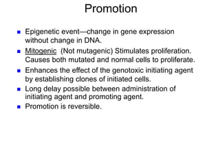
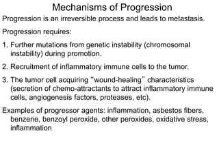
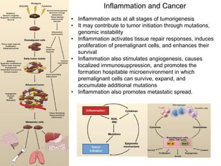
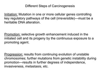
4. Different Steps of Carcinogenesis
Initiation: Mutation in one or more cellular genes controlling
key regulatory pathways of the cell (irreversible)—must be a
heritable DNA alteration.
Promotion: selective growth enhancement induced in the
initiated cell and its progeny by the continuous exposure to a
promoting agent.
Progression: results from continuing evolution of unstable
chromosomes; further mutations from genetic instability during
promotion—results in further degrees of independence,
invasiveness, metastasis, etc.
6. • Initiation is the induction of a mutation in a critical gene
involved in the control of cell proliferation.
•As with mutational events, initiation requires one or more
rounds of cell division for the “fixation” of the process.
• The metabolism of initiating agents to non-reactive forms
and the high efficiency of DNA repair of the tissue can alter
the process of initiation.
• Initiation is irreversible although the initiated cell may
eventually die during the development of the neoplasm.
7. Types of mutations
Chemical carcinogens can cause:
1. Point mutations- the replacement of a single nucleotide base
with another nucleotide.
2. Frameshift mutations- addition or deletion of a nucleotide such
that the protein sequence from that point onward is altered.
3. Chromosomal aberrations- any change in the normal structure
or number of chromosomes
4. Aneuploidy- chromosome number is not a multiple of the
normal haploid (23)
5. Polyploidy- more than twice the haploid number of
chromosomes
8. Mechanisms of DNA Repair
The persistence of chemically-induced DNA adducts is
predominantly the result of failure of DNA repair, due to either:
• carcinogen-induced mutational inactivation of DNA repair
enzymes.
• failure of the DNA repair mechanisms to recognize
carcinogen-induced mutation.
9.
10. Targets of Initiation
Chemical carcinogens initiate cells via:
1. Mutational activation of oncogenic (proliferative) pathways (e.g.
growth factor receptors and downstream signaling proteins,
proteins involved in cell cycle checkpoints.
2. Mutational inactivation of apoptotic (cell death) pathways (e.g.
growth inhibitory receptors, proteins involved in apoptosis,
tumor suppressors).
3. Mutational inactivation of DNA repair mechanisms (e.g. BER,
NER, etc).
4. Mutational inactivation of antioxidant response (e.g. SOD).
11. Tumor suppressor p53 signaling
• p53 is a an important tumor suppressor (transcriptional factor) that
controls cell cycle, apoptosis, DNA repair mechanisms.
• Mdm2 is a negative regulator of p53 that functions both as an E3
ubiquitin ligase and an inhibitor of p53 transcriptional activation.
unstressed cell
p53 binds Mdm2
nuclear export of p53
p53 gets ubiquinated
and degraded by proteosome
cell survival
mitogen
MAP kinase
AP1/Ets binds
Mdm2 promoter
mitogen
PKB/Akt
phosphorylates Mdm2
12. DNA damage, cell damage
ATM kinase activated
increased p53
(tetrameric TF)
increased Apaf
(apoptosis)
increased p21 (G1
arrest)
binds tandem sequence of
PuPuPuC A/t T/a G PyPyPy
increased IGF-BP3
IGF-1
IGF-1 receptor
PKB/Akt activation
cell survival
increased Bax
Bax dimer
depolarizes mitochondrial
membrane
cyt c released into cytosol
cytc, Apaf-1, caspase 9
form apoptosome
increased Fas receptor
Fas ligand
FADD/DISC complex
activates caspase 8 + 10
apoptosis
activates executioner caspases 3,6,7
phosphorylates
p53 so it can't bind Mdm2
phosphorylates Mdm2
prevents ubiquitination of p53
increased E2F
(uncontrolled cell cycle)
from mutated Rb
increased
p14ARF
sequesters
Mdm2
p53—tumor suppressor:
Mutated in most cancers.
Carcinogens often
mutationally inactivate p53 as
well as proteins that control
p53 function (e.g. Mdm2, p14)
13. growth factor
(PDGF, IGF, EGF, NGF)
binds receptor tyrosine kinase
and dimerizes to autophosphorylate
cytosolic Tyr on receptor
recruits Grb2/Sos to phospho-Tyr
Ras-GDP Ras-GTP
PI3-Kinase
binds Ras (active)
PI-4,5-P2
PIP3
binds Akt/PKB and is
activated by
phosphorylation by PDK
P-bad
(inactive)
P-p21, P-p27
(inactive)
P-Mdm2
(sequesters p53)
inhibits
TSC1/2
(mTor active)
activates
protein
synthesis
apoptosis suppressed
Raf Ral-GDS
MEK
ERK
p90
CREB
Fos
Rac
PAK
MEK
MMK4
JNK
Jun
AP1
increased cyclin D
cell cycle progression
PKC
DAG
IP3
Ca2+
CaMK
c-Myc
cyclin D, E2F1-3
CDK4
decreases p21, p15
PLC
norepinephrine
serotonin, etc
binds G-protein coupled
receptor
MAP
Kinase pathway
Ras oncogene: involved in control of cell cycle progression and apoptosis
14. CYP/PHS
O
EH
HO
OH
CYP/PHS
HO
OH
O
benzo[a]pyrene (+) benzo[a]pyrene
7,8-oxide (-) benzo[a]pyrene
7,8-dihydrodiol
(+) benzo[a]pyrene
7,8-dihydrodiol-9,10-epoxide
ULTIMATE CARCINOGEN
HN
N
N
N
O
HN
DNA
HO
OH
HO
BaP-N2
-dG DNA adduct
DNA
GST/GSH
OH
GS
inactive (excreted)
O
CYP/PHS
OH
OH
inactive
Phase II
Phase II and
excretion
15. Benzopyrene Leads to Mutations in K-Ras and p53 in the Genomic
Loci Found to be Mutated in Smoking-Induced Lung Cancers
• K-Ras and p53 are the two oncogenes most frequently mutated in
smoking-related lung cancers
• If not corrected by the cell’s DNA repair mechanism, this guanine “adduct” is
misread as a thymine by the DNA polymerase that copies chromosomes
during replication
• Ultimately, the original G—C base pair may be replaced by a T—A base pair,
a mutation called a traversion
• Cells treated with Benzopyrene show the same spectrum of G—T
transversions as found in the K-RAS and p53 of smokers.
• These mutational “hot spots” map well to the guanine binding sites of BaP
epoxide
19. Epigenetic event—change in gene expression
without change in DNA.
Mitogenic (Not mutagenic) Stimulates proliferation.
Causes both mutated and normal cells to proliferate.
Enhances the effect of the genotoxic initiating agent
by establishing clones of initiated cells.
Long delay possible between administration of
initiating agent and promoting agent.
Promotion is reversible.
Promotion
20. Promoters
1. Reactive Oxygen Species (ROS) and redox active
xenobiotics and metals
2. Phorbol esters (e.g. TPA)
3. Polycyclic aromatic compounds (e.g. Dioxin)
4. Peroxisome Proliferators (oxidized fats)
5. Endocrine Disruptors (estradiol, DES)
21. Structures of Representative Promoters
TPA and other phorbol esters
activate protein kinase C,
which leads to signal
transduction pathways that
increase DNA replication, cell
division
TCDD (dioxin) activates aryl
hydrocarbon receptor (AhR) and
induces the expression of
cytochrome P450increases
oxidative stresscan oxidatively
activate oncogenic pathways (e.g.
RAS)
22. Endocrine Receptors and Carcinogenesis
Endocrine disruptors are involved in breast, ovarian, colon,
prostate cancers.
1. ERβ/ERα (estrogen receptors) ratio is decreased in cancers
(ligands include estradiol); ERs are transcription factors.
2. ERβ inhibits ERα
a.ERα-ERα dimerization (homodimer) leads to mitogenic
activation.
b.ERβ-ERα dimerization (heterodimer) leads to an inactivation.
3. Androgen Receptor (prostate) (AR) can also homodimerize with
AR leading to mitogenic activation; AR can heterodimerize with
ERβ to cause growth arrest (prostate also dependent on
estrogenic signals).
26. Mechanisms of Progression
Progression is an irreversible process and leads to metastasis.
Progression requires:
1. Further mutations from genetic instability (chromosomal
instability) during promotion.
2. Recruitment of inflammatory immune cells to the tumor.
3. The tumor cell acquiring “wound-healing” characteristics
(secretion of chemo-attractants to attract inflammatory immune
cells, angiogenesis factors, proteases, etc).
Examples of progressor agents: inflammation, asbestos fibers,
benzene, benzoyl peroxide, other peroxides, oxidative stress,
inflammation
27.
28. Cellular Necrosis
Activation of Resident
Macrophages
Recruitment and Activation
of More Macrophages
Chronic Toxicant Exposure
Cytokines, chemokines,
Eicosanoids (TNFa, IL1b, PGE2)
Decreased ATP, increased Ca2+,
increased oxidative stress
Fibroblast proliferation,
differentiation
Intracellular contents
(e.g. ATP, dsDNA)
Growth factors
(e.g. TGFb, IGF1,
PDGF, ROS)
Cell proliferation
TGFb
Epithelial-to-mesenchymal
transition (EMT)
Leakier basement
membrane
Infiltration of more
immune cells into damaged
tissues
TGFb, IGF1,
PDGF, TNFa
Excessive formation of
hardened extracellular
matrix (ECM)
TNFa,
ROS
Tissue Cells
And Macrophage
Cellular Necrosis
fibrosis
angiogenesis
Growth factors
(e.g. TGFb, IGF1,
PDGF, ROS)
Genetic instability
Mutations
Cell proliferation
Cellular transformation
VEGF
Malignant progression
of cancer cells
Growth factors
(e.g. TGFb, IGF1,
PDGF, ROS)
Cytokines, chemokines,
Eicosanoids (TNFa, IL1b, PGE2)
TGFb Recruitment and Activation
of More Macrophages
Epithelial-to-mesenchymal
transition (EMT)
EMT and breakdown of ECM
proteases
Proteases,
TGFb
breakdown
of ECM (invasion)
Cancer cells
extravagate with
macrophages and
blood supply into
circulation
metastasis
Tissue
dysfunction,
tissue damage,
degeneration,
organ failure
29. Inflammation and Cancer
• Inflammation acts at all stages of tumorigenesis
• It may contribute to tumor initiation through mutations,
genomic instability
• Inflammation activates tissue repair responses, induces
proliferation of premalignant cells, and enhances their
survival
• Inflammation also stimulates angiogenesis, causes
localized immunosuppression, and promotes the
formation hospitable microenvironment in which
premalignant cells can survive, expand, and
accumulate additional mutations
• Inflammation also promotes metastatic spread.