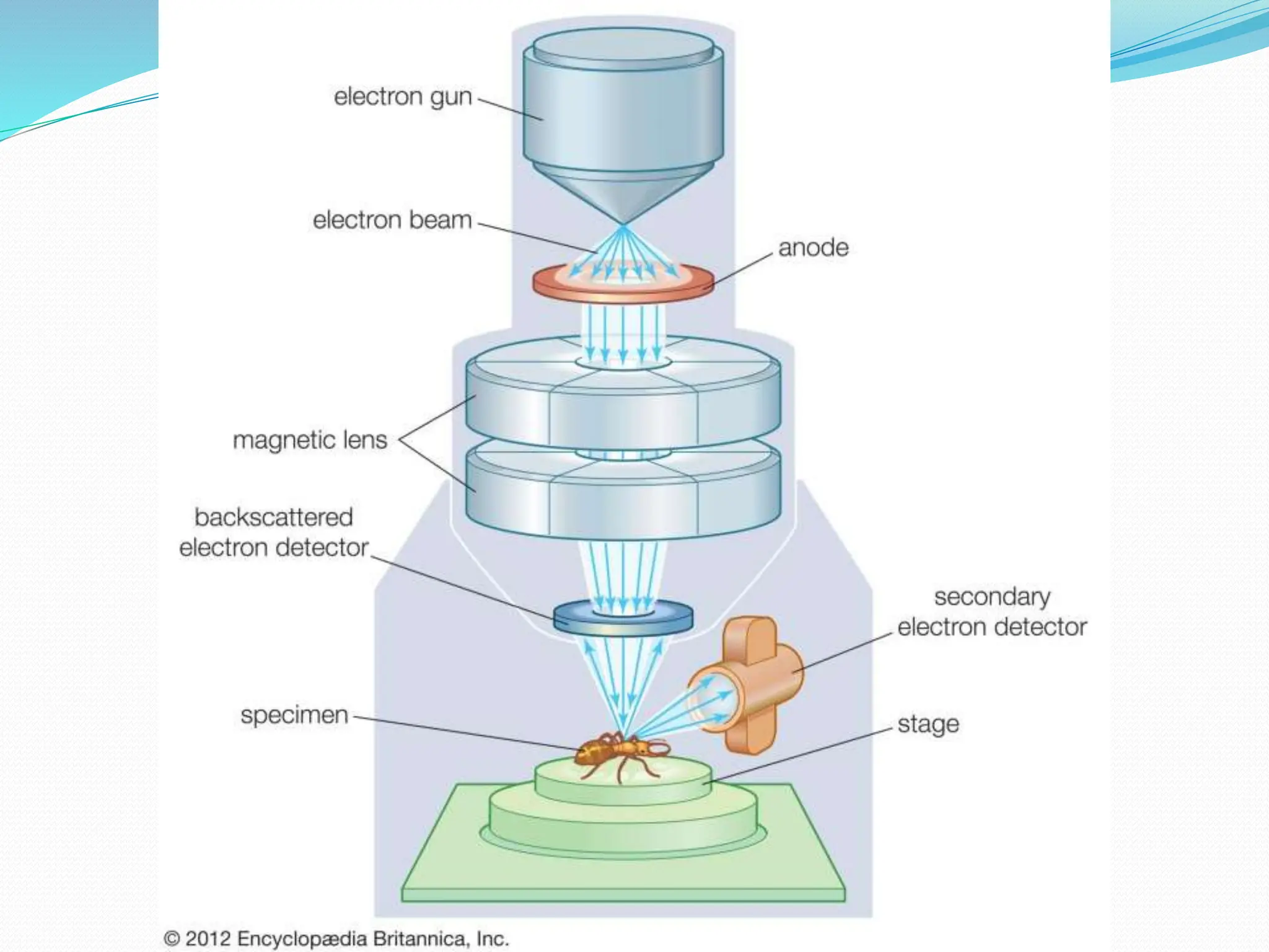
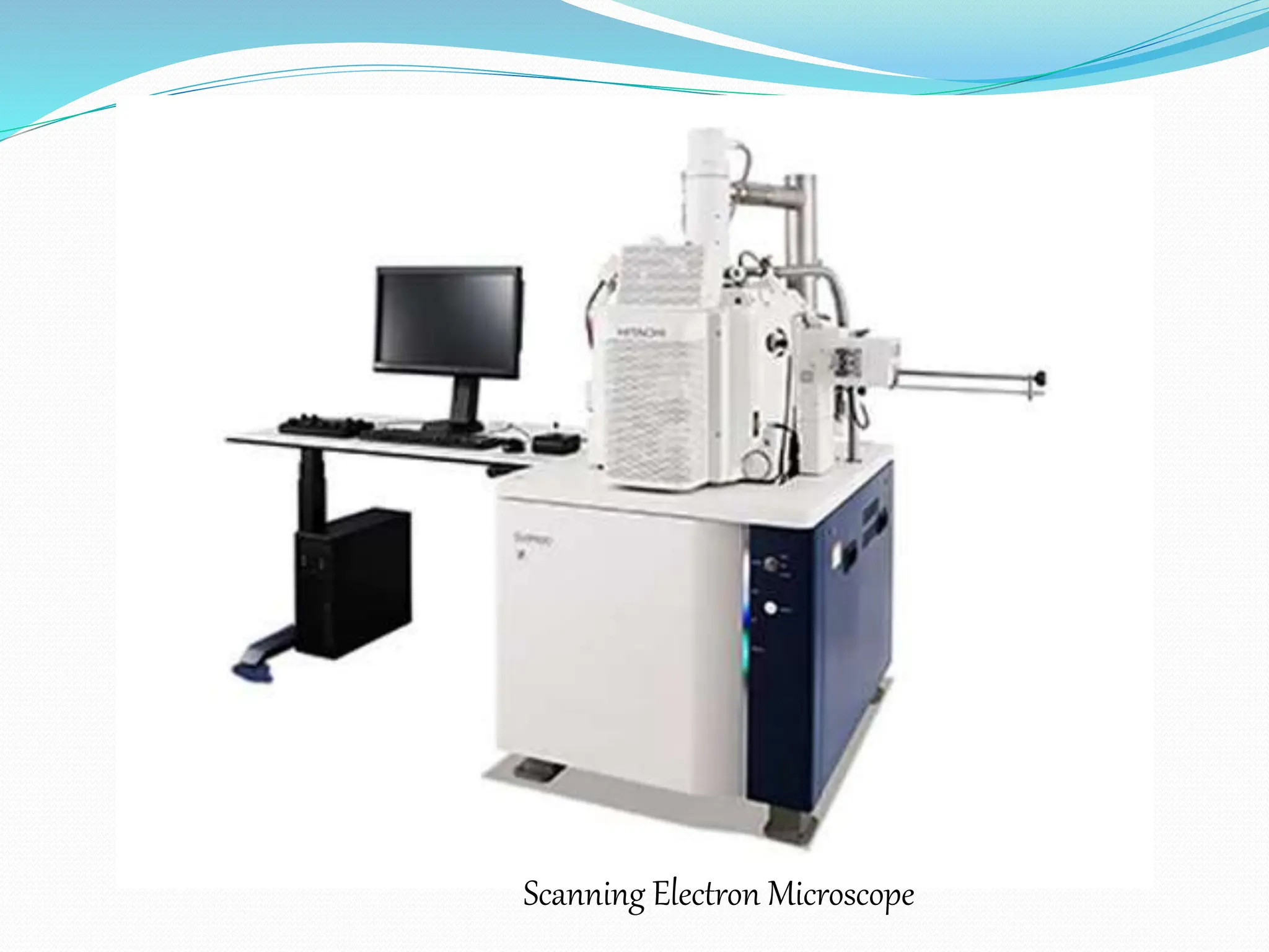
Electron Microscopy
Presented By
Mr. Lalit V. Gabhane
B.Pharm (3rd Semester)
Subject:-Pharmaceutical Microbiology
Anurag College of Pharmacy,
Warthi, Bhandara
INTRODUCTION
 An electron microscope (EM) is an instrument, which utilises
short wavelength of electron as a source of illumination for
observing objects at a greater magnification .
 The major significance of an electron microscope is that it has
the highest resolution and magnification .
 The first electron microscope was developed by Max Knoll and
Ernest Ruska in 1931.
 Staining is not required in the EM.
 ELECTRON MICROSCOPE is classified into two types:
1. Transmission electron microscope (TEM).
2. Scanning electron microscope (SEM).
Scanning Electron Microscope
 The scanning electron microscope (SEM) was built by Van
Ardene in 1938.
 SEM is primarily used for visualizing the surface architecture of
the specimen rather than the internal details.
 It provides striking three dimensional views of specimens.
 Working:-
 An accelerated beam of electrons is produced from the Electron
Gun and is focused on the specimen by the condenser lens.
 It is a basically V-shaped tungsten wire
 Provide high electricity and generate the electrons.
 Anode Attracts the negative charge particles (Electron)
 The magnetic lenses of a SEM is responsible to produce an
extremely thin beam of electrons called the primary electron
beam.
 These electron pass through the electromagnetic lenses and
directed over the surface of the specimen.
 The primary electron beam knocks electron out of the surface of the
specimen.
 This causes the release of secondary electron from the specimen
surface.
 The intensity of these secondary electrons depends on the shape and
the chemical composition of the irradiated object.
 The secondary electron are collected by a detector which generate an
electron signal.
 The signal are then scanned in the manner of a television system to
produce an image on Cathode Ray Tube(CRT).
 During this process, some of primary electron are also reflected and
transmitted to the collector but their number is less than the secondary
electron.
 As a result, the Image signal is developed more by the seconday
electron than the primary electron.
 The secondary electron deflected out of the specimen will be replica
of the refractive index of the surface and thus produce an image on
CRT screen revealing all the topographical details.
 Image contrast is mainly dependent on surface topography which
determines the number of secondary electron reaching the detector.
 SEM also has resolution equal to that of TEM.
 A resolution from 1-10 nm is possible with a corresponding
magnification from 10000-100000.
Scanning Electron Microscope
Advantages of Electron Microscopy:-
 This microscope is especially useful in studying the surface structure
of intact cells and viruses.
 In pharmaceutical field, SEM is very useful in study associated with
the surface characteristics of the drug particles and morphological
studies of antibiotic producing microorganism and their spores.
Limitations Of Electron Microscopy:-
 Tremendous resolution and magnification.
 The specimen being examined is in the chamber that is under a
very high vaccum. Thus, cells cannot be examined in living state.
 Drying process may change some morphological characteristics.
 Low penetration power of electron beam, it is necessary to use of
thin section to reveal the internal structure of the cell.
 Numerical aperture of an electron microscope lens is very small.
Electron_Microscope-Microbiology_3rd_sem_B.pharm.pptx

Electron_Microscope-Microbiology_3rd_sem_B.pharm.pptx

  • 1.
    Electron Microscopy Presented By Mr.Lalit V. Gabhane B.Pharm (3rd Semester) Subject:-Pharmaceutical Microbiology Anurag College of Pharmacy, Warthi, Bhandara
  • 3.
    INTRODUCTION  An electronmicroscope (EM) is an instrument, which utilises short wavelength of electron as a source of illumination for observing objects at a greater magnification .  The major significance of an electron microscope is that it has the highest resolution and magnification .  The first electron microscope was developed by Max Knoll and Ernest Ruska in 1931.  Staining is not required in the EM.  ELECTRON MICROSCOPE is classified into two types: 1. Transmission electron microscope (TEM). 2. Scanning electron microscope (SEM).
  • 4.
    Scanning Electron Microscope The scanning electron microscope (SEM) was built by Van Ardene in 1938.  SEM is primarily used for visualizing the surface architecture of the specimen rather than the internal details.  It provides striking three dimensional views of specimens.  Working:-  An accelerated beam of electrons is produced from the Electron Gun and is focused on the specimen by the condenser lens.  It is a basically V-shaped tungsten wire  Provide high electricity and generate the electrons.  Anode Attracts the negative charge particles (Electron)  The magnetic lenses of a SEM is responsible to produce an extremely thin beam of electrons called the primary electron beam.  These electron pass through the electromagnetic lenses and directed over the surface of the specimen.
  • 6.
     The primaryelectron beam knocks electron out of the surface of the specimen.  This causes the release of secondary electron from the specimen surface.  The intensity of these secondary electrons depends on the shape and the chemical composition of the irradiated object.  The secondary electron are collected by a detector which generate an electron signal.  The signal are then scanned in the manner of a television system to produce an image on Cathode Ray Tube(CRT).  During this process, some of primary electron are also reflected and transmitted to the collector but their number is less than the secondary electron.  As a result, the Image signal is developed more by the seconday electron than the primary electron.
  • 7.
     The secondaryelectron deflected out of the specimen will be replica of the refractive index of the surface and thus produce an image on CRT screen revealing all the topographical details.  Image contrast is mainly dependent on surface topography which determines the number of secondary electron reaching the detector.  SEM also has resolution equal to that of TEM.  A resolution from 1-10 nm is possible with a corresponding magnification from 10000-100000.
  • 8.
  • 9.
    Advantages of ElectronMicroscopy:-  This microscope is especially useful in studying the surface structure of intact cells and viruses.  In pharmaceutical field, SEM is very useful in study associated with the surface characteristics of the drug particles and morphological studies of antibiotic producing microorganism and their spores. Limitations Of Electron Microscopy:-  Tremendous resolution and magnification.  The specimen being examined is in the chamber that is under a very high vaccum. Thus, cells cannot be examined in living state.  Drying process may change some morphological characteristics.  Low penetration power of electron beam, it is necessary to use of thin section to reveal the internal structure of the cell.  Numerical aperture of an electron microscope lens is very small.