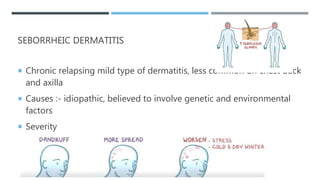
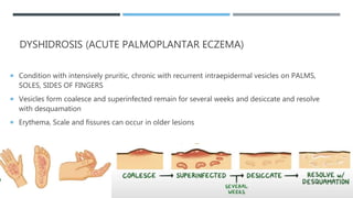
ECZEMA/
DERMATITIS
ECZEMA -
 Characterized by redness, itching, and sometimes oozing vesicular lesions,
which may become scaly, crusted, or hardened.
Eczema
Atopic
Dermatitis
Contact
Dermatitis
Dyshidrosis
Nummular
(Discoid)
Seborrheic Stasis
DIFFERENTIATION AND DIAGNOSIS
 Every type – additional symptoms
 Area of skin affected – Different
 Diagnosis - History and Physical examination
 Patch test – test any allergies
 Skin biopsy
ATOPIC DERMATITIS OR ECZEMA
 Chronic inflammatory condition common in young children last into adulthood
 Results from allergy
 As well as exposed skin surfaces like on the face the hands and the feet
 Aggravating factors- Allergens like cigarette, mold, dust mites, changes in
weather, emotional stress
 Signs and symptoms - dry itchy rash on flexor surfaces like creases of wrist,
insides of elbow, back of knees
 Red itchy skin, blister that peel, lichenified skin
 Clinical diagnosis
 Based on itchy skin
 >3 criteria
 1) History of dermatitis –Skin creases
 2) Visible dermatitis – Flexural surfaces, personal or family history of Asthma, hay
fever
 3) Presence of dry skin since past year
 4) Symptoms - Age <2yrs
 Age <4 yrs with dermatitis on cheeks, dorsal aspect of extremities
TREATMENT
 To decrease inflammation and prevent exacerbations
 Identify the triggers and avoid them – Prevent overheating, manage stress and use
of soft fabrics
 Dry skin treatment - Frequent Moisturization after lukewarm bath
 Oral H1 Antihistamines to reduce itching
 For Inflammation - Topical Corticosteroids
 If uncontrolled by corticosteroids then Topical Calcineurin inhibitors Ex Tacrolimus,
Pimecrolimus
 If uncontrolled then Phototherapy or systemic immunosuppressants (contra in
infants and young children)
SEBORRHEIC DERMATITIS
 Chronic relapsing mild type of dermatitis, less common on chest back
and axilla
 Causes :- idiopathic, believed to involve genetic and environmental
factors
 Severity
TREATMENT
 Mild with only dandruff – Antifungal agents – shampoo or creams
until remission is achieved
 Moderate to severe with Scale, Inflammation, pruritis – Antifungal
agent with topical corticosteroid
DYSHIDROSIS (ACUTE PALMOPLANTAR ECZEMA)
 Condition with intensively pruritic, chronic with recurrent intraepidermal vesicles on PALMS,
SOLES, SIDES OF FINGERS
 Vesicles form coalesce and superinfected remain for several weeks and desiccate and resolve
with desquamation
 Erythema, Scale and fissures can occur in older lesions
TREATMENT
 Causes are unknown, but potential triggers are allergens and frequent hand
washing
 Treatment :- Avoid Potential triggers
 Reduce irritation – Lukewarm water, soap free cleansers, emollients,
antihistamines
 Mild to moderate – topical corticosteroids
 Severe – Systemic corticosteroids
NUMMULAR DERMATITIS (DISCOID ECZEMA)
 Chronic, Relapsing dermatitis with PRURITIC, ROUND, ECZEMATOUS PLAQUES
(dry and scaly coin shaped 1-10cm diameter on extremities
 Causes – Not understood but the implicated factors are – DRY SKIN,
STAPHYLOCOCCUS AURES, METAL CONTACT, ENVIRONMENTAL ALLERGENS
TREATMENT
 Frist line – Topical corticosteroids
 Extensive or unresponsive – PHOTOTHERAPY with NARROWED UV B
PHOTOTHERAPY
 SYSTEMIC IMUNOSUPPRESSANTS – Corticosteroids, Methotrexate,
cyclosporine
STASIS DERMATITIS
 Common inflammatory dermatosis occurring in Legs in individuals with chronic
venous insufficiency (blood pooling)
 Presents with
TYPES AND TREATMENT
 Acute – Inflamed, weeping Plaques, Vesiculation, Crusting, Pruritis leading to
Lichenification
 Chronic – Hyperpigmentation due to :- Dermal Hemosiderin, Scaling,
Lipodermatosclerosis
 Treatment – venous insufficiency – Manual compression increases blood flow,
compression bandages and stocking, elevating legs above heart
 Surgical – Vein transplant, vein repair or removal
 Skin dryness and pruritis – Gentle skin cleansing and emollients
 Skin inflammation – Topical corticosteroids, in failure use systemically
CONTACT DERMATITIS
• Contact dermatitis, a type IV delayed hypersensitivity reaction, is an acute or chronic skin
inflammation that results from direct skin contact with chemicals or allergens.
• Often sharply demarcated inflammation and irritation of the skin caused by contact to substances by
which the skin is sensitive.
• Allergic dermatitis. Allergic dermatitis results from direct contact with substances called allergens.
• Irritant contact dermatitis. Irritant contact dermatitis develops when your skin comes into contact
with an irritating substance.
• Phototoxic contact dermatitis. Phototoxic contact dermatitis is a sunburn-like skin disorder
resulting from direct tissue damage following the ultraviolet light-induced activation of a phototoxic
agent.
• Photoallergic contact dermatitis. Photoallergic contact dermatitis is a delayed-type
hypersensitivity cutaneous reaction in response to a photo antigen applies to the skin in subjects
previously sensitized to the same substance.
PATHOPHYSIOLOGY
• Binding. The hapten (small hydrophobic molecules)-protein
complex enters the stratum corneum and binds to epidermal antigen-
presenting Langerhans cells.
• Deception. These cells process the antigen and travel to
regional lymph nodes where they present the antigen to naive CD4 T cells.
• Proliferation. These T cells then proliferate into memory and effector T cells,
which elicit contact dermatitis within 48 to 96 hours of re exposure to the
allergen.
STATISTICS AND INCIDENCES
 Contact dermatitis incidences are widespread around the world.
• 80% of cases are caused by excessive exposure to or additive effects of
irritants.
• The most common type of dermatitis is irritant contact dermatitis, which
accounts for about 80% of all cases of contact dermatitis.
• In occupational irritant contact dermatitis, the incidence of confirmed cases is 5
per 100,000 workers.
CAUSES
 If there is a history of suffering from allergic conditions, then the skin must be
sensitive and is more likely to develop contact dermatitis.
• Water. You may be surprised, but water can aggravate contact dermatitis,
through frequent hand washing and prolonged contact with water.
• Soaps. All kinds of soaps, detergents, shampoos and other cleaning agents
have harmful substances that could possibly irritate the skin.
• Solvents. Solvents such as turpentine, kerosene, fuel, and thinners are strong
substances that are harmful to the sensitive skin.
• Extremes of temperature. There are people who are really sensitive even when
exposed to extremes of temperature and could cause contact dermatitis.
CLINICAL MANIFESTATIONS
 Usually, there are no systemic symptoms unless the eruption is widespread.
• Itching. Once the patient is exposed to an irritating substance, severe itching would
occur.
• Erythema. The skin turns red as a result of the irritation.
• Skin lesions. Vesicles are a common manifestation of contact dermatitis.
• Weeping. Weeping refers to the oozing of the contents of the vesicles, which can be
pus or a watery substance.
• Crusting. The vesicles start to form a crust as it slowly becomes dry.
• Drying. The skin finally becomes dry and peels off.
COMPLICATIONS
 Contact dermatitis could lead to the following complications:
• Chronic itchy, scaly skin. A skin condition
called neurodermatitis starts with a patch of itchy skin, which, when
scratched habitually, may result in a thick leathery, and discolored
skin.
• Infection. If a rash is scratched habitually, it may turn into an open
wound wherein bacteria could enter and cause infection.
ASSESSMENT AND DIAGNOSTIC FINDINGS
 The location of the skin eruption and the history of exposure aid in
determining the condition.
• Patch test. Patch test on the skin with suspected offending agents
may clarify the diagnosis.
• Thin-layer Rapid Use Epicutaneous (TRUE) test. The patch test
most commonly used is the TRUE test.
MEDICAL MANAGEMENT
 The most important step in the medical management of dermatitis is to
recognize the causative factor so that it could be avoided.
• Avoiding the irritant. The key is to identify the substance that causes
the rash so that it could be avoided.
• Phototherapy. There are patients that need light therapy to calm their
immune system, and the method is called phototherapy.
• Medicated baths. Medicated baths are prescribed for larger areas of
dermatitis.
PHARMACOLOGIC THERAPY
 Drug therapy for contact dermatitis usually consists of lotion, creams, and oral
medications.
• Hydrocortisone, a corticosteroid, may be prescribed to combat inflammation in a
localized area.
• Antihistamines. Prescription antihistamines may be given if non-prescription
strength is inadequate.
• Barrier cream. These products can provide a protective layer for the skin.
• Antibiotics. Topical or oral antibiotics may be used to treat secondary infection.
NURSING MANAGEMENT
NURSING ASSESSMENT
 Skin assessment should be the focus in a patient with contact
dermatitis.
• Skin characteristics. Assess skin, noting color, moisture, texture,
and temperature.
• Lesions. Note erythema, edema, tenderness, presence of erosions,
excoriations, fissures, and thickening.
• Appearance. Assess the patient’s perception of and behavior
related to changed appearance.
NURSING DIAGNOSIS
 Based on the assessment data, the major nursing diagnoses are:
• Impaired skin integrity related to contact with irritants or allergens.
• Disturbed body image related to visible skin lesions.
• Risk for infection related to excoriations and breaks in the skin.
• Risk for impaired skin integrity related to frequent scratching and
dry skin.
NURSING CARE PLANNING & GOALS
 The major goals for the patient are:
• Patient maintains optimal skin integrity within limits of the disease, as
evidenced by intact skin.
• Patient verbalizes feeling about lesions and continues daily activities
and interactions.
• Patient remains free of secondary infection.
• Patient reports increased comfort level and skin remains intact.
NURSING INTERVENTIONS
 Nursing interventions appropriate for the patient include:
• Skin care. Encourage the patient to bathe in warm water using a mild soap, then
air dry the skin and gently pat to dry.
• Topical application. Usual application of topical steroid creams and ointments is
twice a day, spread thinly and sparingly.
• Phototherapy preparation. Prepare the patient for phototherapy, because this
method uses ultraviolet A or B light waves to promote healing of the skin.
• Acknowledge patient’s feelings. Allow patient to verbalize feelings regarding
their skin condition.
• Proper hygiene. Encourage the patient to keep the skin clean, dry, and well
lubricated to reduce skin trauma and risk for infection.
EVALUATION
 Expected patient outcomes include:
• Patient maintained optimal skin integrity within limits of the disease,
as evidenced by intact skin.
• Patient verbalized feeling about lesions and continues daily activities
and interactions.
• Patient remained free of secondary infection.
• Patient reported increased comfort level and skin remains intact.
DISCHARGE AND HOME CARE GUIDELINES
 To help reduce itching and soothe inflamed skin, the following should be
followed:
• Avoid the irritant. Avoid allowing the reaction-causing substance
to touch the skin
• Anti-itch creams. Apply anti-itch creams or calamine lotion to the affected
area.
• Cold application. Moisten soft cloths and hold them against the rash to
soothe the skin for 15 to 30 minutes.
• Avoid fragrance-containing substances. Choose soaps, powders, and
other personal products that are fragrance-free, as it could irritate the
affected area.
DOCUMENTATION GUIDELINES
• Characteristics of lesions or condition.
• Causative and contributing factors.
• Impact of condition on personal image and lifestyle.
• Observations, presence of maladaptive behaviors, emotional changes, level of independence.
• Support system available.
• Recent or current antibiotic therapy.
• Signs and symptoms of infectious process.
• Plan of care.
• Teaching plan.
• Responses to interventions, teaching, and actions performed.
• Attainment or progress toward desired outcomes.
• Modifications to plan of care.
Eczema/ Dermatitis

Eczema/ Dermatitis

  • 1.
  • 2.
    ECZEMA -  Characterizedby redness, itching, and sometimes oozing vesicular lesions, which may become scaly, crusted, or hardened.
  • 3.
  • 4.
    DIFFERENTIATION AND DIAGNOSIS Every type – additional symptoms  Area of skin affected – Different  Diagnosis - History and Physical examination  Patch test – test any allergies  Skin biopsy
  • 5.
    ATOPIC DERMATITIS ORECZEMA  Chronic inflammatory condition common in young children last into adulthood  Results from allergy  As well as exposed skin surfaces like on the face the hands and the feet  Aggravating factors- Allergens like cigarette, mold, dust mites, changes in weather, emotional stress  Signs and symptoms - dry itchy rash on flexor surfaces like creases of wrist, insides of elbow, back of knees  Red itchy skin, blister that peel, lichenified skin
  • 6.
     Clinical diagnosis Based on itchy skin  >3 criteria  1) History of dermatitis –Skin creases  2) Visible dermatitis – Flexural surfaces, personal or family history of Asthma, hay fever  3) Presence of dry skin since past year  4) Symptoms - Age <2yrs  Age <4 yrs with dermatitis on cheeks, dorsal aspect of extremities
  • 7.
    TREATMENT  To decreaseinflammation and prevent exacerbations  Identify the triggers and avoid them – Prevent overheating, manage stress and use of soft fabrics  Dry skin treatment - Frequent Moisturization after lukewarm bath  Oral H1 Antihistamines to reduce itching  For Inflammation - Topical Corticosteroids  If uncontrolled by corticosteroids then Topical Calcineurin inhibitors Ex Tacrolimus, Pimecrolimus  If uncontrolled then Phototherapy or systemic immunosuppressants (contra in infants and young children)
  • 9.
    SEBORRHEIC DERMATITIS  Chronicrelapsing mild type of dermatitis, less common on chest back and axilla  Causes :- idiopathic, believed to involve genetic and environmental factors  Severity
  • 10.
    TREATMENT  Mild withonly dandruff – Antifungal agents – shampoo or creams until remission is achieved  Moderate to severe with Scale, Inflammation, pruritis – Antifungal agent with topical corticosteroid
  • 11.
    DYSHIDROSIS (ACUTE PALMOPLANTARECZEMA)  Condition with intensively pruritic, chronic with recurrent intraepidermal vesicles on PALMS, SOLES, SIDES OF FINGERS  Vesicles form coalesce and superinfected remain for several weeks and desiccate and resolve with desquamation  Erythema, Scale and fissures can occur in older lesions
  • 12.
    TREATMENT  Causes areunknown, but potential triggers are allergens and frequent hand washing  Treatment :- Avoid Potential triggers  Reduce irritation – Lukewarm water, soap free cleansers, emollients, antihistamines  Mild to moderate – topical corticosteroids  Severe – Systemic corticosteroids
  • 13.
    NUMMULAR DERMATITIS (DISCOIDECZEMA)  Chronic, Relapsing dermatitis with PRURITIC, ROUND, ECZEMATOUS PLAQUES (dry and scaly coin shaped 1-10cm diameter on extremities  Causes – Not understood but the implicated factors are – DRY SKIN, STAPHYLOCOCCUS AURES, METAL CONTACT, ENVIRONMENTAL ALLERGENS
  • 14.
    TREATMENT  Frist line– Topical corticosteroids  Extensive or unresponsive – PHOTOTHERAPY with NARROWED UV B PHOTOTHERAPY  SYSTEMIC IMUNOSUPPRESSANTS – Corticosteroids, Methotrexate, cyclosporine
  • 15.
    STASIS DERMATITIS  Commoninflammatory dermatosis occurring in Legs in individuals with chronic venous insufficiency (blood pooling)  Presents with
  • 16.
    TYPES AND TREATMENT Acute – Inflamed, weeping Plaques, Vesiculation, Crusting, Pruritis leading to Lichenification  Chronic – Hyperpigmentation due to :- Dermal Hemosiderin, Scaling, Lipodermatosclerosis  Treatment – venous insufficiency – Manual compression increases blood flow, compression bandages and stocking, elevating legs above heart  Surgical – Vein transplant, vein repair or removal  Skin dryness and pruritis – Gentle skin cleansing and emollients  Skin inflammation – Topical corticosteroids, in failure use systemically
  • 17.
    CONTACT DERMATITIS • Contactdermatitis, a type IV delayed hypersensitivity reaction, is an acute or chronic skin inflammation that results from direct skin contact with chemicals or allergens. • Often sharply demarcated inflammation and irritation of the skin caused by contact to substances by which the skin is sensitive. • Allergic dermatitis. Allergic dermatitis results from direct contact with substances called allergens. • Irritant contact dermatitis. Irritant contact dermatitis develops when your skin comes into contact with an irritating substance. • Phototoxic contact dermatitis. Phototoxic contact dermatitis is a sunburn-like skin disorder resulting from direct tissue damage following the ultraviolet light-induced activation of a phototoxic agent. • Photoallergic contact dermatitis. Photoallergic contact dermatitis is a delayed-type hypersensitivity cutaneous reaction in response to a photo antigen applies to the skin in subjects previously sensitized to the same substance.
  • 18.
    PATHOPHYSIOLOGY • Binding. Thehapten (small hydrophobic molecules)-protein complex enters the stratum corneum and binds to epidermal antigen- presenting Langerhans cells. • Deception. These cells process the antigen and travel to regional lymph nodes where they present the antigen to naive CD4 T cells. • Proliferation. These T cells then proliferate into memory and effector T cells, which elicit contact dermatitis within 48 to 96 hours of re exposure to the allergen.
  • 19.
    STATISTICS AND INCIDENCES Contact dermatitis incidences are widespread around the world. • 80% of cases are caused by excessive exposure to or additive effects of irritants. • The most common type of dermatitis is irritant contact dermatitis, which accounts for about 80% of all cases of contact dermatitis. • In occupational irritant contact dermatitis, the incidence of confirmed cases is 5 per 100,000 workers.
  • 20.
    CAUSES  If thereis a history of suffering from allergic conditions, then the skin must be sensitive and is more likely to develop contact dermatitis. • Water. You may be surprised, but water can aggravate contact dermatitis, through frequent hand washing and prolonged contact with water. • Soaps. All kinds of soaps, detergents, shampoos and other cleaning agents have harmful substances that could possibly irritate the skin. • Solvents. Solvents such as turpentine, kerosene, fuel, and thinners are strong substances that are harmful to the sensitive skin. • Extremes of temperature. There are people who are really sensitive even when exposed to extremes of temperature and could cause contact dermatitis.
  • 21.
    CLINICAL MANIFESTATIONS  Usually,there are no systemic symptoms unless the eruption is widespread. • Itching. Once the patient is exposed to an irritating substance, severe itching would occur. • Erythema. The skin turns red as a result of the irritation. • Skin lesions. Vesicles are a common manifestation of contact dermatitis. • Weeping. Weeping refers to the oozing of the contents of the vesicles, which can be pus or a watery substance. • Crusting. The vesicles start to form a crust as it slowly becomes dry. • Drying. The skin finally becomes dry and peels off.
  • 22.
    COMPLICATIONS  Contact dermatitiscould lead to the following complications: • Chronic itchy, scaly skin. A skin condition called neurodermatitis starts with a patch of itchy skin, which, when scratched habitually, may result in a thick leathery, and discolored skin. • Infection. If a rash is scratched habitually, it may turn into an open wound wherein bacteria could enter and cause infection.
  • 23.
    ASSESSMENT AND DIAGNOSTICFINDINGS  The location of the skin eruption and the history of exposure aid in determining the condition. • Patch test. Patch test on the skin with suspected offending agents may clarify the diagnosis. • Thin-layer Rapid Use Epicutaneous (TRUE) test. The patch test most commonly used is the TRUE test.
  • 24.
    MEDICAL MANAGEMENT  Themost important step in the medical management of dermatitis is to recognize the causative factor so that it could be avoided. • Avoiding the irritant. The key is to identify the substance that causes the rash so that it could be avoided. • Phototherapy. There are patients that need light therapy to calm their immune system, and the method is called phototherapy. • Medicated baths. Medicated baths are prescribed for larger areas of dermatitis.
  • 25.
    PHARMACOLOGIC THERAPY  Drugtherapy for contact dermatitis usually consists of lotion, creams, and oral medications. • Hydrocortisone, a corticosteroid, may be prescribed to combat inflammation in a localized area. • Antihistamines. Prescription antihistamines may be given if non-prescription strength is inadequate. • Barrier cream. These products can provide a protective layer for the skin. • Antibiotics. Topical or oral antibiotics may be used to treat secondary infection.
  • 26.
    NURSING MANAGEMENT NURSING ASSESSMENT Skin assessment should be the focus in a patient with contact dermatitis. • Skin characteristics. Assess skin, noting color, moisture, texture, and temperature. • Lesions. Note erythema, edema, tenderness, presence of erosions, excoriations, fissures, and thickening. • Appearance. Assess the patient’s perception of and behavior related to changed appearance.
  • 27.
    NURSING DIAGNOSIS  Basedon the assessment data, the major nursing diagnoses are: • Impaired skin integrity related to contact with irritants or allergens. • Disturbed body image related to visible skin lesions. • Risk for infection related to excoriations and breaks in the skin. • Risk for impaired skin integrity related to frequent scratching and dry skin.
  • 28.
    NURSING CARE PLANNING& GOALS  The major goals for the patient are: • Patient maintains optimal skin integrity within limits of the disease, as evidenced by intact skin. • Patient verbalizes feeling about lesions and continues daily activities and interactions. • Patient remains free of secondary infection. • Patient reports increased comfort level and skin remains intact.
  • 29.
    NURSING INTERVENTIONS  Nursinginterventions appropriate for the patient include: • Skin care. Encourage the patient to bathe in warm water using a mild soap, then air dry the skin and gently pat to dry. • Topical application. Usual application of topical steroid creams and ointments is twice a day, spread thinly and sparingly. • Phototherapy preparation. Prepare the patient for phototherapy, because this method uses ultraviolet A or B light waves to promote healing of the skin. • Acknowledge patient’s feelings. Allow patient to verbalize feelings regarding their skin condition. • Proper hygiene. Encourage the patient to keep the skin clean, dry, and well lubricated to reduce skin trauma and risk for infection.
  • 30.
    EVALUATION  Expected patientoutcomes include: • Patient maintained optimal skin integrity within limits of the disease, as evidenced by intact skin. • Patient verbalized feeling about lesions and continues daily activities and interactions. • Patient remained free of secondary infection. • Patient reported increased comfort level and skin remains intact.
  • 31.
    DISCHARGE AND HOMECARE GUIDELINES  To help reduce itching and soothe inflamed skin, the following should be followed: • Avoid the irritant. Avoid allowing the reaction-causing substance to touch the skin • Anti-itch creams. Apply anti-itch creams or calamine lotion to the affected area. • Cold application. Moisten soft cloths and hold them against the rash to soothe the skin for 15 to 30 minutes. • Avoid fragrance-containing substances. Choose soaps, powders, and other personal products that are fragrance-free, as it could irritate the affected area.
  • 32.
    DOCUMENTATION GUIDELINES • Characteristicsof lesions or condition. • Causative and contributing factors. • Impact of condition on personal image and lifestyle. • Observations, presence of maladaptive behaviors, emotional changes, level of independence. • Support system available. • Recent or current antibiotic therapy. • Signs and symptoms of infectious process. • Plan of care. • Teaching plan. • Responses to interventions, teaching, and actions performed. • Attainment or progress toward desired outcomes. • Modifications to plan of care.