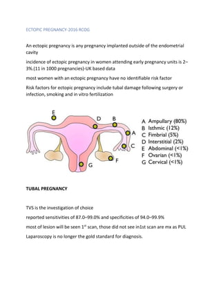
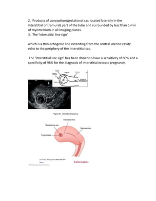
ECTOPIC PREGNANCY-2016 RCOG
An ectopic pregnancy is any pregnancy implanted outside of the endometrial
cavity
incidence of ectopic pregnancy in women attending early pregnancy units is 2–
3%.(11 in 1000 pregnancies)-UK based data
most women with an ectopic pregnancy have no identifiable risk factor
Risk factors for ectopic pregnancy include tubal damage following surgery or
infection, smoking and in vitro fertilization
TUBAL PREGNANCY
TVS is the investigation of choice
reported sensitivities of 87.0–99.0% and specificities of 94.0–99.9%
most of lesion will be seen 1st
scan, those did not see in1st scan are mx as PUL
Laparoscopy is no longer the gold standard for diagnosis.
***False-negative laparoscopies (3.0–4.5%) have been reported when the
procedure is performed too early in the development of an ongoing ectopic
pregnancy.
USS features
Inhomogeneous/non-cystic adnexal mass – 50-60% most common
Empty extrauterine sac 20-40%
Yolk sac+/- fetal pole+/- FHB 15-20%
Pseudo sac seen in 20% but no specific ET or appearance related to tubal ectopic
The key is to distinguish this from an early intrauterine gestational sac.
The intra decidual and double decidual signs can be used to diagnose an early
intrauterine pregnancy.
The intra decidual sign is described as a fluid collection with an echogenic rim
located ‘within a markedly thickened decidua on one side of the uterine cavity’.
The double decidual sign is described as an intrauterine fluid collection
surrounded by ‘two concentric echogenic rings
The presence of a ‘pseudosac’ alone cannot be used to diagnose an ectopic
pregnancy and in fact, a small anechoic cystic structure is more likely to be an
early sac rather than a‘pseudosac’.
A study27has shown that a woman with a positive pregnancy test, an
intrauterine smooth-walled anechoic cystic structure and no adnexal mass has a
0.02% probability of ectopic pregnancy, whilethe probability of intrauterine
pregnancy in such a patient is 99.98
Free fluid is often seen on ultrasound but is not diagnostic of ectopic pregnancy. A
small amount of anechoic fluid in the pouch of Douglas may be found in both
intrauterine and ectopic pregnancies. Echogenic fluid has been
reported in 28–56% of ectopic pregnancies.28,29It may signify
tubal rupture, but most commonly is due to blood leaking from the fimbrial end
of the fallopian tube
Biochemical investigation for Tubal ectopic
Serum beta HCG is for plan mx
Single value can not predict ectopic
<1000 iu of beta HCGs may be seen in ectopic
The initial serum HCG level is a key prognostic indicator for the success of
conservative management (expectant and medical) in cases of ultrasound
visualized tubal ectopic pregnancies.
CERVICAL ECTOPIC
Cervical pregnancies are rare, accounting for less than 1% of all ectopic
gestations.
USS features
1. Empty uterine cavity.
2. A barrel-shaped cervix.
3. A gestational sac present below the level of the internal cervical os.
4. The absence of the ‘sliding sign’.
5. Blood flow around the gestational sac using color Doppler.
‘sliding sign’ enables cervical ectopic pregnancies to be distinguished from
miscarriages that are within the cervical canal.
When pressure is applied to the cervix using the probe, in a miscarriage, the
gestational sac slides against the endocervical canal, but it does not in an
implanted cervical pregnancy
Biochemical Investigation
Only a single HCG
To plan mx , level> 10000 iu-MTX is less successful
LSCS SCAR PEGNANCY
Caesarean scar pregnancy is defined as implantation into the myometrial defect
occurring at the site of the previous uterine incision.
The prevalence of caesarean scar pregnancy is estimated to be approximately1 in
2000 pregnancies
these pregnancies may be ongoing potentially viable pregnancies or miscarriages
within the scar
USS is the investigation of choice
MRI is 2nd
line
TVS criteria
1. Empty uterine cavity.
2. Gestational sac or solid mass of trophoblast located anteriorly at the level
of the internal os embedded at the site of the previous caesarean section
scar
3. Thin or absent layer of myometrium between the gestational sac and the
bladder.
4. Evidence of prominent trophoblastic/placental circulation on Doppler
examination
5. Empty endocervical canal
there are two different types of pregnancies implanted in a caesarean scar:
the first type progressing into the uterine cavity as the gestational sac
grows and develops, so with the potential to reach a viable gestational age,
but with the risk of massive bleeding from the implantation site
the second with progression deeper towards the serosal surface of the
uterus with the risk of 1st
trimester rupture and hemorrhage.
diagnostic criteria have not been subject to validation and are derived from
descriptive case series, so to minimize the risk of false-positive diagnosis,
we recommend that all nonemergency cases of suspected scar pregnancy
are referred to a regional center to confirm the diagnosis.
MRI features are like of TVS. But costly & not easy to find MRI
HCG HAS NO ROLE IN DIAGNOSIS, but can try if plan for conservative mx
INTERSTITIAL PREGNANCY
Interstitial pregnancy occurs when the ectopic pregnancy implants in the
interstitial part of the fallopian tube.
The reported incidence varies between 1.0% and 6.3% of ectopic
pregnancies.
The interstitial part of the fallopian tube is about 1–2 cm in length and
traverses the muscular myometrium of the uterine wall, opening via the
tubal ostium into the uterine cavity
USS Criteria
*if available use 3D scan to better visualization & avoid misunderstand of
ANGULAR PREGNANCIES
1. Empty uterine cavity.
2. Products of conception/gestational sac located laterally in the
interstitial (intramural) part of the tube and surrounded by less than 5 mm
of myometrium in all imaging planes.
3. The ‘interstitial line sign’
which is a thin echogenic line extending from the central uterine cavity
echo to the periphery of the interstitial sac.
The ‘interstitial line sign’ has been shown to have a sensitivity of 80% and a
specificity of 98% for the diagnosis of interstitial ectopic pregnancy.
On MRI examination, a gestational sac-like structure is seen lateral to the
cornua surrounded by the myometrium. The presence of the intact
junctional zone (endo-myometrial junction) between the uterine cavity and
the gestational sac-like structure also supports the diagnosis
BIOCHEMICAL INVESTIGATIONS
if there is suspicion of interstitial pregnancy, a single serum b-hCG should
be carried out.
This does not contribute to the diagnosis, but can be useful in deciding
management options, such as surgical, medical or expectant treatment.
Sometimes repeat 48hrs HCG also used to plan mx.
CORNUAL PREGNANCIES
Defined cornual pregnancy as being ‘in one horn of a bicornuate uterus, or,
by extension of meaning, in one lateral half of a uterus of bifid tendency’
cornual pregnancy is the rarest form of ectopic pregnancy with a reported
incidence of 1 in 76 000 pregnancies
USS features
1. Visualization of a single interstitial portion of fallopian tube in the main
uterine body.
2. Gestational sac/products of conception seen mobile and separate from
the uterus and surrounded by myometrium.
3. A vascular pedicle adjoining the gestational sac to the unicornuate
uterus
BIOCHEMICAL INVESTIGATIONS
if there is suspicion of interstitial pregnancy, a single serum b-HCG should
be carried out.
This does not contribute to the diagnosis, but can be useful in deciding
management options, such as surgical, medical or expectant treatment.
Sometimes repeat 48hrs HCG also used to plan mx.
OVARIAN ECTOPIC
No specific agreed criteria for diagnosis by USS
an empty uterus
a wide echogenic ring with an internal anechoic area on the ovary
yolk sac or embryo is seen less commonly
Color Doppler may aid detection of a fetal heart pulsation
FF may represent rupture
*** It is not possible to separate the cystic structure or gestational sac from the
ovary on gentle palpation (negative sliding organ sign)
Biochemical Investigation
Single Beta HCG is required. Repeat value can consider 48Hrs after
Often diagnosis is difficult & histo sample will confirm this.
ABDOMINAL PREGNANCY
The following ultrasound criteria have been suggested by Gerli et al as
being diagnostic of an early abdominal pregnancy:
1. Absence of an intrauterine gestational sac.
2. Absence of both an evident dilated tube and a complex adnexal mass.
3. A gestational cavity surrounded by loops of bowel and separated from
them by peritoneum.
4. A wide mobility like fluctuation of the sac, particularly evident with
pressure of the transvaginal probe toward the posterior cul-de-sac

ECTOPIC PREGNANCY.docx

  • 1.
    ECTOPIC PREGNANCY-2016 RCOG Anectopic pregnancy is any pregnancy implanted outside of the endometrial cavity incidence of ectopic pregnancy in women attending early pregnancy units is 2– 3%.(11 in 1000 pregnancies)-UK based data most women with an ectopic pregnancy have no identifiable risk factor Risk factors for ectopic pregnancy include tubal damage following surgery or infection, smoking and in vitro fertilization TUBAL PREGNANCY TVS is the investigation of choice reported sensitivities of 87.0–99.0% and specificities of 94.0–99.9% most of lesion will be seen 1st scan, those did not see in1st scan are mx as PUL Laparoscopy is no longer the gold standard for diagnosis.
  • 2.
    ***False-negative laparoscopies (3.0–4.5%)have been reported when the procedure is performed too early in the development of an ongoing ectopic pregnancy. USS features Inhomogeneous/non-cystic adnexal mass – 50-60% most common Empty extrauterine sac 20-40% Yolk sac+/- fetal pole+/- FHB 15-20% Pseudo sac seen in 20% but no specific ET or appearance related to tubal ectopic The key is to distinguish this from an early intrauterine gestational sac. The intra decidual and double decidual signs can be used to diagnose an early intrauterine pregnancy. The intra decidual sign is described as a fluid collection with an echogenic rim located ‘within a markedly thickened decidua on one side of the uterine cavity’. The double decidual sign is described as an intrauterine fluid collection surrounded by ‘two concentric echogenic rings
  • 4.
    The presence ofa ‘pseudosac’ alone cannot be used to diagnose an ectopic pregnancy and in fact, a small anechoic cystic structure is more likely to be an early sac rather than a‘pseudosac’. A study27has shown that a woman with a positive pregnancy test, an intrauterine smooth-walled anechoic cystic structure and no adnexal mass has a 0.02% probability of ectopic pregnancy, whilethe probability of intrauterine pregnancy in such a patient is 99.98 Free fluid is often seen on ultrasound but is not diagnostic of ectopic pregnancy. A small amount of anechoic fluid in the pouch of Douglas may be found in both intrauterine and ectopic pregnancies. Echogenic fluid has been reported in 28–56% of ectopic pregnancies.28,29It may signify
  • 5.
    tubal rupture, butmost commonly is due to blood leaking from the fimbrial end of the fallopian tube Biochemical investigation for Tubal ectopic Serum beta HCG is for plan mx Single value can not predict ectopic <1000 iu of beta HCGs may be seen in ectopic The initial serum HCG level is a key prognostic indicator for the success of conservative management (expectant and medical) in cases of ultrasound visualized tubal ectopic pregnancies. CERVICAL ECTOPIC Cervical pregnancies are rare, accounting for less than 1% of all ectopic gestations. USS features 1. Empty uterine cavity. 2. A barrel-shaped cervix. 3. A gestational sac present below the level of the internal cervical os.
  • 6.
    4. The absenceof the ‘sliding sign’. 5. Blood flow around the gestational sac using color Doppler. ‘sliding sign’ enables cervical ectopic pregnancies to be distinguished from miscarriages that are within the cervical canal. When pressure is applied to the cervix using the probe, in a miscarriage, the gestational sac slides against the endocervical canal, but it does not in an implanted cervical pregnancy Biochemical Investigation Only a single HCG To plan mx , level> 10000 iu-MTX is less successful
  • 8.
    LSCS SCAR PEGNANCY Caesareanscar pregnancy is defined as implantation into the myometrial defect occurring at the site of the previous uterine incision. The prevalence of caesarean scar pregnancy is estimated to be approximately1 in 2000 pregnancies these pregnancies may be ongoing potentially viable pregnancies or miscarriages within the scar USS is the investigation of choice MRI is 2nd line TVS criteria 1. Empty uterine cavity. 2. Gestational sac or solid mass of trophoblast located anteriorly at the level of the internal os embedded at the site of the previous caesarean section scar 3. Thin or absent layer of myometrium between the gestational sac and the bladder. 4. Evidence of prominent trophoblastic/placental circulation on Doppler examination 5. Empty endocervical canal
  • 9.
    there are twodifferent types of pregnancies implanted in a caesarean scar: the first type progressing into the uterine cavity as the gestational sac grows and develops, so with the potential to reach a viable gestational age, but with the risk of massive bleeding from the implantation site the second with progression deeper towards the serosal surface of the uterus with the risk of 1st trimester rupture and hemorrhage.
  • 10.
    diagnostic criteria havenot been subject to validation and are derived from descriptive case series, so to minimize the risk of false-positive diagnosis, we recommend that all nonemergency cases of suspected scar pregnancy are referred to a regional center to confirm the diagnosis. MRI features are like of TVS. But costly & not easy to find MRI HCG HAS NO ROLE IN DIAGNOSIS, but can try if plan for conservative mx INTERSTITIAL PREGNANCY Interstitial pregnancy occurs when the ectopic pregnancy implants in the interstitial part of the fallopian tube. The reported incidence varies between 1.0% and 6.3% of ectopic pregnancies. The interstitial part of the fallopian tube is about 1–2 cm in length and traverses the muscular myometrium of the uterine wall, opening via the tubal ostium into the uterine cavity USS Criteria *if available use 3D scan to better visualization & avoid misunderstand of ANGULAR PREGNANCIES 1. Empty uterine cavity.
  • 11.
    2. Products ofconception/gestational sac located laterally in the interstitial (intramural) part of the tube and surrounded by less than 5 mm of myometrium in all imaging planes. 3. The ‘interstitial line sign’ which is a thin echogenic line extending from the central uterine cavity echo to the periphery of the interstitial sac. The ‘interstitial line sign’ has been shown to have a sensitivity of 80% and a specificity of 98% for the diagnosis of interstitial ectopic pregnancy.
  • 12.
    On MRI examination,a gestational sac-like structure is seen lateral to the cornua surrounded by the myometrium. The presence of the intact junctional zone (endo-myometrial junction) between the uterine cavity and the gestational sac-like structure also supports the diagnosis BIOCHEMICAL INVESTIGATIONS if there is suspicion of interstitial pregnancy, a single serum b-hCG should be carried out. This does not contribute to the diagnosis, but can be useful in deciding management options, such as surgical, medical or expectant treatment. Sometimes repeat 48hrs HCG also used to plan mx. CORNUAL PREGNANCIES Defined cornual pregnancy as being ‘in one horn of a bicornuate uterus, or, by extension of meaning, in one lateral half of a uterus of bifid tendency’ cornual pregnancy is the rarest form of ectopic pregnancy with a reported incidence of 1 in 76 000 pregnancies
  • 13.
    USS features 1. Visualizationof a single interstitial portion of fallopian tube in the main uterine body. 2. Gestational sac/products of conception seen mobile and separate from the uterus and surrounded by myometrium. 3. A vascular pedicle adjoining the gestational sac to the unicornuate uterus BIOCHEMICAL INVESTIGATIONS if there is suspicion of interstitial pregnancy, a single serum b-HCG should be carried out. This does not contribute to the diagnosis, but can be useful in deciding management options, such as surgical, medical or expectant treatment. Sometimes repeat 48hrs HCG also used to plan mx. OVARIAN ECTOPIC No specific agreed criteria for diagnosis by USS
  • 14.
    an empty uterus awide echogenic ring with an internal anechoic area on the ovary yolk sac or embryo is seen less commonly Color Doppler may aid detection of a fetal heart pulsation FF may represent rupture *** It is not possible to separate the cystic structure or gestational sac from the ovary on gentle palpation (negative sliding organ sign) Biochemical Investigation Single Beta HCG is required. Repeat value can consider 48Hrs after Often diagnosis is difficult & histo sample will confirm this. ABDOMINAL PREGNANCY
  • 15.
    The following ultrasoundcriteria have been suggested by Gerli et al as being diagnostic of an early abdominal pregnancy: 1. Absence of an intrauterine gestational sac. 2. Absence of both an evident dilated tube and a complex adnexal mass. 3. A gestational cavity surrounded by loops of bowel and separated from them by peritoneum. 4. A wide mobility like fluctuation of the sac, particularly evident with pressure of the transvaginal probe toward the posterior cul-de-sac