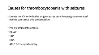
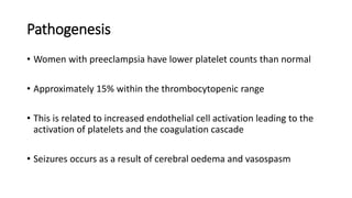
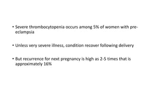
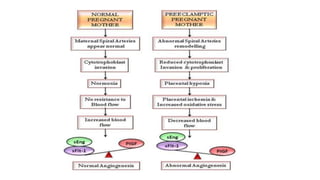
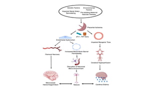
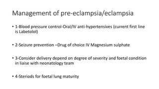
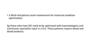
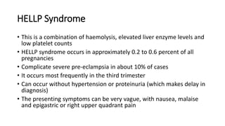
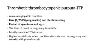
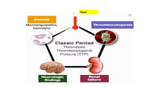
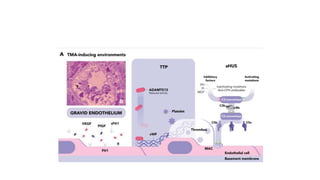
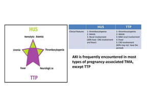
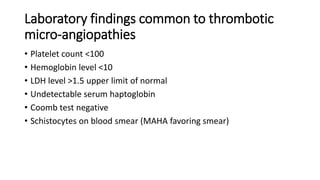
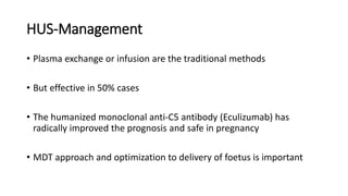
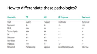
Thrombocytopenia & Seizures
in
Pregnancy
Dr.Madura Jayawardane(MBBS,MD,MRCOG)
Senior Lecturer
University of Sri Jayawardenapura
Platelets
Non nucleated cells
Originates from megakaryocytes
Half life of 8-10 days in peripheral blood
Critical for initiation of haemostasis when activated
Normally levels vary between 150-400×109/L
Physiological changes to platelets in pregnancy
• Number of circulating platelets declines (as POG advances) to lower
normal level-150×109 /L.
• Platelet aggregation ability increases
• Platelet lifespan declines
• Platelet volume minimally increase
Reasons for physiological changes to platelets
• Increased consumption of platelets in the utero-placental circulation
• Hemodilution
Thrombocytopenia in pregnancy
• A platelet count of less than 150×10 9/L is known as
thrombocytopenia
• A platelet count below the normal range is found in 8–10% of
pregnancies
• Most cases are mild and have no significance for mother or foetus
• Occasionally complex pathologies with severe morbidity and
mortality can be seen
Causes for thrombocytopenia in pregnancy
Is every thrombocytopenia bad?
• 75% of cases are due to a benign process of gestational
thrombocytopenia
• 15–20% related to hypertensive disorders in pregnancy
• 3-4% related to immunological disorders
• 1-2% of all due to infections, malignancies or rare platelet disorders
Causes for thrombocytopenia with seizures
• Unless no ICH or infective origin causes very few pregnancy related
events can cause this presentation.
• Pre-eclampsia/Eclampsia
• HELLP
• TTP
• HUS
• AFLP & Encephalopathy
Eclampsia/Pre-eclampsia
• Eclampsia - convulsive condition associated with pre-eclampsia.
• Pre-eclampsia- de novo hypertension(140/90mmHg) after POG 20
weeks with 1 or more following conditions
Proteinuria-UPCR>30 or ACR>8 or Dipstick +2 (>1g/L)
Utero-placenta insufficiency
Maternal organ dysfunction (blood-low platelets, CNS-fits, liver or
renal problems)
Pathogenesis
• Women with preeclampsia have lower platelet counts than normal
• Approximately 15% within the thrombocytopenic range
• This is related to increased endothelial cell activation leading to the
activation of platelets and the coagulation cascade
• Seizures occurs as a result of cerebral oedema and vasospasm
• Severe thrombocytopenia occurs among 5% of women with pre-
eclampsia
• Unless very severe illness, condition recover following delivery
• But recurrence for next pregnancy is high as 2-5 times that is
approximately 16%
Why condition resolves after delivery?
• The utero-placental ischemia causes the release of various vasoactive
molecules (Endoglin, Soluble FMS like tyrosine kinases)
• After placenta is expulsed, abrupt decline of molecules cease
pathology and platelets recover over about next 7-10 days.
Management of pre-eclampsia/eclampsia
• 1-Blood pressure control-Oral/IV anti-hypertensives (current first line
is Labetolol)
• 2-Seizure prevention –Drug of choice IV Magnesium sulphate
• 3-Consider delivery depend on degree of severity and foetal condition
in liaise with neonatology team
• 4-Steriods for foetal lung maturity
• 5-Multi disciplinary team involvement for maternal condition
optimization
Eg-those who have DIC need to be optimized with haematologists and
transfusion specialists input in a ICU. Those patients require blood and
blood products.
HELLP Syndrome
• This is a combination of haemolysis, elevated liver enzyme levels and
low platelet counts
• HELLP syndrome occurs in approximately 0.2 to 0.6 percent of all
pregnancies
• Complicate severe pre-eclampsia in about 10% of cases
• It occurs most frequently in the third trimester
• Can occur without hypertension or proteinuria (which makes delay in
diagnosis)
• The presenting symptoms can be very vague, with nausea, malaise
and epigastric or right upper quadrant pain
Pathophysiology
• Pathogenesis of HELLP syndrome is not well understood
• This multisystem disease is attributed to abnormal vascular tone,
vasospasm and coagulation defects
• Syndrome seems to be the final manifestation of an insult that leads to
micro vascular endothelial damage and intravascular platelet activation
• All patients with HELLP syndrome may have an underlying coagulopathy
that is usually undetectable. Most patients show no abnormalities on
coagulation studies.
• The haemolysis in HELLP syndrome is a microangiopathic haemolytic
anaemia
• Red blood cells become fragmented as they pass through small blood
vessels with endothelial damage and fibrin deposits
• The elevated liver enzyme levels is due to obstruction of hepatic
blood flow by fibrin deposits in the sinusoids
• The thrombocytopenia has been attributed to increased consumption
and/or destruction of platelets.
• Disseminated intravascular coagulation may be present in
approximately 20% of cases
• Abruption occurs in approximately 16%
• The central nervous and renal systems are usually unaffected by this
condition, in contrast to TTP
• Depend on liver enzymes and platelet levels, incomplete forms of
HELLP syndrome has been described.
Management
• Delivery is the treatment for the mother
• Steroids should be given (only to help mature the baby’s lungs)-but NICE
guideline do not support high dose steroids to mother in order to improve
HELLP syndrome.
• The platelet count should be maintained at >50 and MDT approach is often
improve care
• condition usually improves quite quickly after delivery, although it may
worsen during the first 24–48 hours postpartum
Thrombotic thrombocytopenic purpura-TTP
• A microangiopathic condition
• Rare (1/25000 pregnancies) and life-threatening
• Pentad of symptoms and signs
• The time of onset in pregnancy is variable
• Mostly occurs in 2nd trimester
• Highest mortality is when condition starts de novo in pregnancy and
co-exist with pre-eclampsia
Fever
Pathogenesis
• Due to a severe deficiency of von Willebrand’s factor-cleaving protein
(ADAMTS 13)
• leads to persistence of ultra-large multimers of von Willebrand’s
factor that unfold and react with platelet receptors
• Generates microthrombi in many organs,particularly in the kidneys,
brain and heart, and causing microangiopathic hemolytic anemia and
thrombocytopenia
• Condition is difficult to diagnose
• Most commonly an acquired deficiency caused by an autoantibody
• rarely, a primary congenital deficiency caused by a genetic defect
• The two types can be distinguished by measurement of ADAMTS 13
antigen activity and inhibitor
• Inhibitor is absent in the congenital form.
Management
• Plasmaparesis-to remove antibodies is the cornerstone of
management
• 1–1.5 l fresh frozen plasma containing the absent enzyme is infused
daily until the platelet count & LDH normalize.
• Number of treatment cycle is variable
• Immune suppression with high dose steroids or use of Rituximab
against CD20 cells are known alternative options when condition
resists.
• Recurrence for subsequent pregnancy is high as 1 in 4 (25%)
• When the condition is congenital, Plasmaparesis is not effective as
there is no specific antibody forms to remove
• These patients are best treated with plasma infusion only
• Platelet transfusion is contraindicated as it aggravates CNS
symptoms
• The risk of bleeding is low in this condition
• Relapses can be predicted by previous clinical manifestation pattern
and low level of ADAMTS 13 during remission
HUS-Haemolytic uremic syndrome
• Similar to TTP-microangiopathic condition
• Triad of symptoms and signs
• Renal failure is marked
Non-immune,
MAHA
• Typical HUS-associated to shiga toxin and seen in children
• Atypical HUS-seen in adults and related to complement dysregulation
• Pregnancy is linked to Atypical-HUS
• High morbidity and mortality
Pathogenesis
• Acquired or constitutional complement alternative pathway
dysregulation
• leading to complement-induced endothelial cell damage in atypical
HUS
• Various patterns of endothelial cell lesions in the heterogeneous
group of secondary HUS associated with autoimmune diseases, drugs,
infections, and pregnancy.
AKI is frequently encountered in most
types of pregnancy-associated TMA,
except TTP
Laboratory findings common to thrombotic
micro-angiopathies
• Platelet count <100
• Hemoglobin level <10
• LDH level >1.5 upper limit of normal
• Undetectable serum haptoglobin
• Coomb test negative
• Schistocytes on blood smear (MAHA favoring smear)
HUS-Management
• Plasma exchange or infusion are the traditional methods
• But effective in 50% cases
• The humanized monoclonal anti-C5 antibody (Eculizumab) has
radically improved the prognosis and safe in pregnancy
• MDT approach and optimization to delivery of foetus is important
Microangiopathies and neonatal issues
• prognosis for the baby in all the Microangiopathies described is poor
because of extensive placental ischemia
How to differentiate these pathologies?
References
• 1) D L W Dasanayake, Y Costa, A Weerawardana.Management of thrombocytopenia in
pregnancy.Sri Lanka Journal of Obstetrics and Gynaecology 2021; 43: 259-
268(DOI:http://doi.org/10.4038/sljog.v43i3.8020)
• 2) Mayers B.Thrombocytopenia in pregnancy.TOG;2009;11:177–183.
• 3) Paddon MO. HELLP Syndrome: Recognition and Perinatal Management. Am Fam Physician.
1999;60(3):829-836
• 4) Management of thrombotic microangiopathy in pregnancy and postpartum: report from an
international working group. https://doi.org/10.1182/blood.2020005221
• 5) Gernsheimer T, James AH, Stasi R. How I treat thrombocytopenia in pregnancy. Blood. 2013 Jan
03;121(1):38-47
• 6) George JN, Nester CM. Syndromes of thrombotic microangiopathy. N Engl J Med. 2014 Nov
06;371(19):1847-8
In summery
• Not all thrombocytopenia's are bad or fatal
• When it coexist with seizures-need to rule out pregnancy related
unique conditions
• Chronic epilepsy with low platelet causing medical conditions to be
considered as well
• Microangiopathies make diagnosis more complicated
• MDT approach and optimization for delivery is often safe for mother
and baby
Thrombocytopenia & Seizures.pptx

Thrombocytopenia & Seizures.pptx

  • 1.
    Thrombocytopenia & Seizures in Pregnancy Dr.MaduraJayawardane(MBBS,MD,MRCOG) Senior Lecturer University of Sri Jayawardenapura
  • 2.
    Platelets Non nucleated cells Originatesfrom megakaryocytes Half life of 8-10 days in peripheral blood Critical for initiation of haemostasis when activated Normally levels vary between 150-400×109/L
  • 3.
    Physiological changes toplatelets in pregnancy • Number of circulating platelets declines (as POG advances) to lower normal level-150×109 /L. • Platelet aggregation ability increases • Platelet lifespan declines • Platelet volume minimally increase
  • 4.
    Reasons for physiologicalchanges to platelets • Increased consumption of platelets in the utero-placental circulation • Hemodilution
  • 5.
    Thrombocytopenia in pregnancy •A platelet count of less than 150×10 9/L is known as thrombocytopenia • A platelet count below the normal range is found in 8–10% of pregnancies • Most cases are mild and have no significance for mother or foetus • Occasionally complex pathologies with severe morbidity and mortality can be seen
  • 6.
  • 7.
    Is every thrombocytopeniabad? • 75% of cases are due to a benign process of gestational thrombocytopenia • 15–20% related to hypertensive disorders in pregnancy • 3-4% related to immunological disorders • 1-2% of all due to infections, malignancies or rare platelet disorders
  • 8.
    Causes for thrombocytopeniawith seizures • Unless no ICH or infective origin causes very few pregnancy related events can cause this presentation. • Pre-eclampsia/Eclampsia • HELLP • TTP • HUS • AFLP & Encephalopathy
  • 9.
    Eclampsia/Pre-eclampsia • Eclampsia -convulsive condition associated with pre-eclampsia. • Pre-eclampsia- de novo hypertension(140/90mmHg) after POG 20 weeks with 1 or more following conditions Proteinuria-UPCR>30 or ACR>8 or Dipstick +2 (>1g/L) Utero-placenta insufficiency Maternal organ dysfunction (blood-low platelets, CNS-fits, liver or renal problems)
  • 10.
    Pathogenesis • Women withpreeclampsia have lower platelet counts than normal • Approximately 15% within the thrombocytopenic range • This is related to increased endothelial cell activation leading to the activation of platelets and the coagulation cascade • Seizures occurs as a result of cerebral oedema and vasospasm
  • 11.
    • Severe thrombocytopeniaoccurs among 5% of women with pre- eclampsia • Unless very severe illness, condition recover following delivery • But recurrence for next pregnancy is high as 2-5 times that is approximately 16%
  • 14.
    Why condition resolvesafter delivery? • The utero-placental ischemia causes the release of various vasoactive molecules (Endoglin, Soluble FMS like tyrosine kinases) • After placenta is expulsed, abrupt decline of molecules cease pathology and platelets recover over about next 7-10 days.
  • 15.
    Management of pre-eclampsia/eclampsia •1-Blood pressure control-Oral/IV anti-hypertensives (current first line is Labetolol) • 2-Seizure prevention –Drug of choice IV Magnesium sulphate • 3-Consider delivery depend on degree of severity and foetal condition in liaise with neonatology team • 4-Steriods for foetal lung maturity
  • 16.
    • 5-Multi disciplinaryteam involvement for maternal condition optimization Eg-those who have DIC need to be optimized with haematologists and transfusion specialists input in a ICU. Those patients require blood and blood products.
  • 17.
    HELLP Syndrome • Thisis a combination of haemolysis, elevated liver enzyme levels and low platelet counts • HELLP syndrome occurs in approximately 0.2 to 0.6 percent of all pregnancies • Complicate severe pre-eclampsia in about 10% of cases • It occurs most frequently in the third trimester • Can occur without hypertension or proteinuria (which makes delay in diagnosis) • The presenting symptoms can be very vague, with nausea, malaise and epigastric or right upper quadrant pain
  • 18.
  • 19.
    • Pathogenesis ofHELLP syndrome is not well understood • This multisystem disease is attributed to abnormal vascular tone, vasospasm and coagulation defects • Syndrome seems to be the final manifestation of an insult that leads to micro vascular endothelial damage and intravascular platelet activation • All patients with HELLP syndrome may have an underlying coagulopathy that is usually undetectable. Most patients show no abnormalities on coagulation studies.
  • 20.
    • The haemolysisin HELLP syndrome is a microangiopathic haemolytic anaemia • Red blood cells become fragmented as they pass through small blood vessels with endothelial damage and fibrin deposits • The elevated liver enzyme levels is due to obstruction of hepatic blood flow by fibrin deposits in the sinusoids • The thrombocytopenia has been attributed to increased consumption and/or destruction of platelets.
  • 21.
    • Disseminated intravascularcoagulation may be present in approximately 20% of cases • Abruption occurs in approximately 16% • The central nervous and renal systems are usually unaffected by this condition, in contrast to TTP
  • 22.
    • Depend onliver enzymes and platelet levels, incomplete forms of HELLP syndrome has been described.
  • 23.
    Management • Delivery isthe treatment for the mother • Steroids should be given (only to help mature the baby’s lungs)-but NICE guideline do not support high dose steroids to mother in order to improve HELLP syndrome. • The platelet count should be maintained at >50 and MDT approach is often improve care • condition usually improves quite quickly after delivery, although it may worsen during the first 24–48 hours postpartum
  • 24.
    Thrombotic thrombocytopenic purpura-TTP •A microangiopathic condition • Rare (1/25000 pregnancies) and life-threatening • Pentad of symptoms and signs • The time of onset in pregnancy is variable • Mostly occurs in 2nd trimester • Highest mortality is when condition starts de novo in pregnancy and co-exist with pre-eclampsia
  • 25.
  • 26.
  • 27.
    • Due toa severe deficiency of von Willebrand’s factor-cleaving protein (ADAMTS 13) • leads to persistence of ultra-large multimers of von Willebrand’s factor that unfold and react with platelet receptors • Generates microthrombi in many organs,particularly in the kidneys, brain and heart, and causing microangiopathic hemolytic anemia and thrombocytopenia • Condition is difficult to diagnose
  • 28.
    • Most commonlyan acquired deficiency caused by an autoantibody • rarely, a primary congenital deficiency caused by a genetic defect • The two types can be distinguished by measurement of ADAMTS 13 antigen activity and inhibitor • Inhibitor is absent in the congenital form.
  • 29.
    Management • Plasmaparesis-to removeantibodies is the cornerstone of management • 1–1.5 l fresh frozen plasma containing the absent enzyme is infused daily until the platelet count & LDH normalize. • Number of treatment cycle is variable • Immune suppression with high dose steroids or use of Rituximab against CD20 cells are known alternative options when condition resists.
  • 30.
    • Recurrence forsubsequent pregnancy is high as 1 in 4 (25%) • When the condition is congenital, Plasmaparesis is not effective as there is no specific antibody forms to remove • These patients are best treated with plasma infusion only • Platelet transfusion is contraindicated as it aggravates CNS symptoms
  • 31.
    • The riskof bleeding is low in this condition • Relapses can be predicted by previous clinical manifestation pattern and low level of ADAMTS 13 during remission
  • 32.
    HUS-Haemolytic uremic syndrome •Similar to TTP-microangiopathic condition • Triad of symptoms and signs • Renal failure is marked Non-immune, MAHA
  • 33.
    • Typical HUS-associatedto shiga toxin and seen in children • Atypical HUS-seen in adults and related to complement dysregulation • Pregnancy is linked to Atypical-HUS • High morbidity and mortality
  • 34.
    Pathogenesis • Acquired orconstitutional complement alternative pathway dysregulation • leading to complement-induced endothelial cell damage in atypical HUS • Various patterns of endothelial cell lesions in the heterogeneous group of secondary HUS associated with autoimmune diseases, drugs, infections, and pregnancy.
  • 36.
    AKI is frequentlyencountered in most types of pregnancy-associated TMA, except TTP
  • 37.
    Laboratory findings commonto thrombotic micro-angiopathies • Platelet count <100 • Hemoglobin level <10 • LDH level >1.5 upper limit of normal • Undetectable serum haptoglobin • Coomb test negative • Schistocytes on blood smear (MAHA favoring smear)
  • 38.
    HUS-Management • Plasma exchangeor infusion are the traditional methods • But effective in 50% cases • The humanized monoclonal anti-C5 antibody (Eculizumab) has radically improved the prognosis and safe in pregnancy • MDT approach and optimization to delivery of foetus is important
  • 39.
    Microangiopathies and neonatalissues • prognosis for the baby in all the Microangiopathies described is poor because of extensive placental ischemia
  • 40.
    How to differentiatethese pathologies?
  • 41.
    References • 1) DL W Dasanayake, Y Costa, A Weerawardana.Management of thrombocytopenia in pregnancy.Sri Lanka Journal of Obstetrics and Gynaecology 2021; 43: 259- 268(DOI:http://doi.org/10.4038/sljog.v43i3.8020) • 2) Mayers B.Thrombocytopenia in pregnancy.TOG;2009;11:177–183. • 3) Paddon MO. HELLP Syndrome: Recognition and Perinatal Management. Am Fam Physician. 1999;60(3):829-836 • 4) Management of thrombotic microangiopathy in pregnancy and postpartum: report from an international working group. https://doi.org/10.1182/blood.2020005221 • 5) Gernsheimer T, James AH, Stasi R. How I treat thrombocytopenia in pregnancy. Blood. 2013 Jan 03;121(1):38-47 • 6) George JN, Nester CM. Syndromes of thrombotic microangiopathy. N Engl J Med. 2014 Nov 06;371(19):1847-8
  • 42.
    In summery • Notall thrombocytopenia's are bad or fatal • When it coexist with seizures-need to rule out pregnancy related unique conditions • Chronic epilepsy with low platelet causing medical conditions to be considered as well • Microangiopathies make diagnosis more complicated • MDT approach and optimization for delivery is often safe for mother and baby