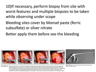
COLPOSCOPY
CLINICAL DISCUSSION AND
TRAINING SESSION
WHAT IS COLPOSCOPY?
• Inroduced by Hinselman in late 1925
• Magnification and illumination of female lower
genital tract with various stains
Lugol’s iodine and Acetic acid(3% or 5%)
Except illumination, magnification and Green filter
to enhance visualization of vascularity, no much
advanced over the last century
Old Versus New
INDICATIONS
1)one cervical sample with borderline changes in high risk HPV+
2)one sample with mild dyskaryosis in high risk HPV+
3)One sample with mild dyskaryosis with inadequate HPV test
4)one sample with borderline changes involving endocervix
5)one sample with moderate – severe dyskaryosis
6)One sample with possible invasive disease
7)One sample with glandular neoplasia
Cont.
8)One sample with glandular neoplasia
9) Three consecutive inadequate samples
10)Any grade of dyskaryosis after CIN treatment prior next recall
11)Three abnormal sample of any grade over 10 years period
12)Suspicious symptoms and abnormal cervix
If there is no HPV DNA test facilities, consider colposcopy if,
13)Three samples with borderline changes in squamous cells
14)One sample with mild dyskaryosis
• Accuracy of colposcopy improved if entire
squamo-columnar junction is seen and upper
limit of any lesion is seen
• Size and topography of lesion are taken
seriously when extend into cervical canal or
vagina
• This is a subjective assessment with intra
observer variability and commonly produce
inconclusive findings
• Inter observer variability affect low grade
lesions more often than high grade lesions
BJOG 2012-AUGEST/13-VOL119/ISSUE11
• Accuracy of colposcopy targeted biopsies in
systematic review and Metanalysis
TYPE SENSITIVITY SPECIFICITY
CIN 1 92% 25%
CIN 2 80% 64%
CIN 3 83% 44%
Colposcopy rather than the biopsy itself limit the
detection, because we can not take targeted
biopsy from place which is not visible
Lets see the STEPS !!!
1. Explain the procedure and get consent-some
times cryotherapy or Loop excision can do
simultaneously to reduce re-visits.
2. Put on modified lithotomy position and use
medium sized warm clean water lubricated
Cusco speculum insert gently.
3. Note the changes seen in cervix and gently
wipe secretion on cervix using saline soaked
cotton balls without eroding cervix
4.Avoid rough traumatic handling of cervix
5.If colposcopist is interested in assessing repeat
smear, it can be taken with spatula prior to
apply any solution. This is a debatable step and
yet people practice.
Smears induced bleeding settles once acetic acid
apply
6)Cervical cells for HPV DNA, and bacteriological
studies also can take before apply acid-iodine
7)Apply green filter and 15 power magnification to
see vascularity before acid-iodine apply as they
cause swelling of vessels themselves
Other value of this pre assessment is to see whole
transformation zone. If inner border of
transformation zone as S-C junction was failed to
recognize in entire 360’ view-procedure is
inadequate or unsatisfactory
Distal limit or original S-C junction is demarcated at
imaginary line along most distal glandular crypts or
nabothian follicles
8) Apply 5% acetic acid as it is more faster and
obviously stains. Confirm strength of solution.
Liberally apply even on ectocervix.
This enables to find entire new SC junction and
detect abnormal transformation zones
Aceto white areas forms over 1 min and
thereafter dissapear.Hence need repetitive
application.
9)Apply Lugol’s iodine
Normal squamous cells contain glycogen that
stain brown with iodine. But not the columnar
cells as they have minimal or no glycogen.
Similarly immature squamous metaplasia,
inflammatory and regenerating epithelium and
congenital transformation zone contain very
little or no glycogen and either do not or only
partially stain with iodine.
cont.
Abnormal transformation zones, such as those with CIN
or invasive cancer, contain very little or no glycogen. The
degree of differentiation of the cells in a preneoplastic
squamous lesion determines the amount of intracellular
glycogen and thus the degree of staining observed.
Therefore, one would expect to see a range of staining
from partially brown to mustard yellow across the
spectrum from low- to high-grade CIN. Usually high-grade
CIN takes up less of the stain, appearing as mustard or
saffron yellow areas. In the case of high-grade CIN,
vigorous or repeated application of iodine may
occasionally peel off the abnormal epithelium and the
underlying tissue stroma may appear pale, as it lacks
glycogen.
10)If necessary, perform biopsy from site with
worst features and multiple biopsies to be taken
while observing under scope
Bleeding sites cover by Monsel paste (ferric
subsulfate) or silver nitrate
Better apply them before see the bleeding
11)Consider endocervical curate if ectocervix
shows no abnormality but abnormal cytology,
glandular lesions evidenced in cytology or
colposcopy examination is unsatisfactory
ECC is frequently associated with inadequate
sampling so that negative ECC do not rule out
ABSENCE OF ENDOCERVICAL NEOPLASIA
Before ECC,posterior fornix should not have
acetic acid to protect sample
12) Before end, as speculum withdraws, inspect
rest of genital tract and do bimanual& digital
rectal examination to complete clinical
assessment.
Thank patient, admire for enthusiasm, notes
complete and arrange follow-up with histology
report
If the woman is pregnant!!!
• The steps in the colposcopic procedure for a pregnant
woman are similar to those for a non-pregnant woman,
but extra care must be taken not to injure any tissues
when a digital examination or speculum insertion is
performed. If a repeat cytology smear is needed, this
may be performed using a spatula, by applying gentle
pressure to avoid bleeding. Some may prefer to obtain
a cytology sample at the end of the colposcopic
procedure, in order to avoid inducing bleeding that
may obscure the colposcopic field, but this may result
in a poor hypocellular sample, as cells might have been
washed away during the different steps of the
colposcopic procedure.
• increased vascularity leads to easily induced
bleeding; the blood vessel pattern in cervical
pseudo-decidual tissue tends to mimic
invasive cancer; and CIN tends to appear as a
more severe grade than it actually is (due to
increased size, increased oedema and
vasculature pattern). Thus considerable
experience is required for colposcopy in
pregnancy.
• As pregnancy progresses, cervical biopsy is associated with an
increased probability and degree of bleeding, which may often be
difficult to control. The risk of biopsy should always be weighed
against the risk of missing an early invasive cancer. All lesions
suspicious of invasive cancer must be biopsied or wedge excised.
Sharp biopsy forceps should be used, as they will produce less
tearing of tissue. Biopsy should always be carried out under
colposcopic vision to control depth. The prompt application of
Monsel’s paste or silver nitrate to the biopsy site, immediate bed
rest for 15 to 30 minutes, and the use of a tampon or other
haemostatic packing to put pressure on the biopsy site are helpful
to minimize bleeding. Some women may need an injection of
pitressin into the cervix or suturing for haemostasis. To avoid a large
amount of tissue slough, due to the effect of Monsel’s paste,
haemostatic packs should not be left in place for more than a few
hours after the paste has been applied. Alternatively, cervical biopsy
in a pregnant woman may be performed with diathermy loop. If
colposcopy is inadequate, and cytology suggests invasive cancer, a
conization must be performed, ideally in the second trimester. Non-
invasive lesions may be evaluated post-partum
Colposcopy

Colposcopy

  • 1.
  • 2.
    WHAT IS COLPOSCOPY? •Inroduced by Hinselman in late 1925 • Magnification and illumination of female lower genital tract with various stains Lugol’s iodine and Acetic acid(3% or 5%) Except illumination, magnification and Green filter to enhance visualization of vascularity, no much advanced over the last century
  • 3.
  • 4.
    INDICATIONS 1)one cervical samplewith borderline changes in high risk HPV+ 2)one sample with mild dyskaryosis in high risk HPV+ 3)One sample with mild dyskaryosis with inadequate HPV test 4)one sample with borderline changes involving endocervix 5)one sample with moderate – severe dyskaryosis 6)One sample with possible invasive disease 7)One sample with glandular neoplasia
  • 5.
    Cont. 8)One sample withglandular neoplasia 9) Three consecutive inadequate samples 10)Any grade of dyskaryosis after CIN treatment prior next recall 11)Three abnormal sample of any grade over 10 years period 12)Suspicious symptoms and abnormal cervix If there is no HPV DNA test facilities, consider colposcopy if, 13)Three samples with borderline changes in squamous cells 14)One sample with mild dyskaryosis
  • 6.
    • Accuracy ofcolposcopy improved if entire squamo-columnar junction is seen and upper limit of any lesion is seen • Size and topography of lesion are taken seriously when extend into cervical canal or vagina • This is a subjective assessment with intra observer variability and commonly produce inconclusive findings • Inter observer variability affect low grade lesions more often than high grade lesions
  • 7.
    BJOG 2012-AUGEST/13-VOL119/ISSUE11 • Accuracyof colposcopy targeted biopsies in systematic review and Metanalysis TYPE SENSITIVITY SPECIFICITY CIN 1 92% 25% CIN 2 80% 64% CIN 3 83% 44%
  • 8.
    Colposcopy rather thanthe biopsy itself limit the detection, because we can not take targeted biopsy from place which is not visible
  • 9.
    Lets see theSTEPS !!! 1. Explain the procedure and get consent-some times cryotherapy or Loop excision can do simultaneously to reduce re-visits. 2. Put on modified lithotomy position and use medium sized warm clean water lubricated Cusco speculum insert gently. 3. Note the changes seen in cervix and gently wipe secretion on cervix using saline soaked cotton balls without eroding cervix
  • 10.
    4.Avoid rough traumatichandling of cervix 5.If colposcopist is interested in assessing repeat smear, it can be taken with spatula prior to apply any solution. This is a debatable step and yet people practice. Smears induced bleeding settles once acetic acid apply 6)Cervical cells for HPV DNA, and bacteriological studies also can take before apply acid-iodine
  • 11.
    7)Apply green filterand 15 power magnification to see vascularity before acid-iodine apply as they cause swelling of vessels themselves Other value of this pre assessment is to see whole transformation zone. If inner border of transformation zone as S-C junction was failed to recognize in entire 360’ view-procedure is inadequate or unsatisfactory Distal limit or original S-C junction is demarcated at imaginary line along most distal glandular crypts or nabothian follicles
  • 14.
    8) Apply 5%acetic acid as it is more faster and obviously stains. Confirm strength of solution. Liberally apply even on ectocervix. This enables to find entire new SC junction and detect abnormal transformation zones Aceto white areas forms over 1 min and thereafter dissapear.Hence need repetitive application.
  • 16.
    9)Apply Lugol’s iodine Normalsquamous cells contain glycogen that stain brown with iodine. But not the columnar cells as they have minimal or no glycogen. Similarly immature squamous metaplasia, inflammatory and regenerating epithelium and congenital transformation zone contain very little or no glycogen and either do not or only partially stain with iodine.
  • 17.
    cont. Abnormal transformation zones,such as those with CIN or invasive cancer, contain very little or no glycogen. The degree of differentiation of the cells in a preneoplastic squamous lesion determines the amount of intracellular glycogen and thus the degree of staining observed. Therefore, one would expect to see a range of staining from partially brown to mustard yellow across the spectrum from low- to high-grade CIN. Usually high-grade CIN takes up less of the stain, appearing as mustard or saffron yellow areas. In the case of high-grade CIN, vigorous or repeated application of iodine may occasionally peel off the abnormal epithelium and the underlying tissue stroma may appear pale, as it lacks glycogen.
  • 19.
    10)If necessary, performbiopsy from site with worst features and multiple biopsies to be taken while observing under scope Bleeding sites cover by Monsel paste (ferric subsulfate) or silver nitrate Better apply them before see the bleeding
  • 20.
    11)Consider endocervical curateif ectocervix shows no abnormality but abnormal cytology, glandular lesions evidenced in cytology or colposcopy examination is unsatisfactory ECC is frequently associated with inadequate sampling so that negative ECC do not rule out ABSENCE OF ENDOCERVICAL NEOPLASIA Before ECC,posterior fornix should not have acetic acid to protect sample
  • 21.
    12) Before end,as speculum withdraws, inspect rest of genital tract and do bimanual& digital rectal examination to complete clinical assessment. Thank patient, admire for enthusiasm, notes complete and arrange follow-up with histology report
  • 22.
    If the womanis pregnant!!! • The steps in the colposcopic procedure for a pregnant woman are similar to those for a non-pregnant woman, but extra care must be taken not to injure any tissues when a digital examination or speculum insertion is performed. If a repeat cytology smear is needed, this may be performed using a spatula, by applying gentle pressure to avoid bleeding. Some may prefer to obtain a cytology sample at the end of the colposcopic procedure, in order to avoid inducing bleeding that may obscure the colposcopic field, but this may result in a poor hypocellular sample, as cells might have been washed away during the different steps of the colposcopic procedure.
  • 23.
    • increased vascularityleads to easily induced bleeding; the blood vessel pattern in cervical pseudo-decidual tissue tends to mimic invasive cancer; and CIN tends to appear as a more severe grade than it actually is (due to increased size, increased oedema and vasculature pattern). Thus considerable experience is required for colposcopy in pregnancy.
  • 24.
    • As pregnancyprogresses, cervical biopsy is associated with an increased probability and degree of bleeding, which may often be difficult to control. The risk of biopsy should always be weighed against the risk of missing an early invasive cancer. All lesions suspicious of invasive cancer must be biopsied or wedge excised. Sharp biopsy forceps should be used, as they will produce less tearing of tissue. Biopsy should always be carried out under colposcopic vision to control depth. The prompt application of Monsel’s paste or silver nitrate to the biopsy site, immediate bed rest for 15 to 30 minutes, and the use of a tampon or other haemostatic packing to put pressure on the biopsy site are helpful to minimize bleeding. Some women may need an injection of pitressin into the cervix or suturing for haemostasis. To avoid a large amount of tissue slough, due to the effect of Monsel’s paste, haemostatic packs should not be left in place for more than a few hours after the paste has been applied. Alternatively, cervical biopsy in a pregnant woman may be performed with diathermy loop. If colposcopy is inadequate, and cytology suggests invasive cancer, a conization must be performed, ideally in the second trimester. Non- invasive lesions may be evaluated post-partum