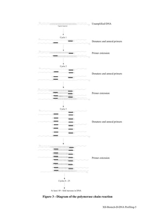
DNA PROFILING IN FORENSIC SCIENCE

   DNA is the chemical code that is found in every cell of an individual's body, and is unique
   to each individual. Because it is unique, the ability to examine DNA found at a crime
   scene is a very useful forensic tool. In New Zealand, two main techniques are used to
   profile (i.e. identify and describe) DNA.

   Restriction fragment length polymorphism (RFLP)
   In RFLP, the DNA is cut into segments of varying lengths by an enzyme, then the
   segments separated out on the basis of size using a technique called electrophoresis.
   Fragments of a particular length are transfered to a nylon membrane. They are matched up
   with radioactively labelled fragments of DNA in such a way that only fragments that are
   identical stick together. The excess radioactive fragments are washed away and an x-ray
   of the remaining fragments taken. This gives a picture of which of the labelled fragments
   were in the original sample.

   Short tandem repeat profiling (STR)
   An enzyme is used to make many copies of a small section of the DNA. This section cut
   into pieces by another enzyme, and separated by electrophoresis. The fragments are then
   visuallised with a silver stain, with the pattern of light and dark bands seen being
   characteristic for an individual.


INTRODUCTION

Deoxyribonucleic acid, or DNA, is the molecule of life. It is the chemical code specifying
our function, appearance and pedigree and is unique for all individuals except identical
twins. An individual’s DNA is formed by combination of DNA from his or her parents with
half coming from the mother and half from the father. For this reason, DNA testing can be
used as evidence of paternity of a child.

DNA is found in most cells of the body, including white blood cells, semen, hair roots and
body tissue. Traces of DNA can be detected in body fluids such as saliva and perspiration.
Mitochondrial DNA, which follows the maternal line of an individual, can be extracted from
hair and bone samples. This can be used to examine relatedness and common ancestry
between individuals, and to verify the identity of buried remains. This technique was used in
the much publicised case of the Romanovs.

Forensic Science utilises the properties of DNA in several ways. The adage “every contact
leaves a trace” indicates the importance of a technique able to type trace amounts of genetic
material left during the commission of a crime. Hairs or saliva left on a balaclava worn
during a robbery, semen located at a rape scene, blood collected from an assault, perspiration
on clothing, traces of assailant’s skin under a victim’s fingernails, can often be DNA profiled.
 This genetic information can then be used to include or exclude suspects as being the source
of the genetic material.

It is not yet possible to test the whole of an individual’s DNA. Forensic analysis involves the
testing of regions of an individuals DNA. Databases have been compiled which list the
abundance of a particular fragment of DNA in the population. From this information, an
estimate of the abundance of combinations of DNA at several regions can be made and
compared to the DNA of victims or suspects. In this way, an individual can be included or

                                                                     XII-Biotech-D-DNA Profiling-1
excluded as a possible source of DNA found in relation to a criminal investigation.
Statistical interpretation of the information can be made to estimate the likelihood of material
coming from a particular individual relative to coming from a random member of the
population. In New Zealand, forensic DNA testing is carried out at the Institute of
Environmental Science and Research Ltd. (ESR), where a Bayesian approach to statistical
interpretation is used.

THE STRUCTURE OF DNA

The DNA molecule is a vast ladder in which the vertical pieces consist of alternating sugar
molecules and phosphate groups, and the rungs are complementary bases. The 4 major bases
available for the deoxyribonucleotides are adenine (A), guanine (G), thymine (T) and
cytosine (C) (Figure 1). The backbone of the molecule consists of phosphodiester bonds
connecting the 3'- hydroxyl of the deoxyribonucleotide to the 5'-hydroxyl of the next sugar.
Two backbone chains, running in opposite directions, are paired complementarily by the
interaction of a purine (A or G) with a pyrimidine (T or C). The pairing of a purine and
pyrimidine results in a regular structure. A can only bond with T, and G can only bond with
C due to steric hindrance, so if the structure of one side of the chain is known, the other can
be determined.


        NH2                             O                    NH2                 O

                                H                                        H              CH3
               N                            N            N                   N
    N                               N

        N      N          H2N           N   N        O       N           O       N
               H                            H                H                   H
                      Figure 1 - The four major bases present in DNA


In vivo, DNA is a template for reproduction of genetic material and cellular information.
The molecule splits along its central axis, providing access to one side of the complementary
chain. Enzymes then add the appropriate bases to each chain, giving a new exact copy of the
complementary sequence. This ability of DNA to repair and replicate itself is exploited in
the DNA profiling techniques used in forensic science.

Parts of the DNA known as genes contain codes to make a specific product, usually a protein.
 These compose only a small part of the total DNA. Most of the remainder has no known
function, and can be referred to as spacer or packer DNA between the genes. Much of this is
highly repetitive sequences of DNA repeated end-on-end a varying number of times. These
regions are referred to as minisatellites. Smaller repetitive sequences of, for example, 4 base
pairs are referred to as microsatellites. The number of times this sequence is repeated is
determined in the type of profiling called Short Tandem Repeats (STR’s).


XII-Biotech-D-DNA Profiling-2
DNA PROFILING TECHNIQUES

Two of the common types of DNA profiling used at ESR for Forensic analyses are described.

Restriction fragment length polymorphism (RFLP)
This technique is outlined in Figure 2. Double stranded DNA is extracted from blood or
semen. The DNA is cut into small pieces by a sequence-specific enzyme, i.e. an enzyme that
cuts the DNA wherever a particular sequence of bases occurs. The fragments are then
separated out by a process called electrophoresis: the sample is put at one end of a bath of a
jelly-like substance called agarose gel and a voltage is applied. The fragments are charged,
and the voltage is applied in such a way as to encourage the fragments to migrate to the other
end of the gel. Small fragments move much faster than large ones, so separation on the basis
of molecular weight occurs.


                                                               Restriction enzyme added


                                          DNA extracted                                       DNA fragments
 Blood or semen samples
                                          from samples
                                                                                                            Electrophoresis


                                                                                           Person Person   Person   Person
                                                                                              1      2        3       4
                                                                                long                                         -
                            Blotting and addtion of radioactive probes       fragments


                                                                             short
                                                                          fragments                                              +
                                                                                      Agarose gel after electrophoresis


                                                    1      2       3     4


            Washing and autoradiography


                                                           X-ray

                          Figure 2 - Diagram of single locus RFLP profiling

After electrophoresis the gel and fragments are exposed to 0.25M HCl to depurinate the DNA
and nick the sugar phosphate backbone, as this assists in fragment transfer. The depurinated
sites are then cleaved by washing in NaOH/NaCl. The denatured DNA is then transferred to
a nylon membrane and the variable minisatellite region of the DNA examined by 32P-
radiolabelled short pieces of single stranded DNA called probes. A probe binds to its
complementary sequence on the membrane. The radiolabelled membrane is exposed to film
to produce an autoradiograph.

Successive regions of the DNA are examined. The distribution of each of the probed regions


                                                                                          XII-Biotech-D-DNA Profiling-3
of DNA within the population is estimated from a population database to give an indication
of the probability that the sample comes from a given suspect.

The RFLP profiling technique is outlined in Figure 2.

Short tandem repeat profiling (STR’s)
This is the new generation of DNA profiling. In this technique microsatellite repeating
regions are examined. The technique is based on the polymerase chain reaction - the cycle of
reactions in which DNA is split, replicated then split again for replication. This amplification
gives an exponential increase in the number of copies of the original template (Figure 3 on
next page). The reaction is under kinetic control, reaching a plateau which is dependent on
competition, the ultimate inactivation of the catalytic enzyme and the original number of
template molecules, primer and dNTPs (deoxynucleotide triphosphates - the building blocks
of DNA).

The DNA is denatured (split) at approximately 94oC. Short strands of DNA, called primers,
attach to the target DNA at a specific site. Bases are added enzymatically to the end of the
primers to form a new complementary strand. Approximately thirty such cycles are carried
out to produce many copies of the original material. Since the amount of original material is
increased, this technique is particularly suited to the analysis of trace amounts of DNA.

The amplified DNA is separated by electrophoresis through an ultra-thin denaturing
polyacrylamide gel. This technique can be performed manually, with repeats visualised
using silver staining (Figure 4), or automatically, with multiple loci visualised
simultaneously using fluorescent dyes. The number of repeats for a particular individual is
determined at several loci. The manual method currently in use examines three loci, with an
extra male/female sex test. The automated method currently examines four loci. Statistical
analysis on the abundance of the observed patterns in the population is carried out.


 Figure 4 - Example of a manual STR analysis. The loci HUMTH01, TPOX, CSF1PO
                     and a sex test are analysed simultaneously.

XII-Biotech-D-DNA Profiling-4
Unamplified DNA
             Targeted sequence


                 Cycle 1
                                            Denature and anneal primers


                                            Primer extension


                 Cycle 2


                                            Denature and anneal primers


                                            Primer extension


                 Cycle 3


                                            Denature and anneal primers


                                            Primer extension


              Cycles 4 - 25


      At least 105 - fold increase in DNA

Figure 3 - Diagram of the polymerase chain reaction


                                                     XII-Biotech-D-DNA Profiling-5
CONCLUSION

DNA technology is constantly evolving. New techniques, new loci and the ability to analyse
smaller samples with increased automation promise faster and more discriminating results for
the presentation of forensic evidence in court.


Article written by Susan Petricevic (ESR), with reference to:

•       Edwards A, Citivello A, Hammond HA and Caskey CT (1991) DNA typing and
        genetic mapping with trimeric and tetrameric repeats Am J Hum Gen 49: 746-756

•       Gill P, Ivanov PL, Kimpton C, Piercy R, Benson N, Tully G, Evett I, Hagelberg E
        (1994) Identification of the remains of the Romanov family by DNA analysis Nature
        genetics 6:130-135

•       Jeffreys AJ, Wilson V, Thein SL (1985) Individual-specific “fingerprints” of human
        DNA Nature 316:76-79

•       Locard E L’enquete criminelle et les methodes scientifiques, Paris 1920

•       Sullivan KM, Pope S, Gill P, Robertson JM (1992) Automated DNA profiling by
        fluorescent labelling of PCR products PCR Methods and Applications 2: 34-40

Summary box and diagrams by Heather Wansbrough.


XII-Biotech-D-DNA Profiling-6

Dna testing

  • 1.
    DNA PROFILING INFORENSIC SCIENCE DNA is the chemical code that is found in every cell of an individual's body, and is unique to each individual. Because it is unique, the ability to examine DNA found at a crime scene is a very useful forensic tool. In New Zealand, two main techniques are used to profile (i.e. identify and describe) DNA. Restriction fragment length polymorphism (RFLP) In RFLP, the DNA is cut into segments of varying lengths by an enzyme, then the segments separated out on the basis of size using a technique called electrophoresis. Fragments of a particular length are transfered to a nylon membrane. They are matched up with radioactively labelled fragments of DNA in such a way that only fragments that are identical stick together. The excess radioactive fragments are washed away and an x-ray of the remaining fragments taken. This gives a picture of which of the labelled fragments were in the original sample. Short tandem repeat profiling (STR) An enzyme is used to make many copies of a small section of the DNA. This section cut into pieces by another enzyme, and separated by electrophoresis. The fragments are then visuallised with a silver stain, with the pattern of light and dark bands seen being characteristic for an individual. INTRODUCTION Deoxyribonucleic acid, or DNA, is the molecule of life. It is the chemical code specifying our function, appearance and pedigree and is unique for all individuals except identical twins. An individual’s DNA is formed by combination of DNA from his or her parents with half coming from the mother and half from the father. For this reason, DNA testing can be used as evidence of paternity of a child. DNA is found in most cells of the body, including white blood cells, semen, hair roots and body tissue. Traces of DNA can be detected in body fluids such as saliva and perspiration. Mitochondrial DNA, which follows the maternal line of an individual, can be extracted from hair and bone samples. This can be used to examine relatedness and common ancestry between individuals, and to verify the identity of buried remains. This technique was used in the much publicised case of the Romanovs. Forensic Science utilises the properties of DNA in several ways. The adage “every contact leaves a trace” indicates the importance of a technique able to type trace amounts of genetic material left during the commission of a crime. Hairs or saliva left on a balaclava worn during a robbery, semen located at a rape scene, blood collected from an assault, perspiration on clothing, traces of assailant’s skin under a victim’s fingernails, can often be DNA profiled. This genetic information can then be used to include or exclude suspects as being the source of the genetic material. It is not yet possible to test the whole of an individual’s DNA. Forensic analysis involves the testing of regions of an individuals DNA. Databases have been compiled which list the abundance of a particular fragment of DNA in the population. From this information, an estimate of the abundance of combinations of DNA at several regions can be made and compared to the DNA of victims or suspects. In this way, an individual can be included or XII-Biotech-D-DNA Profiling-1
  • 2.
    excluded as apossible source of DNA found in relation to a criminal investigation. Statistical interpretation of the information can be made to estimate the likelihood of material coming from a particular individual relative to coming from a random member of the population. In New Zealand, forensic DNA testing is carried out at the Institute of Environmental Science and Research Ltd. (ESR), where a Bayesian approach to statistical interpretation is used. THE STRUCTURE OF DNA The DNA molecule is a vast ladder in which the vertical pieces consist of alternating sugar molecules and phosphate groups, and the rungs are complementary bases. The 4 major bases available for the deoxyribonucleotides are adenine (A), guanine (G), thymine (T) and cytosine (C) (Figure 1). The backbone of the molecule consists of phosphodiester bonds connecting the 3'- hydroxyl of the deoxyribonucleotide to the 5'-hydroxyl of the next sugar. Two backbone chains, running in opposite directions, are paired complementarily by the interaction of a purine (A or G) with a pyrimidine (T or C). The pairing of a purine and pyrimidine results in a regular structure. A can only bond with T, and G can only bond with C due to steric hindrance, so if the structure of one side of the chain is known, the other can be determined. NH2 O NH2 O H H CH3 N N N N N N N N H2N N N O N O N H H H H Figure 1 - The four major bases present in DNA In vivo, DNA is a template for reproduction of genetic material and cellular information. The molecule splits along its central axis, providing access to one side of the complementary chain. Enzymes then add the appropriate bases to each chain, giving a new exact copy of the complementary sequence. This ability of DNA to repair and replicate itself is exploited in the DNA profiling techniques used in forensic science. Parts of the DNA known as genes contain codes to make a specific product, usually a protein. These compose only a small part of the total DNA. Most of the remainder has no known function, and can be referred to as spacer or packer DNA between the genes. Much of this is highly repetitive sequences of DNA repeated end-on-end a varying number of times. These regions are referred to as minisatellites. Smaller repetitive sequences of, for example, 4 base pairs are referred to as microsatellites. The number of times this sequence is repeated is determined in the type of profiling called Short Tandem Repeats (STR’s). XII-Biotech-D-DNA Profiling-2
  • 3.
    DNA PROFILING TECHNIQUES Twoof the common types of DNA profiling used at ESR for Forensic analyses are described. Restriction fragment length polymorphism (RFLP) This technique is outlined in Figure 2. Double stranded DNA is extracted from blood or semen. The DNA is cut into small pieces by a sequence-specific enzyme, i.e. an enzyme that cuts the DNA wherever a particular sequence of bases occurs. The fragments are then separated out by a process called electrophoresis: the sample is put at one end of a bath of a jelly-like substance called agarose gel and a voltage is applied. The fragments are charged, and the voltage is applied in such a way as to encourage the fragments to migrate to the other end of the gel. Small fragments move much faster than large ones, so separation on the basis of molecular weight occurs. Restriction enzyme added DNA extracted DNA fragments Blood or semen samples from samples Electrophoresis Person Person Person Person 1 2 3 4 long - Blotting and addtion of radioactive probes fragments short fragments + Agarose gel after electrophoresis 1 2 3 4 Washing and autoradiography X-ray Figure 2 - Diagram of single locus RFLP profiling After electrophoresis the gel and fragments are exposed to 0.25M HCl to depurinate the DNA and nick the sugar phosphate backbone, as this assists in fragment transfer. The depurinated sites are then cleaved by washing in NaOH/NaCl. The denatured DNA is then transferred to a nylon membrane and the variable minisatellite region of the DNA examined by 32P- radiolabelled short pieces of single stranded DNA called probes. A probe binds to its complementary sequence on the membrane. The radiolabelled membrane is exposed to film to produce an autoradiograph. Successive regions of the DNA are examined. The distribution of each of the probed regions XII-Biotech-D-DNA Profiling-3
  • 4.
    of DNA withinthe population is estimated from a population database to give an indication of the probability that the sample comes from a given suspect. The RFLP profiling technique is outlined in Figure 2. Short tandem repeat profiling (STR’s) This is the new generation of DNA profiling. In this technique microsatellite repeating regions are examined. The technique is based on the polymerase chain reaction - the cycle of reactions in which DNA is split, replicated then split again for replication. This amplification gives an exponential increase in the number of copies of the original template (Figure 3 on next page). The reaction is under kinetic control, reaching a plateau which is dependent on competition, the ultimate inactivation of the catalytic enzyme and the original number of template molecules, primer and dNTPs (deoxynucleotide triphosphates - the building blocks of DNA). The DNA is denatured (split) at approximately 94oC. Short strands of DNA, called primers, attach to the target DNA at a specific site. Bases are added enzymatically to the end of the primers to form a new complementary strand. Approximately thirty such cycles are carried out to produce many copies of the original material. Since the amount of original material is increased, this technique is particularly suited to the analysis of trace amounts of DNA. The amplified DNA is separated by electrophoresis through an ultra-thin denaturing polyacrylamide gel. This technique can be performed manually, with repeats visualised using silver staining (Figure 4), or automatically, with multiple loci visualised simultaneously using fluorescent dyes. The number of repeats for a particular individual is determined at several loci. The manual method currently in use examines three loci, with an extra male/female sex test. The automated method currently examines four loci. Statistical analysis on the abundance of the observed patterns in the population is carried out. Figure 4 - Example of a manual STR analysis. The loci HUMTH01, TPOX, CSF1PO and a sex test are analysed simultaneously. XII-Biotech-D-DNA Profiling-4
  • 5.
    Unamplified DNA Targeted sequence Cycle 1 Denature and anneal primers Primer extension Cycle 2 Denature and anneal primers Primer extension Cycle 3 Denature and anneal primers Primer extension Cycles 4 - 25 At least 105 - fold increase in DNA Figure 3 - Diagram of the polymerase chain reaction XII-Biotech-D-DNA Profiling-5
  • 6.
    CONCLUSION DNA technology isconstantly evolving. New techniques, new loci and the ability to analyse smaller samples with increased automation promise faster and more discriminating results for the presentation of forensic evidence in court. Article written by Susan Petricevic (ESR), with reference to: • Edwards A, Citivello A, Hammond HA and Caskey CT (1991) DNA typing and genetic mapping with trimeric and tetrameric repeats Am J Hum Gen 49: 746-756 • Gill P, Ivanov PL, Kimpton C, Piercy R, Benson N, Tully G, Evett I, Hagelberg E (1994) Identification of the remains of the Romanov family by DNA analysis Nature genetics 6:130-135 • Jeffreys AJ, Wilson V, Thein SL (1985) Individual-specific “fingerprints” of human DNA Nature 316:76-79 • Locard E L’enquete criminelle et les methodes scientifiques, Paris 1920 • Sullivan KM, Pope S, Gill P, Robertson JM (1992) Automated DNA profiling by fluorescent labelling of PCR products PCR Methods and Applications 2: 34-40 Summary box and diagrams by Heather Wansbrough. XII-Biotech-D-DNA Profiling-6