1) Researchers developed a new method to directly functionalize poly(dimethylsiloxane) (PDMS) surfaces with DNA probes by taking advantage of the hydrosilylation reaction that occurs during PDMS polymerization.
2) They synthesized a 20-mer DNA sequence modified at the 5' end with a vinyl group. This vinyl group could participate in the hydrosilylation reaction and covalently graft the DNA probe to the PDMS structure.
3) DNA spots were arrayed on a Teflon surface and covered with liquid PDMS during curing. Upon peeling, this generated a PDMS microarray with oriented DNA probes at the surface. The vinyl-modified probes showed higher hybridization
![1252 Chem. Mater., Vol. 20, No. 4, 2008 Communications
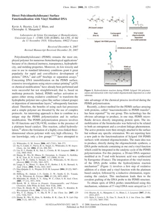
Figure 3. Analytical performances of biochips prepared with immobilized
(]) 5′-vinyl DNA probes and (0) unmodified DNA probes. Biotin labeled
DNA were incubated 60 min at 37 °C in buffer containing BSA 1% and
0.1% Tween 20. Error bars are the standard deviation of four experiments,
and curves are provided as a guide for the eyes.
The spos characteristics were previously described for
protein arrays.14 Particularly, their specific area was shown
to be highly dependent on the composition of the spotting
Figure 2. (a) Overview of the “macromolecules to PDMS transfer”. (b) solution. The use of carbonate buffer (Na2CO3, 0.1 M, pH
Schematic representation of the chemiluminescent labeling of DNA probe/ 9) instead of water as spotting carrier was shown to increase
target hybridization. (c) Chemiluminescent image of a hybridized 5′-vinyl the specific area of the spot and then lead to higher spot
DNA microarray.
signal. Indeed, the salt charged spotting solution crystallizes
nL drops) at the surface of a 3D Teflon master (Figure 2a). during the drying step, leading to highly textured surfaces.
Liquid PDMS was then poured onto the Teflon substrate, Thus, during the PDMS pouring and drying steps, these
covering the 5′-vinyl DNA spots, and cured at 90 °C for 20 surfaces were used as masters to produce PDMS replica
min. Peeling off the polymer generates a PDMS microarray entrapping the biomolecules and having a high specific area.
exhibiting spots of orientated DNA probe at the PDMS-air A similar effect was experienced for the present 5′-vinyl
interface. The ability of these immobilized probes to be DNA spots (AFM images shown in the Supporting Informa-
hybridized with complementary oligonucleotide was studied tion). A drastic 940% increase of the spot chemiluminescent
using a chemiluminescent labeling of a biotinylated target signal, related to this increased specific area, was recorded
sequence (Figure 2b). A special arraying pattern was easily when using carbonate buffer instead of water as a spotting
produced for this purpose by spotting our institute logo carrier.
ICBMS with 5′-vinyl DNA (25 µM) out of a 625 spots To demonstrate the importance of the vinyl residue in the
matrix. The chemiluminescent image obtained after hybrid- actual DNA immobilization reaction, unmodified DNA
ization with a complementary biotinylated target sequence sequence probes were immobilized using the “macromol-
(1 nM), and its labeling using peroxidase-streptavidin ecules to PDMS transfer”. After hybridization with the
conjugate is presented in Figure 2c. An intense and specific corresponding target sequence, a specific signal was obtained,
signal was obtained from the interactions between the demonstrating the possibility of trapping accessible unmodi-
immobilized 5′-vinyl DNA probe and the target sequence, fied DNA strand during the PDMS polymerization and
while no detectable signal was obtained from the nonspecific underlining the importance of the interactions between DNA
interactions with the bare PDMS surface. and PDMS, mainly due to Van der Waals interactions and
As a control experiment, 5′-modified DNA molecules were hydrophobic effects.16
spotted directly onto already polymerized PDMS and hybrid- Nevertheless, as can be seen in Figure 3, the analytical
ized with complementary probes. No measurable signal was performances of the biochips prepared with either the 5′-
obtained, suggesting the importance of the interactions vinyl DNA or the unmodified DNA were really dissimilar.
between PDMS and biomolecules during the polymerization The vinyl based biochip exhibits a broad detection range from
process. Obtaining such a PDMS DNA array with a so 0.2 pM (3.75 mol in 25 µL) to 1 nM with a detection limit
straightforward protocol was only possible because of the three decades lower than using the biochip prepared with
use on the one hand of the “Macromolecules to PDMS unmodified DNA. This difference of hybridization ability is
transfer” technique and on the other hand of 5′-vinyl DNA believed to be related to (i) the high reactivity of the vinyl
probes. Indeed, most of the current technologies for DNA modification toward PDMS chains during polymerization and
based biochips rely on different separated steps for the
preparation of the chip which are cost intensive and time- (16) Quist, A. P.; Pavlovic, E.; Oscarsson, S. Anal. Bioanal. Chem. 2005,
consuming. 381 (3), 591–600.](https://image.slidesharecdn.com/directpolydimethylsiloxanesurface-12565561346415-phpapp01/85/Direct-Poly-Dimethylsiloxane-Surface-2-320.jpg)