2. B Langmuir Heyries et al.
depositions.19,20 Photoinduced polymer grafting21 or photografting
polymer using benzophenone22 were also used to create an
intermediate layer on PDMS surfaces.
All of these methods suffer from several drawbacks which for
the most critical are the chemical instability of the surface
modification obtained and the lack of a simple process for the
immobilization of biomolecules. Developing such direct modi-
fication of PDMS surfaces with biomolecules, in a microarray
format, has been the main objective of our group. Thus, a method
was proposed for the direct entrapment in PDMS of micro (120-1
µm)23,24 and nano (330-50 nm)25,26 beads, bearing biological
molecules such as enzymes, antibodies, oligonucleotides, and
peptides. The beads were then spotted and dried on a 3D master,
covered with Sylgard 184, cured, and recovered, after peeling
off, as spots of beads entrapped at the surface of the bare PDMS.
We propose herein to push forward this methodology to
demonstrate the possibility of functionalizing the PDMS surface
by direct entrapment of biomolecules. Thus, the present work
will demonstrate the surface incorporation, during the PDMS
curing, of molecules as small as 3000 Da (dextran polymer) or
as fragile as proteins (antibodies).
The mechanism of this immobilization will be studied, and a
model will be proposed based on experimental evidence.
Morphological studies through atomic force microscopy of the
spots obtained in different conditions will also be proposed and
discussed according to the analytical signal measured.
Experimental Section
Materials. Arachis hypogaea lectin (from peanut), anti-Arachis
hypogaea lectin antibodies developed in rabbit, human IgG,
luminol (3-aminophthalhydrazide), and peroxidase-labeled strepta-
vidin were purchased from Sigma (France). Peroxidase-labeled
polyclonal anti-human Ig(G, A, M) antibodies developed in goat
and peroxidase-labeled polyclonal anti-rabbit IgG(H+L) anti-
bodies developed in mouse were supplied by Jackson Immuno-
Research (USA). Biotin-labeled dextran (3 and 500 kDa, lysine
fixable) and biotin-labeled dextran (3 kDa) were obtained from
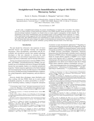
Molecular Probes (The Netherlands). Immunoglobulins from Figure 1. Overview of the technique highlighting the four main
rabbit serum (rabbit IgG) were obtained from Life Line Lab steps leading to the achievement of protein spots directly entrapped
(Pomezia, Italy). The PDMS precursor and curing agent (Sylgard at the PDMS interface.
184) were supplied by Dow Corning (France). All buffers and
aqueous solutions were made with distilled, demineralized water. Biochip Preparation (Figure 1). The biochips were prepared
by arraying 1.3 nL drops of spotting solutions with a BioChip
(18) Yang, T.; Baryshnikova, O. K.; Mao, H.; Holden, M. A.; Cremer, P. S. Arrayer BCA1 (Perkin-Elmer). Spotting solutions were prepared,
Investigations of Bivalent Antibody Binding on Fluid-Supported Phospholipid
Membranes: The Effect of Hapten Density. J. Am. Chem. Soc. 2003, 125 (16),
when not mentioned, in carbonate buffer 0.1 M pH 9. Proteins
4779-4784. (human IgG, rabbit IgG, and peanut lectin) spotting solutions
(19) Makamba, H.; Hsieh, Y.-Y.; Sung, W.-C.; Chen, S.-H. Stable Permanently were prepared at 1 mg/mL. Biotin modified dextran spotting
Hydrophilic Protein-Resistant Thin-Film Coatings on Poly(dimethylsiloxane)
Substrates by Electrostatic Self-Assembly and Chemical Cross-Linking. Anal. solutions were prepared to contain a constant concentration of
Chem. 2005, 77, 3971-3978. biotin of 232 µmol/L. Each array was composed of 16 spots
(20) Liu, Y.; Fanguy, J. C.; Bledsoe, J. M.; Henry, C. S. Dynamic Coating (identical or not, diameter ) 150 µm) that were deposited on the
Using Polyelectrolyte Multilayers for Chemical Control of Electroosmotic Flow
in Capillary Electrophoresis Microchips. Anal. Chem. 2000, 72, 5939-5944. surface of a 3D Teflon master composed of 24 rectangular
(21) Goda, T.; Konno, T.; Takai, M.; Moro, T.; Ishihara, K. Biomimetic structures (w ) 5 mm, l ) 5 mm, h ) 1 mm). Teflon was chosen
phosphorylcholine polymer grafting from polydimethylsiloxane surface using
photo-induced polymerization. Biomaterials 2006, 27 (30), 5151-5160.
as the deposition material according to its hydrophobicity and
(22) Wang, Y.; Lai, H.-H.; Bachman, M.; Sims, C. E.; Li, G. P.; Allbritton, its convenience for 3D micromachining. After spotting, the drops
N. L. Covalent Micropatterning of Poly(dimethylsiloxane) by Photografting through were dried, and the arrays were transferred to the PDMS interface,
a Mask. Anal. Chem. 2005, 77, 7539-7546.
(23) Marquette, C. A.; Blum, L. J. Direct immobilization in poly(dimethyl- by pouring a mixture of precursor and curing agent (10:1) onto
siloxane) for DNA, protein and enzyme fluidic biochips. Anal. Chim. Acta 2004, the Teflon substrate and curing for 20 min at 90 °C. Peeling off
506 (2), 127-132. the PDMS polymer then terminated the biochip preparation. Prior
(24) Marquette, C. A.; Blum, L. J. Conducting elastomer surface texturing:
a path to electrode spotting: Application to the biochip production. Biosens. to any further use, the biochips were saturated with VBSTA
Bioelectron. 2004, 20 (2), 197-203. (Veronal buffer 30 mM, NaCl 0.2 M, pH 8.5 with addition of
(25) Marquette, C. A.; Degiuli, A.; Imbert-Laurenceau, E.; Mallet, F.; Chaix,
C.; Mandrand, B.; Blum, L. J. Latex bead immobilisation in PDMS matrix for
tween 20 0.1% v/v and BSA 1% w/v) for 20 min at 37 °C.
the detection of p53 gene point mutation and anti-HIV-1 capsid protein antibodies. Immobilized Molecules Detection. The immobilized mol-
Anal. Bioanal. Chem. 2005, 381 (5), 1019-1024. ecules (i.e., biotinylated dextran, Arachis hypogaea lectin, rabbit
(26) Marquette, C. A.; Cretich, M.; Blum, L. J.; Chiari, M. Protein microarrays
enhanced performance using nanobeads arraying and polymer coating. Talanta IgG, or human IgG) were detected through chemiluminescent
2006, in press, corrected proof. labeling using peroxidase-labeled streptavidine, anti-lectin,
3. Protein Immobilization on Sylgard 184 Langmuir C
Table 1. Analytical Characteristics of Rabbit IgG Microarrays
Prepared Using Different Immobilization Procedures
immobilization signala (SD) LOD,b
method ng/mL
latex 1 µm26 20420 a.u. (9.4%) 100
silica 330 nm26 8748 a.u. (13.5%) 50
direct entrapment 20240 a.u. (8.2%) 10
of 10-15 nm (Supporting Information 1)
proteins
a
Calculated from three microarrays incubated with 1 µg/mL of anti-
rabbit IgG. b Limit of detection (LOD) calculated for a signal-to-noise
ratio of 3.
peroxidase-labeled anti-rabbit, or anti-human IgG, respectively.
The different labeled proteins were incubated (20 µL) on the
saturated microarrays for 1 h at 37 °C and the excess reagents
washed out with a 20 min incubation in VBS (Veronal buffer
Figure 2. Proposed mechanisms for the interactions between protein
30 mM, NaCl 0.2 M, pH 8.5). and PDMS during the elastomer curing step.
The microarrays were then placed in the CCD camera’s (Las-
1000 Plus, Intelligent Dark Box II, FUJIFILM) measurement such as human IgG/anti-human IgG and peanut allergen (Arachis
chamber for light integration for 10 min (measuring solution: hypogaea lectin)/anti-allergen were studied. In every case, a very
VBS containing in addition 220 µM of luminol, 200 µM of good recognition of the immobilized protein was experienced,
p-iodophenol and 500 µM of hydrogen peroxide). The numeric with no possibility of removing the immobilized entities, even
micrographs obtained were quantified with a FUJIFILM image in very harsh conditions. Indeed, proteins/PDMS microarrays
analysis program (Image Gauge 3.122). were submitted to a vigorous washing under stirring in 100 mL
of VBSTA buffer for 18 h. Protein spots morphologies before
Results
and after immersion were compared using optical microscopy,
The immobilization of biomolecules and particularly proteins and no major change was noticed. Moreover, the variation of the
has been one of the major targets of our group for the last 15 immobilized protein reactivity before and after immersion was
years.27 The last 3 years have been particularly devoted to the found to be 10%. The proteins were then firmly immobilized at
development of innovative immobilization methods, compatible the PDMS interface.
with the spotting technology widely used for microarrays. Thus, The effect of the polymerization process (drying and heating
technological solutions for spotting beads bearing protein were 20 min at 90 °C), which submits proteins to denaturing conditions,
proposed based on the entrapment of those beads at the PDMS/ was investigated. Indeed, these uncommon conditions are not
air interface.23-26 In the present work, we highlight an interesting supposed to be compatible with the proteins used for immu-
phenomenon leading to the transfer to the elastomer/air interface nodetection. However, protein drying for immunoassay develop-
of proteins not preimmobilized on carrier beads. Table 1 ments has been already extensively used, particularly within the
summarizes the results obtained with previously described micro-contact printing field,32,33 demonstrating the protein
microarrays prepared with 1 µm latex beads bearing rabbit IgG stability following such treatment.34 To evaluate the effect of the
or 330 nm silica beads bearing rabbit IgG and with the actual curing step (90 °C) on the integrity of the dried proteins,
free rabbit IgG system. The three different microarrays were microarrays were prepared by polymerizing PDMS at room
prepared using a similar protocol (i.e., spotting, drying, molding temperature (25 ( 2°C) onto protein spots for 48 h. The analytical
of PDMS, curing, and peeling off) and incubated with peroxidase- performances of these microarrays were found to compare well
labeled anti-rabbit IgG antibodies. Surprisingly, the direct with the ones prepared at 90 °C, evidencing the low effect of the
entrapment was found to be more effective than the beads-based elevated temperature on the subsequent immobilized antigen-
format previously developed. antibody recognition.
These results suggest that lowering the size of the entrapped
Different mechanisms could be involved in the direct im-
entity, from a 1 µm bead to 10-15 nm immunoglobulin protein,28,29
mobilization of accessible proteins during the PDMS curing
does not fail the immobilization of accessible rabbit IgGs. The
(Figure 2). First a molding effect, comparable to the key/lock
analytical signal obtained is then really convincing with high
couples observed within the molecular imprinting research
chemiluminescent intensities obtained with a relatively low SD
field.35-37 Then, hydrophobic interactions, as shown by Bartzoka
value, giving the best limit of detection (LOD) of the three systems.
Since rabbit IgG/anti-rabbit IgG are model proteins with well-
(32) Bernard, A.; Delamarche, E.; Schmid, H.; Michel, B.; Bosshard, H. R.;
known high affinity and stability,30,31 weaker recognition systems Biebuyck, H. Printing Patterns of Proteins. Langmuir 1998, 14 (9), 2225-2229.
(33) Arjan, P. Q.; Elisabeth, P.; Sven, O. Recent advances in microcontact
(27) Blum, L. J.; Coulet, P. R. Biosensor Principles and Applications; Marcel printing. Anal. Bioanal. Chem. 2005, 381 (3), 591-600.
Dekker: New York, 1991; p 357. (34) LaGraff, J. R.; Chu-LaGraff, Q. Scanning force microscopy and
(28) Godoy, S.; Chauvet, J. P.; Boullanger, P.; Blum, L. J.; Girard-Egrot, A. fluorescence microscopy of microcontact printed antibodies and antibody
P. New Functional Proteo-glycolipidic Molecular Assembly for Biocatalysis fragments. Langmuir 2006, 22 (10), 4685-4693.
Analysis of an Immobilized Enzyme in a Biomimetic Nanostructure. Langmuir (35) Turner, N. W.; Jeans, C. W.; Brain, K. R.; Allender, C. J.; Hlady, V.; Britt,
2003, 19 (13), 5448-5456. D. W. From 3D to 2D: A Review of the Molecular Imprinting of Proteins.
(29) Godoy, S.; Violot, S.; Boullanger, P.; Bouchu, M.-N.; Leca-Bouvier, B. Biotechnol. Prog. 2006, in press.
D.; Blum, L. J.; Girard-Egrot, A. P. Kinetics Study of Bungarus fasciatus Venom (36) Alexander, C.; Andersson, H. S.; Andersson, L. I.; Ansell, R. J.; Kirsch,
Acetylcholinesterase Immobilised on a Langmuir-Blodgett Proteo-Glycolipidic N.; Nicholls, I. A.; O’Mahony, J.; Whitcombe, M. J. Molecular imprinting science
Bilayer. ChemBioChem 2005, 6 (2), 395-404. and technology: a survey of the literature for the years up to and including 2003.
(30) Deshpande, S. S. Enzyme Immunoassays, Chapman & Hall ed.; ITP: J. Mol. Recognit. 2006, 19 (2), 106-180.
New York, 1996; p 464. (37) Marty, J. D.; Mauzac, M. Molecular imprinting: State of the art and
(31) The Immunoassay Handbook, 3rd ed.; Elsevier: The Netherlands, 2005; perspectives. Microlithography - Molecular Imprinting; Springer: Berlin, 2005;
p 930. Vol. 172, pp 1-35.
4. D Langmuir Heyries et al.
Figure 3. Effect of different amino acids on the chemiluminescent
signals obtained from microarrays composed of directly immobilized
lysine-modified biotinylated dextran. The immobilized dextran was
detected through peroxidase-labeled streptavidin 1 µg/mL (30 min,
37 °C).
Figure 4. Atomic force microscopy (NT-MDT, tapping mode)
and co-worker,38,39 could also be involved between proteins and images of spots of lysine modified biotinylated dextran obtained in
water (A) or in 0.1 M carbonate buffer, pH 9 (B). The arrows indicate
uncured PDMS. Finally, covalent bindings between the protein the edge of each spot. The straight lines correspond to the presented
and the polymer could occur while PDMS is curing, mainly profiles.
through poisoning of the Kardstedt catalyst40-42 by the amino
or thiol groups of the protein lateral chains (as lone pair electron have little effect on the immobilization of the dextran molecules,
donors; Supporting Information 2). even at high concentration (50 mM). On the contrary, lysine and
Dextran chains bearing biotin and lysine residues were chosen cysteine were shown to inhibit strongly and with a dose
as model molecules to study this direct entrapment. The presence dependence relation the immobilization of the biotinylated
of the accessible immobilized molecule was then evidenced using polymer. These results are in agreement with the involvement
peroxidase labeled streptavidine and chemiluminescent imaging. of a poisoning of the Kardstedt catalyst in the immobilization
500 kDa and 3 kDa dextran chains were thus successfully process.
immobilized at the PDMS/air interface. The very large size
A drastic effect of the cysteine, which is known as a very
difference between the two polymers did not appear to critically
efficient Kardstedt catalyst poison,43 was observed. Indeed, 78%
influence the immobilization efficiency, proving that molecules
and 40% of the immobilization of the 3 kDa dextran was inhibited
as small as 3000 Da could be trapped and accessible at the
by the presence of the maximum concentration of cysteine and
elastomer surface.
lysine, respectively. The 60% of remaining immobilization in
Within the dextran chains used, only the amino group of the
the presence of 50 mM lysine could then be attributed to the
lysine lateral chains could be involved in a chemical reaction
others proposed mechanisms (i.e., molding effect and hydrophobic
with the Kardstedt catalyst during the Sylgard 184 curing. As
interactions). Regarding the poor hydrophobicity of the dextran
a control experiment, dextran chains not bearing any lysine
backbone, hydrophobic interactions were believed to be minimum.
residues were spotted and transferred to the elastomer. The
Potential interactions through the carbohydrate moiety of the
chemiluminescent signal obtained was then 45% of the initial
lysine-dextran were then also considered. Thus, immobilization
signal, demonstrating the implication of the amino group in the
inhibition tests were performed by spotting dextran in the presence
immobilization process but also evidencing the molding effect
of maltose (a subunit of dextran). No effect on the lysine-
implicated in 45% of the immobilization efficiency.
dextran immobilization was observed for maltose concentrations
Further studies were performed to complete this theoretical
up to 100 mM.
immobilization mechanism. 3 kD dextran molecules bearing
According to the results presented above, two mechanisms
lysine residues were spotted in the presence of different
appeared to be preponderant in the actual lysine-dextran
concentrations of free amino acids: glycine, lysine, and cysteine
immobilization on PDMS: chemical bounding through poisoning
(Figure 3). Glycine, with only its R-amino group, was found to
of the Kardstedt catalyst by the primary amine of the lysine
(38) Bartzoka, V.; Brook, M. A.; McDermott, M. R. Silicone-Protein Films: residue and a molding effect, similar to a key/lock mechanism.
Establishing the Strength of the Protein-Silicone Interaction. Langmuir 1998, 14 Our previous works on bead assisted protein immobilization
(7), 1892-1898.
(39) Bartzoka, V.; Brook, M. A.; McDermott, M. R. Protein-Silicone on microarrays25,26 have demonstrated the usefulness of increasing
Interactions: How Compatible Are the Two Species? Langmuir 1998, 14 (7), the specific area of the spots. Indeed, increasing this area while
1887-1891. keeping constant the geometrical one enables the immobilization
(40) Perutz, S.; Kramer, E. J.; Baney, J.; Hui, C. Y.; Cohen, C. Investigation
of adhesion hysteresis in poly(dimethylsiloxane) networks using the JKR technique. of a higher amount of proteins per spot and then the increase of
J. Polym. Sci. Part B: Polym. Phys. 1998, 36 (12), 2129-2139. the microarray performances. Herein, since the proteins are spotted
(41) Quirk, R. P.; Kim, H.; Polce, M. J.; Wesdemiotis, C. Anionic Synthesis
of Primary Amine Functionalized Polystyrenes via Hydrosilation of Allylamines
and transferred directly to the PDMS interface, an original way
with Silyl Hydride Functionalized Polystyrenes. Macromolecules 2005, 38, 7895- to increase this specific area has been to use spot solutions with
7906. relatively high salt concentration. Indeed, the salt charged protein
(42) Katsuhiko Kishi, T. I.; Ozono, M.; Tomita, I.: Endo, T. Development and
application of a latent hydrosilylation catalyst. IX. Control of the catalytic activity
of a platinum catalyst by polymers bearing amine moieties. J. Polym. Sci. Part (43) Marian, M.; Winter, H. H. Relaxation patterns of endlinking polydim-
A: Polym. Chem. 2000, 38 (5), 804-809. ethylsiloxane near the gel point. Polym. Bull. 1998, 40 (2), 267-274.
5. Protein Immobilization on Sylgard 184 PAGE EST: 4.8 Langmuir E
a concentration of 1 mg/mL in carbonate buffer (pH 9) and
transferred to the PDMS interface. The spotting pattern appeared
on the upper part of Figure 5. Arachis hypogaea lectin, rabbit
IgG, and human IgG were spotted as eight replicas. Bovine serum
albumin (BSA) was used as a negative control for all of the
tested antibodies (i.e., anti-rabbit IgG, anti-human IgG, and anti-
lectin). As can be seen (Figure 5), absolutely no nonspecific
signal was detected outside of the area delimited by the specific
protein spots. Classical dose response curves can be observed
(Supporting Information 1) from each range of antibody
concentrations tested, giving detection limits of 20 ng/mL for
anti-rabbit IgG and anti-human IgG and 10 ng/mL for anti-
lectin. These concentrations correspond, in the 20 µL incubation
volume, to amounts of antibody in the fmol range (2.6 and 1.3
fmol), which are considered low enough for most of the major
immunoassay applications.31,44
Conclusion
In summary, we have developed a new approach to directly
modify Sylgard 184 surfaces with spots of protein and dem-
onstrated its use for microarray-based immunoassays. The
mechanisms of this direct protein immobilization have been
investigated, and clear experimental results suggested that both
chemical bonding and molding effects were implicated in the
observed phenomenon. Chemical bonding between the protein
and the PDMS elastomer structure was believed to be mainly
due to a poisoning of the Kardstedt catalyst used during the
polymer curing. This poisoning was demonstrated to be related,
with reference to previous works,38,40,41 to the presence of free
Figure 5. Spotting pattern and chemiluminescent image of the
microarray prepared by spotting Arachis hypogaea lectin, rabbit amino or thiol groups in the immobilized molecules. Moreover,
IgG, and human IgG in 0.1M carbonate buffer (pH 9). one interesting point is that Si-H functions have been reported
to be sensitive to hydrolysis45 and, regarding the influence of
solutions crystallize during the drying step, leading to highly nucleophilic groups such as primary amino groups, it could be
textured surfaces. Thus, during the PDMS pouring and drying postulated that interactions could also occurr between protein
steps, these surfaces were used as a master to produce PDMS lateral chains and Si-H functions.
replica entrapping proteins, having a high specific area. Future work includes the integration of such microarrays in
Two examples of this technique are illustrated by the AFM microfluidic systems, thanks to the use of PDMS as immobiliza-
images of protein spots obtained in pure water and in the presence tion support.
of carbonate buffer 0.1 M (Figure 4). The two spots obviously
exhibit very different surfaces as evidenced by the spot profiles.
Acknowledgment. Published with the support of the European
Calculated from the AFM images, the specific area increasing Commission, Sixth Framework Program, Information Society
between both spots was found to be 1 order of magnitude. This Technologies. NANOSPAD (No. 016610).
difference of the surface geometry has a direct repercussion on
the chemiluminescent signal obtained from those spots. Indeed,
Supporting Information Available: Dose response curves and
a more than 200% increase of the signal was observed when the kardstedt catalyst reaction cycle. This material is available free of
carbonate was added to the spotting solution, and this was charge via the Internet at http://pubs.acs.org.
irrespective of the buffer pH used (7, 9, and 11). This enhancement
of the signal is then not linked to an increase of the reactivity LA070018O
of the lysine chains amino group at high pH but to the actual
increase of the specific area of the spot. (44) Wu, A. H. B. A selected history and future of immunoassay development
and applications in clinical chemistry. Clin. Chim. Acta 2006, 369 (2), 119-124.
In order to fully characterize the analytical possibilities of the (45) Brook, M. A. Silicon in Organic, Organometallic, and Polymer Chemistry;
developed microarray, three different proteins were spotted at John Wiley & Sons: New York, 2000.