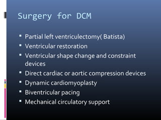
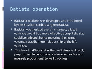
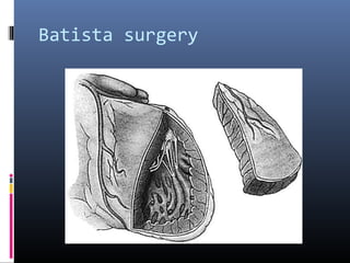
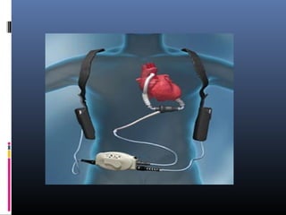
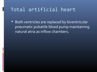
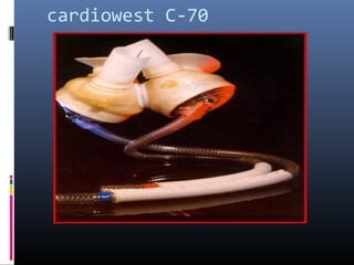
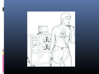
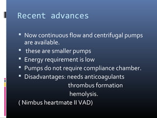
Surgery for DCM and RCM


        Dr Amjad Shaikh
Introduction

 Main treatment is for heart failure which is
  the end result of DCM and RCM.
 Most of the surgeries still are under
  development.
 Best treatment is heart transplant.
Surgery for DCM

 Partial left ventriculectomy( Batista)
 Ventricular restoration
 Ventricular shape change and constraint
    devices
   Direct cardiac or aortic compression devices
   Dynamic cardiomyoplasty
   Biventricular pacing
   Mechanical circulatory support
Batista operation

 Batista procedure, was developed and introduced
  by the Brazilian cardiac surgeon Batista.
 Batista hypothesized that an enlarged, dilated
  ventricle would be a more effective pump if the size
  could be reduced, hence restoring the normal
  volume/mass/diameter relationship of the left
  ventricle.
 The law of LaPlace states that wall stress is directly
  proportional to ventricular pressure and radius and
  inversely proportional to wall thickness.
 removing a triangular wedge of the lateral wall of the left
  ventricle, which typically weighs more than 100 g . The
  incision begins at the apex of the left ventricle and
  extends to the atrioventricular groove.
 Typically a posterolateral branch of the left coronary
  artery is removed with the excised specimen. Because of
  the change in geometry and juxtaposition of the papillary
  muscles, the mitral valve is repaired to ensure
  competency.
 Batista performs a mitral valve repair (Alfieri technique),
  in which the anterior and posterior leaflets are sutured
  resulting in a double-orifice mitral valve, which yields the
  characteristic figure-of-eight appearance when the mitral
  valve is viewed in the short-axis echocardiographic view
Batista surgery
2D Echo: figure of eight
 Advantages: it improves systolic function and
  hence cardiac output..

 Limitations:
  It removes functioning though weakened
  myocardium. It may actually decrease net
  ventricular pumping capacity by affecting
  diastolic compliance.
Ventricular restoration

 Anatomical basis:
 - heart is dual spiral helix( torrent – Gausp)
-configuration of muscle fibers at apex is figure
   of eight which provides mechanism for
   ventricular ejection and suction of filling.
Aim of surgery:
   convert spherical heart to normal elliptical
   heart
Ventricular shape change and
constraint devices
 These devices change left ventricular shape
  or to restrain ventricular dilatation of heart.
 McCarthy and schenk used myosplints:
    three of devices are placed perpandicular to
  long axis of left ventricle.
Chaudhary used prosthetic jacket of knitted
  polyster mesh: it prevents progressive left
  ventricular remodelling and abolished
  functional mitral valve regurgitation.
Direct cardiac or aortic
compression devices
 It helps failing heart by direct compression of heart and aorta.
 It avoids interaction between blood and foreign surface of assist
  device.
 Ease of application and ease of removal.


A: The cardio support system:
   it surrounds both ventricles to the AV groove
  - -200 mm Hg pressure for vaccume seal.
 - compression bladder inflated and deflated in synchrony with
    cardiac contraction.
 - short term use for cardiogenic shock.
 B: The heart booster:
  - multiple small parallel compression tubes
 covering both ventricular chembers.
 - hydraulic drive system fills and empties the
 tubes
 - still under development stage
 C: Kantrovitz CARDIOVAD( LVAD):


 Principle: diastolic augmentation like IABP.
- the pump is warn externally and provides the
  stroke volume of up to 60 ml via the tube
  through the skin.
Dynamic Cardiomyoplasty

 Basic: use of the skeletal muscle wrapped
    around heart which is stimulated electrically
    to augment or restore contractility of
    ventricles.
   Kantrovitz and McKinon used first in 1959.
   Latissimus dorsi muscle used.
   Electrodes are implanted in muscle and
    stimuled in synchronization with heart
   Late deaths are seen due to ventricular
    arrhythmias and fibrillation.
Mechanical circulatory support

 It is a means of imparting energy for forward flow of
  blood in the body by man made device.
 It can be
A: - temporary
     - interim
     - permanent
B: - internal (implantable)
    - external( partially implantable, paracrporeal)
Implantable or partialy
implantable
 A: Ventricular Assist Device:
  - VADs are connected to the failing heart in
   parallel.
 - it pumps all or part of stroke volume.
- It generally bypass the failing heart.
- It takes blood from atria in to the pump and
   return to great arteries beyond the ventricle.
- It provides pulsatile blood flow
Paracorporeal VADS

 Abiomed VAD
Thoratec VAD
 It is particularly useful in smaller adults or
  older children.
 Pumping ventricle is mounted on the
  abdomen and canula from ventricle are
  brought out of mediastinum as drainage
  tube.
 Rate is determined automatically.
 The pump is flow limited to about 5l/m.
Implantable VADS

 Thoratec heartmate VAD II used as bridge to
  heart transplant.
 It improves the chance for survival until the
  suitable donor can be located.
 Pump is driven pneumatically or electrically.
 Many newer modifications are available now.
Total artificial heart

 Both ventricles are replaced by biventricular
  pneumatic pulsatile blood pump maintaining
  natural atria as inflow chambers.
cardiowest C-70
Recent advances

 Now continuous flow and centrifugal pumps
   are available.
 these are smaller pumps
 Energy requirement is low
 Pumps do not require compliance chamber.
 Disadvantages: needs anticoagulants
                  thrombus formation
                   hemolysis.
( Nimbus heartmate II VAD)
Last resort….

  Heart transplant…
 Thank you….

Dilated cardiomyopathy

  • 1.
    Surgery for DCMand RCM Dr Amjad Shaikh
  • 2.
    Introduction  Main treatmentis for heart failure which is the end result of DCM and RCM.  Most of the surgeries still are under development.  Best treatment is heart transplant.
  • 3.
    Surgery for DCM Partial left ventriculectomy( Batista)  Ventricular restoration  Ventricular shape change and constraint devices  Direct cardiac or aortic compression devices  Dynamic cardiomyoplasty  Biventricular pacing  Mechanical circulatory support
  • 4.
    Batista operation  Batistaprocedure, was developed and introduced by the Brazilian cardiac surgeon Batista.  Batista hypothesized that an enlarged, dilated ventricle would be a more effective pump if the size could be reduced, hence restoring the normal volume/mass/diameter relationship of the left ventricle.  The law of LaPlace states that wall stress is directly proportional to ventricular pressure and radius and inversely proportional to wall thickness.
  • 5.
     removing atriangular wedge of the lateral wall of the left ventricle, which typically weighs more than 100 g . The incision begins at the apex of the left ventricle and extends to the atrioventricular groove.  Typically a posterolateral branch of the left coronary artery is removed with the excised specimen. Because of the change in geometry and juxtaposition of the papillary muscles, the mitral valve is repaired to ensure competency.  Batista performs a mitral valve repair (Alfieri technique), in which the anterior and posterior leaflets are sutured resulting in a double-orifice mitral valve, which yields the characteristic figure-of-eight appearance when the mitral valve is viewed in the short-axis echocardiographic view
  • 6.
  • 7.
  • 8.
     Advantages: itimproves systolic function and hence cardiac output..  Limitations: It removes functioning though weakened myocardium. It may actually decrease net ventricular pumping capacity by affecting diastolic compliance.
  • 9.
    Ventricular restoration  Anatomicalbasis: - heart is dual spiral helix( torrent – Gausp) -configuration of muscle fibers at apex is figure of eight which provides mechanism for ventricular ejection and suction of filling. Aim of surgery: convert spherical heart to normal elliptical heart
  • 10.
    Ventricular shape changeand constraint devices  These devices change left ventricular shape or to restrain ventricular dilatation of heart.  McCarthy and schenk used myosplints: three of devices are placed perpandicular to long axis of left ventricle. Chaudhary used prosthetic jacket of knitted polyster mesh: it prevents progressive left ventricular remodelling and abolished functional mitral valve regurgitation.
  • 11.
    Direct cardiac oraortic compression devices  It helps failing heart by direct compression of heart and aorta.  It avoids interaction between blood and foreign surface of assist device.  Ease of application and ease of removal. A: The cardio support system: it surrounds both ventricles to the AV groove - -200 mm Hg pressure for vaccume seal. - compression bladder inflated and deflated in synchrony with cardiac contraction. - short term use for cardiogenic shock.
  • 13.
     B: Theheart booster: - multiple small parallel compression tubes covering both ventricular chembers. - hydraulic drive system fills and empties the tubes - still under development stage
  • 16.
     C: KantrovitzCARDIOVAD( LVAD): Principle: diastolic augmentation like IABP. - the pump is warn externally and provides the stroke volume of up to 60 ml via the tube through the skin.
  • 18.
    Dynamic Cardiomyoplasty  Basic:use of the skeletal muscle wrapped around heart which is stimulated electrically to augment or restore contractility of ventricles.  Kantrovitz and McKinon used first in 1959.  Latissimus dorsi muscle used.  Electrodes are implanted in muscle and stimuled in synchronization with heart  Late deaths are seen due to ventricular arrhythmias and fibrillation.
  • 20.
    Mechanical circulatory support It is a means of imparting energy for forward flow of blood in the body by man made device.  It can be A: - temporary - interim - permanent B: - internal (implantable) - external( partially implantable, paracrporeal)
  • 21.
    Implantable or partialy implantable A: Ventricular Assist Device: - VADs are connected to the failing heart in parallel. - it pumps all or part of stroke volume. - It generally bypass the failing heart. - It takes blood from atria in to the pump and return to great arteries beyond the ventricle. - It provides pulsatile blood flow
  • 22.
  • 23.
  • 24.
     It isparticularly useful in smaller adults or older children.  Pumping ventricle is mounted on the abdomen and canula from ventricle are brought out of mediastinum as drainage tube.  Rate is determined automatically.  The pump is flow limited to about 5l/m.
  • 25.
    Implantable VADS  Thoratecheartmate VAD II used as bridge to heart transplant.  It improves the chance for survival until the suitable donor can be located.  Pump is driven pneumatically or electrically.  Many newer modifications are available now.
  • 27.
    Total artificial heart Both ventricles are replaced by biventricular pneumatic pulsatile blood pump maintaining natural atria as inflow chambers.
  • 28.
  • 30.
    Recent advances  Nowcontinuous flow and centrifugal pumps are available.  these are smaller pumps  Energy requirement is low  Pumps do not require compliance chamber.  Disadvantages: needs anticoagulants thrombus formation hemolysis. ( Nimbus heartmate II VAD)
  • 31.
    Last resort…. Heart transplant…
  • 32.