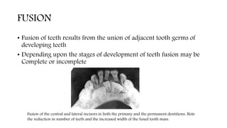
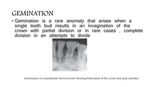
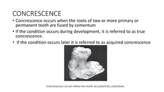
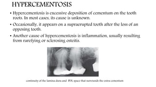
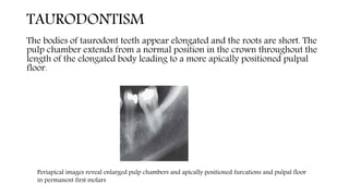
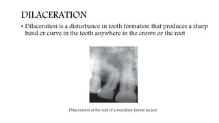
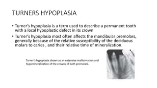
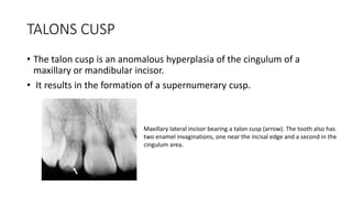
RADIOGRAPHIC
INTERPRETATIONS OF DENTAL
ANOMALIES AND IMPLANTS
CONTENTS
• INTRODUCTION
• INTERPRETATIONS
ANOMALIES
SYSTEMIC CONDITIONS IN RELATION TO PERIODONTITIS
IMPLANTS
PERIMPLATITIS
• CONCLUSION
INTRODUCTION
• The radiographic interpretation is used widely to identify the
presence or absence of disease , to provide information on the nature
and extent of disease and also for differential diagnosis
SUPERNUMERARY TEETH
Supernumerary Teeth shows Supplemental deciduous molar (arrow).
Supernumerary teeth are teeth that develop in addition to the normal
complement as a result of excess dental lamina in the jaws. The teeth that
develop may be morphologically normal or abnormal
MACRODONTIA
• In macrodontia, the teeth are larger than normal. Macrodontia rarely affects the
entire dentition. Often a single tooth, individual contralateral teeth, or a group of
teeth maybe involved
Panoramic image shows the greater mesial/distal widths of the tooth
crowns compared with their respective first premolars.
MICRODONTIA
In microdontia, the teeth are smaller than normal. As with macrodontia,
microdontia may involve all the teeth or be limited to a single tooth or group of
teeth. Often the lateral incisors and third molars may be small
Peg-shaped deformity in microdontia of a maxillary lateral incisor.
FUSION
• Fusion of teeth results from the union of adjacent tooth germs of
developing teeth
• Depending upon the stages of development of teeth fusion may be
Complete or incomplete
Fusion of the central and lateral incisors in both the primary and the permanent dentitions. Note
the reduction in number of teeth and the increased width of the fused tooth mass.
GEMINATION
• Gemination is a rare anomaly that arises when a
single tooth bud results in an invagination of the
crown with partial division or in rare cases , complete
division in an attempts to divide
Gemination of a mandibular lateral incisor showing bifurcation of the crown and pulp chamber
CONCRESCENCE
• Concrescence occurs when the roots of two or more primary or
permanent teeth are fused by cementum
• If the condition occurs during development, it is referred to as true
concrescence.
• If the condition occurs later it is referred to as acquired concrescence
Concrescence occurs when two teeth are joined by cementum.
HYPERCEMENTOSIS
• Hypercementosis is excessive deposition of cementum on the tooth
roots. In most cases, its cause is unknown.
• Occasionally, it appears on a supraerupted tooth after the loss of an
opposing tooth.
• Another cause of hypercementosis is inflammation, usually resulting
from rarefying or sclerosing osteitis.
continuity of the lamina dura and PDL space that surrounds the extra cementum
TAURODONTISM
The bodies of taurodont teeth appear elongated and the roots are short. The
pulp chamber extends from a normal position in the crown throughout the
length of the elongated body leading to a more apically positioned pulpal
floor.
Periapical images reveal enlarged pulp chambers and apically positioned furcations and pulpal floor
in permanent first molars
DILACERATION
• Dilaceration is a disturbance in tooth formation that produces a sharp
bend or curve in the tooth anywhere in the crown or the root
Dilaceration of the root of a maxillary lateral incisor
DENS INVAGINATUS, DENS IN DENTE, AND DILATED ODONTOME
• The three entities dens invaginatus, dens in dente, and dilated odontome all
result from varying degrees of invagination or infolding of the enamel surface
into the interior of a tooth.
• The least severe form of this infolding is dens invaginatus and the most severe
form is dilated odontome.
Radiopaque, inverted teardrop outline of dens invaginatus in seen in a maxillary lateral incisor.
the position of the invagination in seen in the cingulum area of the tooth crown
DENS EVAGINATUS
Dens evaginatus appears as a tubercle of enamel on the occlusal surface of
the affected tooth . Dens evaginatus may occur bilaterally and usually in the
mandible.
The tubercle often has a dentin core and a very slender pulp horn frequently
extends into the evagination
Occlusal tubercle of dens evaginatus as seen in a mandibular premolar.
AMELOGENESIS IMPERFECTA
Amelogenesis imperfecta is a genetic anomaly arising from mutations
that may have occurred in one or more of four candidates
The imaging signs of amelogenesis imperfecta include a square crown,
a relatively thin radiopaque layer of enamel, low or absent cusps, and
multiple open contacts between the teeth. The anterior teeth are said
to have a “picket fence”—type appearance
DENTINOGENESIS IMPERFECTA
• Dentinogenesis imperfecta is a genetic anomaly involving primarily
the dentin, although the enamel may be thinner than normal in this
condition.
Dentinogenesis imperfecta characteristically shows bulbous crowns, constriction
of tooth at the cementoenamel junction, short roots, and a reduced size of the pulp
chamber and root canals
DENTIN DYSPLASIA
• Dentin dysplasia is a genetically inherited autosomal dominant
abnormality that resembles dentinogenesis imperfecta.
• A slight bluish brown translucency is apparent. The teeth are often
misaligned in the arch
short, poorly developed roots, obliterated pulp chambers and root canals, and periapical rarefying
osteitis associated with dentin dysplasia. Half-moon or “demilune” shape of the pulp chambers.
REGIONAL ODONTODYSPLASIA
Regional odontodysplasia is a rare condition in which both enamel and
dentin are hypoplastic and hypocalcified
Teeth affected with regional odontodysplasia are small and mottled
brown as a result of staining of the hypocalcified and hypoplastic enamel
poor mineralization of all of the dental hard tissues is seen in regional odontodysplasia. lack of eruption
and hypoplasia of enamel and dentin expressed mainly as short roots.
TURNERS HYPOPLASIA
• Turner’s hypoplasia is a term used to describe a permanent tooth
with a local hypoplastic defect in its crown
• Turner’s hypoplasia most often affects the mandibular premolars,
generally because of the relative susceptibility of the deciduous
molars to caries , and their relative time of mineralization.
Turner’s hypoplasia shown as an extensive malformation and
hypomineralization of the crowns of both premolars.
TALONS CUSP
• The talon cusp is an anomalous hyperplasia of the cingulum of a
maxillary or mandibular incisor.
• It results in the formation of a supernumerary cusp.
Maxillary lateral incisor bearing a talon cusp (arrow). The tooth also has
two enamel invaginations, one near the incisal edge and a second in the
cingulum area.
HYPOPHOSPHATASIA
• Hypophosphatasia is a rare inherited disorder that is caused by a
reduced activity of alkaline phosphatase, an enzyme that is produced
by osteoblasts and odontoblasts which is required for the normal
mineralization of osteoid and teeth
• Both primary and permanent teeth have a thin enamel layer and large
pulp chambers and root canals
• The teeth may also be hypoplastic and may be lost prematurely
large pulp chambers in the deciduous dentition and the
premature loss of the mandibular incisors.
HYPERTHYROIDISM
• Hyperthyroidism is a syndrome that involves excessive production of
thyroxine in the thyroid gland
• results in an increased rate of bone turnover causes excessive bone
resorption results in decrease in bone density or loss of some areas of
edentulous alveolar bone
• Advanced development of teeth and early eruption with premature
loss of primary teeth
PAPILLION LEFEVRE SYNDROME
• In localized form vertical alveolar bone loss is seen around the
incisors and first molars
• In generalized form bone loss may range from involvement of
two teeth to a maximum number of teeth
• In advanced cases severe alveolar bone loss and teeth appear to
float in air
PLS is an autosomal recessive disorder caused by mutations in the cathepsin C gene
located on chromosome 11q14
An increase in tooth mobility and periodontal abscesses is frequently observed
after the eruption of permanent teeth.
CHEDIAK – HIGASHI SYNDROME
Chediak-Higashi syndrome (CHS) is a rare autosomal recessive genetic
disease.
Clinical image shows tooth malpositioning , generalized swelling of gingival
margins, loss of interdental papillae, and heavy accumulations of dental
plaque
Radiographic examination showed generalized advanced
alveolar bone loss reaching the apical third of most teeth
Several teeth appeared to have no remaining bone support.
DIABETES MELLITUS
• Diabetes mellitus is a metabolic disorder that has two primary forms.
• Type 1, insulin-dependent diabetes mellitus ,results from an absence
or insufficiency of insulin.
• Type 2, non–insulin dependent diabetes mellitus, results from insulin
resistance.
HYPERPARATHYROIDISM
Excessive secretion of parathyroid hormone due to any cause is termed as
hyperparathyroidism
In hyperparathyroidism there will be a radiographic evidence of alveolar
osteoporosis with closely meshed trabeculae ,widening of periodontal space
absence of lamina dura and radiolucent cyst like spaces
PRESURGICAL IMPLANT IMAGING
• 1. Determine bone height(7-9mm) and width(5mm) (bone dimensions) using
CBCT
• 2. Determine bone quality with comparative density analysis in 3D.
• 3. Determine the long axis of alveolar bone.
• 4. Identify and localize internal anatomies, such as nerves and sinus cavities.
• 5. Determine jaw boundaries.
• 6. Identify pathology
• 7. Transfer of radiographic planning information.
• 8. Communicate radiographic diagnostic and planning information
POST IMPLANT IMAGING
• Determine the long term maintenance of implant rigid fixation and
function including crestal bone levels
• To evaluate implant complex
• Assess the bone adjacent to dental implants
RADIOGRAPHIC CLASSIFICATION OF
PERIMPLANT
• Mild lesion (class 1) is characterized by radiographic bone
loss that extends to <25% of implant length from the
implant apex
• Moderate lesions (class2) is characterized by radiographic
bone loss between 25 to 50%of the implant length as
measured from implant apex
• Advanced lesion(class3) is characterized by radiographic
bone loss extending to >50% of the implant length from
implant apex
CONCLUSION
• Successful interpretation of radiograph depends ultimately on
clinician understanding the radiographic image its normal appearance
as well as knowing the salient features of pathological conditions

radiographic interpretations.pptx

  • 1.
  • 2.
    CONTENTS • INTRODUCTION • INTERPRETATIONS ANOMALIES SYSTEMICCONDITIONS IN RELATION TO PERIODONTITIS IMPLANTS PERIMPLATITIS • CONCLUSION
  • 3.
    INTRODUCTION • The radiographicinterpretation is used widely to identify the presence or absence of disease , to provide information on the nature and extent of disease and also for differential diagnosis
  • 4.
    SUPERNUMERARY TEETH Supernumerary Teethshows Supplemental deciduous molar (arrow). Supernumerary teeth are teeth that develop in addition to the normal complement as a result of excess dental lamina in the jaws. The teeth that develop may be morphologically normal or abnormal
  • 5.
    MACRODONTIA • In macrodontia,the teeth are larger than normal. Macrodontia rarely affects the entire dentition. Often a single tooth, individual contralateral teeth, or a group of teeth maybe involved Panoramic image shows the greater mesial/distal widths of the tooth crowns compared with their respective first premolars.
  • 6.
    MICRODONTIA In microdontia, theteeth are smaller than normal. As with macrodontia, microdontia may involve all the teeth or be limited to a single tooth or group of teeth. Often the lateral incisors and third molars may be small Peg-shaped deformity in microdontia of a maxillary lateral incisor.
  • 7.
    FUSION • Fusion ofteeth results from the union of adjacent tooth germs of developing teeth • Depending upon the stages of development of teeth fusion may be Complete or incomplete Fusion of the central and lateral incisors in both the primary and the permanent dentitions. Note the reduction in number of teeth and the increased width of the fused tooth mass.
  • 8.
    GEMINATION • Gemination isa rare anomaly that arises when a single tooth bud results in an invagination of the crown with partial division or in rare cases , complete division in an attempts to divide Gemination of a mandibular lateral incisor showing bifurcation of the crown and pulp chamber
  • 9.
    CONCRESCENCE • Concrescence occurswhen the roots of two or more primary or permanent teeth are fused by cementum • If the condition occurs during development, it is referred to as true concrescence. • If the condition occurs later it is referred to as acquired concrescence Concrescence occurs when two teeth are joined by cementum.
  • 10.
    HYPERCEMENTOSIS • Hypercementosis isexcessive deposition of cementum on the tooth roots. In most cases, its cause is unknown. • Occasionally, it appears on a supraerupted tooth after the loss of an opposing tooth. • Another cause of hypercementosis is inflammation, usually resulting from rarefying or sclerosing osteitis. continuity of the lamina dura and PDL space that surrounds the extra cementum
  • 11.
    TAURODONTISM The bodies oftaurodont teeth appear elongated and the roots are short. The pulp chamber extends from a normal position in the crown throughout the length of the elongated body leading to a more apically positioned pulpal floor. Periapical images reveal enlarged pulp chambers and apically positioned furcations and pulpal floor in permanent first molars
  • 12.
    DILACERATION • Dilaceration isa disturbance in tooth formation that produces a sharp bend or curve in the tooth anywhere in the crown or the root Dilaceration of the root of a maxillary lateral incisor
  • 13.
    DENS INVAGINATUS, DENSIN DENTE, AND DILATED ODONTOME • The three entities dens invaginatus, dens in dente, and dilated odontome all result from varying degrees of invagination or infolding of the enamel surface into the interior of a tooth. • The least severe form of this infolding is dens invaginatus and the most severe form is dilated odontome. Radiopaque, inverted teardrop outline of dens invaginatus in seen in a maxillary lateral incisor. the position of the invagination in seen in the cingulum area of the tooth crown
  • 14.
    DENS EVAGINATUS Dens evaginatusappears as a tubercle of enamel on the occlusal surface of the affected tooth . Dens evaginatus may occur bilaterally and usually in the mandible. The tubercle often has a dentin core and a very slender pulp horn frequently extends into the evagination Occlusal tubercle of dens evaginatus as seen in a mandibular premolar.
  • 15.
    AMELOGENESIS IMPERFECTA Amelogenesis imperfectais a genetic anomaly arising from mutations that may have occurred in one or more of four candidates The imaging signs of amelogenesis imperfecta include a square crown, a relatively thin radiopaque layer of enamel, low or absent cusps, and multiple open contacts between the teeth. The anterior teeth are said to have a “picket fence”—type appearance
  • 16.
    DENTINOGENESIS IMPERFECTA • Dentinogenesisimperfecta is a genetic anomaly involving primarily the dentin, although the enamel may be thinner than normal in this condition. Dentinogenesis imperfecta characteristically shows bulbous crowns, constriction of tooth at the cementoenamel junction, short roots, and a reduced size of the pulp chamber and root canals
  • 17.
    DENTIN DYSPLASIA • Dentindysplasia is a genetically inherited autosomal dominant abnormality that resembles dentinogenesis imperfecta. • A slight bluish brown translucency is apparent. The teeth are often misaligned in the arch short, poorly developed roots, obliterated pulp chambers and root canals, and periapical rarefying osteitis associated with dentin dysplasia. Half-moon or “demilune” shape of the pulp chambers.
  • 18.
    REGIONAL ODONTODYSPLASIA Regional odontodysplasiais a rare condition in which both enamel and dentin are hypoplastic and hypocalcified Teeth affected with regional odontodysplasia are small and mottled brown as a result of staining of the hypocalcified and hypoplastic enamel poor mineralization of all of the dental hard tissues is seen in regional odontodysplasia. lack of eruption and hypoplasia of enamel and dentin expressed mainly as short roots.
  • 19.
    TURNERS HYPOPLASIA • Turner’shypoplasia is a term used to describe a permanent tooth with a local hypoplastic defect in its crown • Turner’s hypoplasia most often affects the mandibular premolars, generally because of the relative susceptibility of the deciduous molars to caries , and their relative time of mineralization. Turner’s hypoplasia shown as an extensive malformation and hypomineralization of the crowns of both premolars.
  • 20.
    TALONS CUSP • Thetalon cusp is an anomalous hyperplasia of the cingulum of a maxillary or mandibular incisor. • It results in the formation of a supernumerary cusp. Maxillary lateral incisor bearing a talon cusp (arrow). The tooth also has two enamel invaginations, one near the incisal edge and a second in the cingulum area.
  • 21.
    HYPOPHOSPHATASIA • Hypophosphatasia isa rare inherited disorder that is caused by a reduced activity of alkaline phosphatase, an enzyme that is produced by osteoblasts and odontoblasts which is required for the normal mineralization of osteoid and teeth • Both primary and permanent teeth have a thin enamel layer and large pulp chambers and root canals • The teeth may also be hypoplastic and may be lost prematurely large pulp chambers in the deciduous dentition and the premature loss of the mandibular incisors.
  • 22.
    HYPERTHYROIDISM • Hyperthyroidism isa syndrome that involves excessive production of thyroxine in the thyroid gland • results in an increased rate of bone turnover causes excessive bone resorption results in decrease in bone density or loss of some areas of edentulous alveolar bone • Advanced development of teeth and early eruption with premature loss of primary teeth
  • 23.
    PAPILLION LEFEVRE SYNDROME •In localized form vertical alveolar bone loss is seen around the incisors and first molars • In generalized form bone loss may range from involvement of two teeth to a maximum number of teeth • In advanced cases severe alveolar bone loss and teeth appear to float in air PLS is an autosomal recessive disorder caused by mutations in the cathepsin C gene located on chromosome 11q14 An increase in tooth mobility and periodontal abscesses is frequently observed after the eruption of permanent teeth.
  • 24.
    CHEDIAK – HIGASHISYNDROME Chediak-Higashi syndrome (CHS) is a rare autosomal recessive genetic disease. Clinical image shows tooth malpositioning , generalized swelling of gingival margins, loss of interdental papillae, and heavy accumulations of dental plaque Radiographic examination showed generalized advanced alveolar bone loss reaching the apical third of most teeth Several teeth appeared to have no remaining bone support.
  • 25.
    DIABETES MELLITUS • Diabetesmellitus is a metabolic disorder that has two primary forms. • Type 1, insulin-dependent diabetes mellitus ,results from an absence or insufficiency of insulin. • Type 2, non–insulin dependent diabetes mellitus, results from insulin resistance.
  • 26.
    HYPERPARATHYROIDISM Excessive secretion ofparathyroid hormone due to any cause is termed as hyperparathyroidism In hyperparathyroidism there will be a radiographic evidence of alveolar osteoporosis with closely meshed trabeculae ,widening of periodontal space absence of lamina dura and radiolucent cyst like spaces
  • 27.
    PRESURGICAL IMPLANT IMAGING •1. Determine bone height(7-9mm) and width(5mm) (bone dimensions) using CBCT • 2. Determine bone quality with comparative density analysis in 3D. • 3. Determine the long axis of alveolar bone. • 4. Identify and localize internal anatomies, such as nerves and sinus cavities. • 5. Determine jaw boundaries. • 6. Identify pathology • 7. Transfer of radiographic planning information. • 8. Communicate radiographic diagnostic and planning information
  • 28.
    POST IMPLANT IMAGING •Determine the long term maintenance of implant rigid fixation and function including crestal bone levels • To evaluate implant complex • Assess the bone adjacent to dental implants
  • 29.
    RADIOGRAPHIC CLASSIFICATION OF PERIMPLANT •Mild lesion (class 1) is characterized by radiographic bone loss that extends to <25% of implant length from the implant apex • Moderate lesions (class2) is characterized by radiographic bone loss between 25 to 50%of the implant length as measured from implant apex • Advanced lesion(class3) is characterized by radiographic bone loss extending to >50% of the implant length from implant apex
  • 31.
    CONCLUSION • Successful interpretationof radiograph depends ultimately on clinician understanding the radiographic image its normal appearance as well as knowing the salient features of pathological conditions