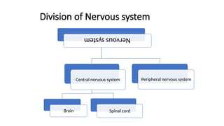
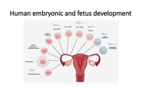
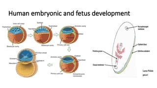
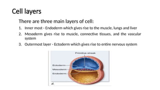
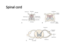
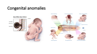
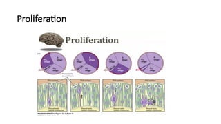
DEVELOPMENT OF CENTRAL NERVOUS SYSTEM
Presenter: Dr. Yagnya Priya
Moderator: Dr. Vijaykumar Biradar
Division of Nervous system
Nervous
system
Central nervous system
Brain Spinal cord
Peripheral nervous system
Human embryonic and fetus development
Cell layers
There are three main layers of cell:
1. Inner most - Endoderm which gives rise to the muscle, lungs and liver
2. Mesoderm gives rise to muscle, connective tissues, and the vascular
system
3. Outermost layer - Ectoderm which gives rise to entire nervous system
Stages of development
During third week of development, the ectoderm on the dorsal surface of
the embryo between the primitive node and the buccopharyngeal
membrane thickens to form the neural plate.
Stages of development
• The plate develops a longitudinal neural groove(either side by neural
fold).
• Further, the neural fold fuse,
converting the neural groove
into a neural tube.
• Fusion starts from midpoint
then extends cranially and caudally.
Stages of development
• The cavity of tube remain in communication anterior and posterior neuropores.
• Anterior neuropore closes first and the posterior neuropore 2 days later.
• Neural tube closure
is complete within 28 days.
• The process of formation of
neural tube is known as neurulation
and the embryo at this stage is called neurula.
Derivatives of neural crest cells:
• Neural crest are bilaterally paired strips of cells arising in the ectoderm
at the margins of the neural tube.
Stages of development
Meanwhile, the proliferation of cells at the cephalic end of the
neural tube causes it to dilate and form three primary vesicles:
i. The forebrain vesicles
ii. Midbrain vesicles
iii. Hindbrain vesicles
The primary division of the developing brain
Formation of neural tube
Neural tube is seen to dilate at the cephalic end into three vesicle and rest of the
tube elongates and remain smaller in diameter; it will form the spinal cord and
ventricular system of the brain.
Spinal cord
• The wall of neural tube consists of neuroepithelial cells.
• They divide rapidly producing more and more neuroepithelial cells.
• Collectively they constitute
the neuroepithelial layer.
Spinal cord
• Neuroepithelial cells give rise to another cell type, these are the primitive nerve
cells, these are neuroblasts which forms mantle layer.
• The mantle layer later forms the gray matter of the spinal cord.
Spinal cord
• The outer layer of the spinal cord, the marginal layer, contains nerve fibers
emerging from neuroblasts in the mantle layer.
• This layer takes white appearance and therefore called the white matter of the
spinal cord.
• As a result of continue addition of neuroblast to the mantle layer, each side of the
neural tube show a ventral and a dorsal thickening.
Spinal cord
• The ventral thickening - the Basal plates - contain ventral motor horn cells - form
the motor areas of the spinal cord.
• The dorsal thickening - the Alar plates - form sensory areas.
Spinal cord
• There will be longitudinal groove, the sulcus limitans, marks the boundary
between two.
• The ventral motor horn and dorsal sensory horn, the group of neurons
accumulates between the two areas and forms a small intermediate horn.
Spinal cord
Congenital anomalies
Radial and Tangential patterns of Neurogenesis
and migration
• Dorsal forebrain give rise to the cerebral cortex.
• The cerebral cortex is the paradigmatic model of inside-to-outside neurogenesis.
• The cell body containing nucleus and other organelles grow and has contact with
inner and outer surface of the cell wall of the brain.
• Outer is called pial surface of the brain and inner surface is inner surface of neural
tube; that goes into differentiation into ependymal lining of ventricles in human
brain.
Proliferation
Radial and Tangential patterns of Neurogenesis
and migration
• The cell body begin to translocate upward in the pial surface.
• Cell enter the stage where DNA replicate and then cell body begin to migrate down
towards the luminal surface of neural tube and cell divide
• Mitosis occurs and cell divide into symmetrical and asymmetrical from which
neuroblast will form.
• Cortical plate will be formed.
• Now, the neuroblast which are in ventricular zone should go to cortical plate and
this required migration.
• From the ganglionic eminence inhibitory neurons are produced and they migrate in
radial and tangential pattern.
Migration
Migration
Inductive signals
Cell death
Developmental cell death:
It is a reproducible, spatially and temporarily restricted death of cells that occurs
during the organism’s development.
Three types of developmental cell death are there:
1. Phylogentic cell death
2. Morphogentic cell death
3. Histogentic cell death
Cell death
Apoptosis:
• Major type, programmed cell death, involve specific molecule that posses
enzymatic activities.
• A failure to inhibit apoptosis is involved in cancers and autoimmune
disease(multiple sclerosis) and excess of apoptosis is seen in
neurodegenerative disease(Alzheimer’s and Parkinson’s disease).
The neurodevelopment basis of psychiatric
disease
• The increasing number of neuropsychiatric condition are considered to originate
during brain development, including schizophrenia, depression, autism, and
attention-deficit hyperactivity disorder.
• Schizophrenia: at the time of onset and diagnosis, the prefrontal cortex and
hippocampus are smaller and ventricles enlarged already at adolescent
presentation.
• In autism spectrum: the amygdala and fusiform gyrus demonstrate abnormal
attribution during facial recognition.
References
• Snell’s clinical neuroanatomy 8th
edition
• Kaplan and Sadock’s Synopsis of psychiatry 11th
edition

Development of Central nervous system ppt

  • 1.
    DEVELOPMENT OF CENTRALNERVOUS SYSTEM Presenter: Dr. Yagnya Priya Moderator: Dr. Vijaykumar Biradar
  • 2.
    Division of Nervoussystem Nervous system Central nervous system Brain Spinal cord Peripheral nervous system
  • 3.
    Human embryonic andfetus development
  • 4.
    Human embryonic andfetus development
  • 5.
    Cell layers There arethree main layers of cell: 1. Inner most - Endoderm which gives rise to the muscle, lungs and liver 2. Mesoderm gives rise to muscle, connective tissues, and the vascular system 3. Outermost layer - Ectoderm which gives rise to entire nervous system
  • 6.
    Stages of development Duringthird week of development, the ectoderm on the dorsal surface of the embryo between the primitive node and the buccopharyngeal membrane thickens to form the neural plate.
  • 7.
    Stages of development •The plate develops a longitudinal neural groove(either side by neural fold). • Further, the neural fold fuse, converting the neural groove into a neural tube. • Fusion starts from midpoint then extends cranially and caudally.
  • 8.
    Stages of development •The cavity of tube remain in communication anterior and posterior neuropores. • Anterior neuropore closes first and the posterior neuropore 2 days later. • Neural tube closure is complete within 28 days. • The process of formation of neural tube is known as neurulation and the embryo at this stage is called neurula.
  • 9.
    Derivatives of neuralcrest cells: • Neural crest are bilaterally paired strips of cells arising in the ectoderm at the margins of the neural tube.
  • 10.
    Stages of development Meanwhile,the proliferation of cells at the cephalic end of the neural tube causes it to dilate and form three primary vesicles: i. The forebrain vesicles ii. Midbrain vesicles iii. Hindbrain vesicles
  • 11.
    The primary divisionof the developing brain
  • 12.
    Formation of neuraltube Neural tube is seen to dilate at the cephalic end into three vesicle and rest of the tube elongates and remain smaller in diameter; it will form the spinal cord and ventricular system of the brain.
  • 13.
    Spinal cord • Thewall of neural tube consists of neuroepithelial cells. • They divide rapidly producing more and more neuroepithelial cells. • Collectively they constitute the neuroepithelial layer.
  • 14.
    Spinal cord • Neuroepithelialcells give rise to another cell type, these are the primitive nerve cells, these are neuroblasts which forms mantle layer. • The mantle layer later forms the gray matter of the spinal cord.
  • 15.
    Spinal cord • Theouter layer of the spinal cord, the marginal layer, contains nerve fibers emerging from neuroblasts in the mantle layer. • This layer takes white appearance and therefore called the white matter of the spinal cord. • As a result of continue addition of neuroblast to the mantle layer, each side of the neural tube show a ventral and a dorsal thickening.
  • 16.
    Spinal cord • Theventral thickening - the Basal plates - contain ventral motor horn cells - form the motor areas of the spinal cord. • The dorsal thickening - the Alar plates - form sensory areas.
  • 17.
    Spinal cord • Therewill be longitudinal groove, the sulcus limitans, marks the boundary between two. • The ventral motor horn and dorsal sensory horn, the group of neurons accumulates between the two areas and forms a small intermediate horn.
  • 18.
  • 19.
  • 20.
    Radial and Tangentialpatterns of Neurogenesis and migration • Dorsal forebrain give rise to the cerebral cortex. • The cerebral cortex is the paradigmatic model of inside-to-outside neurogenesis. • The cell body containing nucleus and other organelles grow and has contact with inner and outer surface of the cell wall of the brain. • Outer is called pial surface of the brain and inner surface is inner surface of neural tube; that goes into differentiation into ependymal lining of ventricles in human brain.
  • 21.
  • 22.
    Radial and Tangentialpatterns of Neurogenesis and migration • The cell body begin to translocate upward in the pial surface. • Cell enter the stage where DNA replicate and then cell body begin to migrate down towards the luminal surface of neural tube and cell divide • Mitosis occurs and cell divide into symmetrical and asymmetrical from which neuroblast will form. • Cortical plate will be formed. • Now, the neuroblast which are in ventricular zone should go to cortical plate and this required migration. • From the ganglionic eminence inhibitory neurons are produced and they migrate in radial and tangential pattern.
  • 23.
  • 24.
  • 25.
  • 26.
    Cell death Developmental celldeath: It is a reproducible, spatially and temporarily restricted death of cells that occurs during the organism’s development. Three types of developmental cell death are there: 1. Phylogentic cell death 2. Morphogentic cell death 3. Histogentic cell death
  • 27.
    Cell death Apoptosis: • Majortype, programmed cell death, involve specific molecule that posses enzymatic activities. • A failure to inhibit apoptosis is involved in cancers and autoimmune disease(multiple sclerosis) and excess of apoptosis is seen in neurodegenerative disease(Alzheimer’s and Parkinson’s disease).
  • 28.
    The neurodevelopment basisof psychiatric disease • The increasing number of neuropsychiatric condition are considered to originate during brain development, including schizophrenia, depression, autism, and attention-deficit hyperactivity disorder. • Schizophrenia: at the time of onset and diagnosis, the prefrontal cortex and hippocampus are smaller and ventricles enlarged already at adolescent presentation. • In autism spectrum: the amygdala and fusiform gyrus demonstrate abnormal attribution during facial recognition.
  • 29.
    References • Snell’s clinicalneuroanatomy 8th edition • Kaplan and Sadock’s Synopsis of psychiatry 11th edition