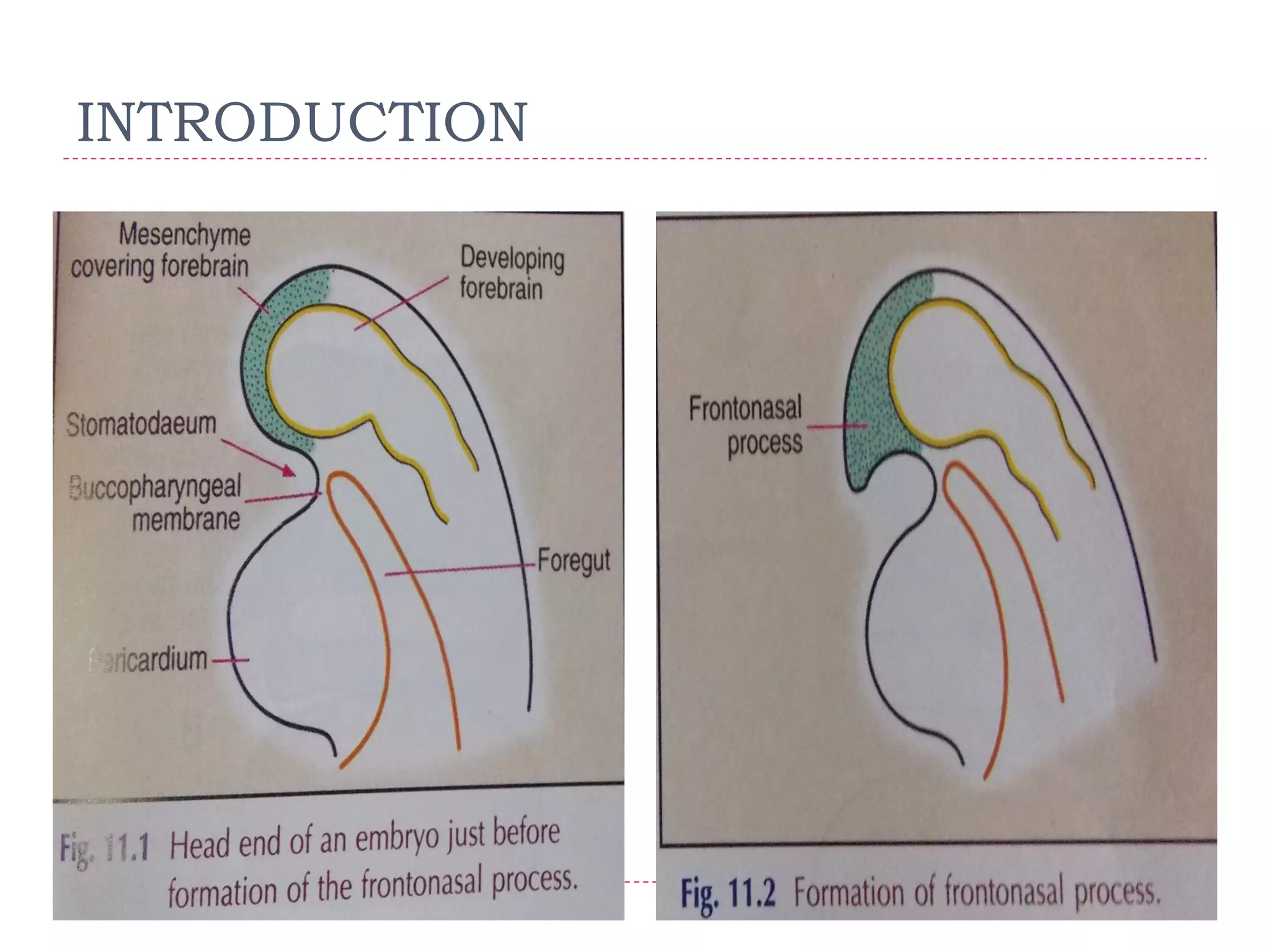
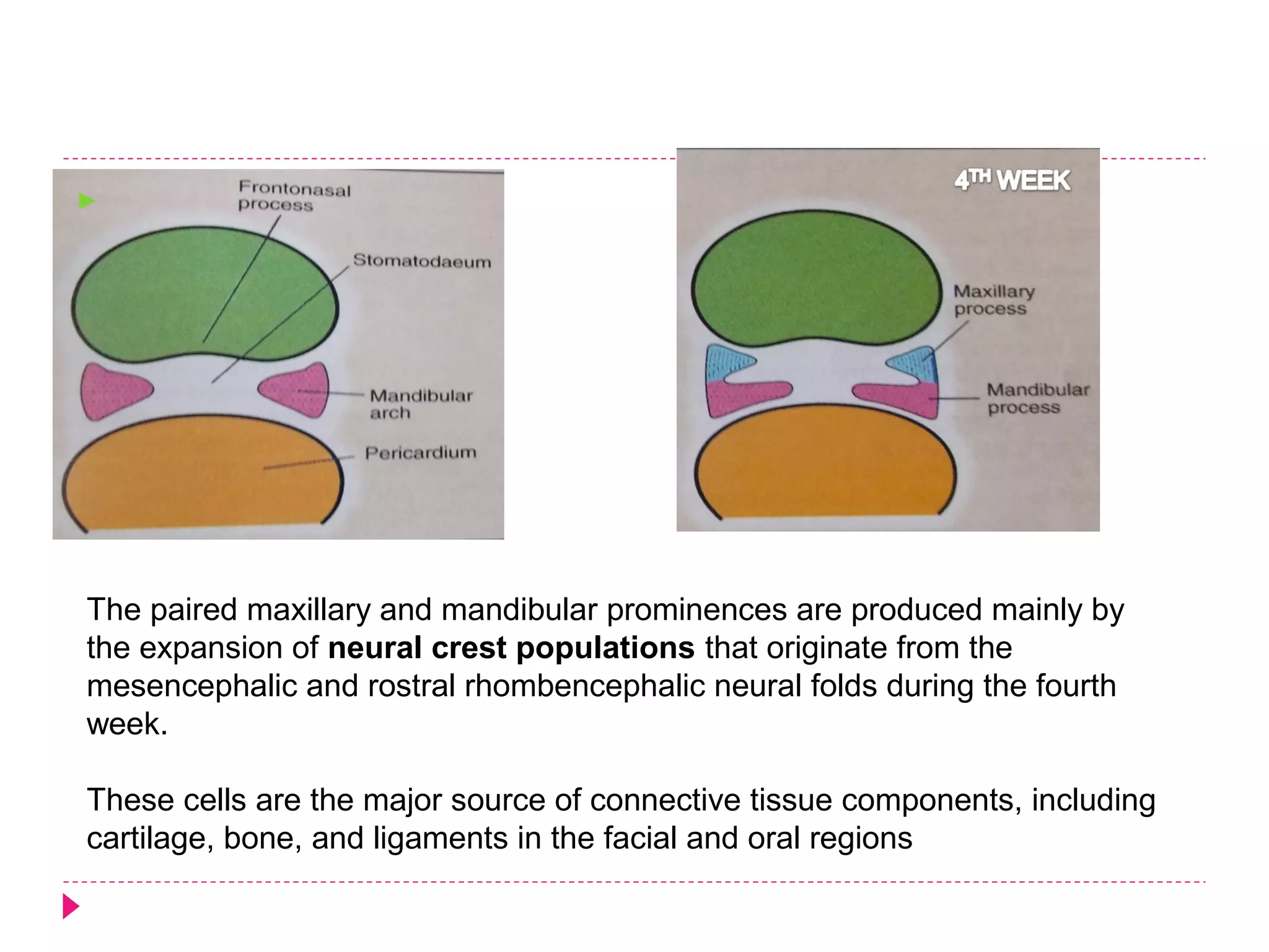
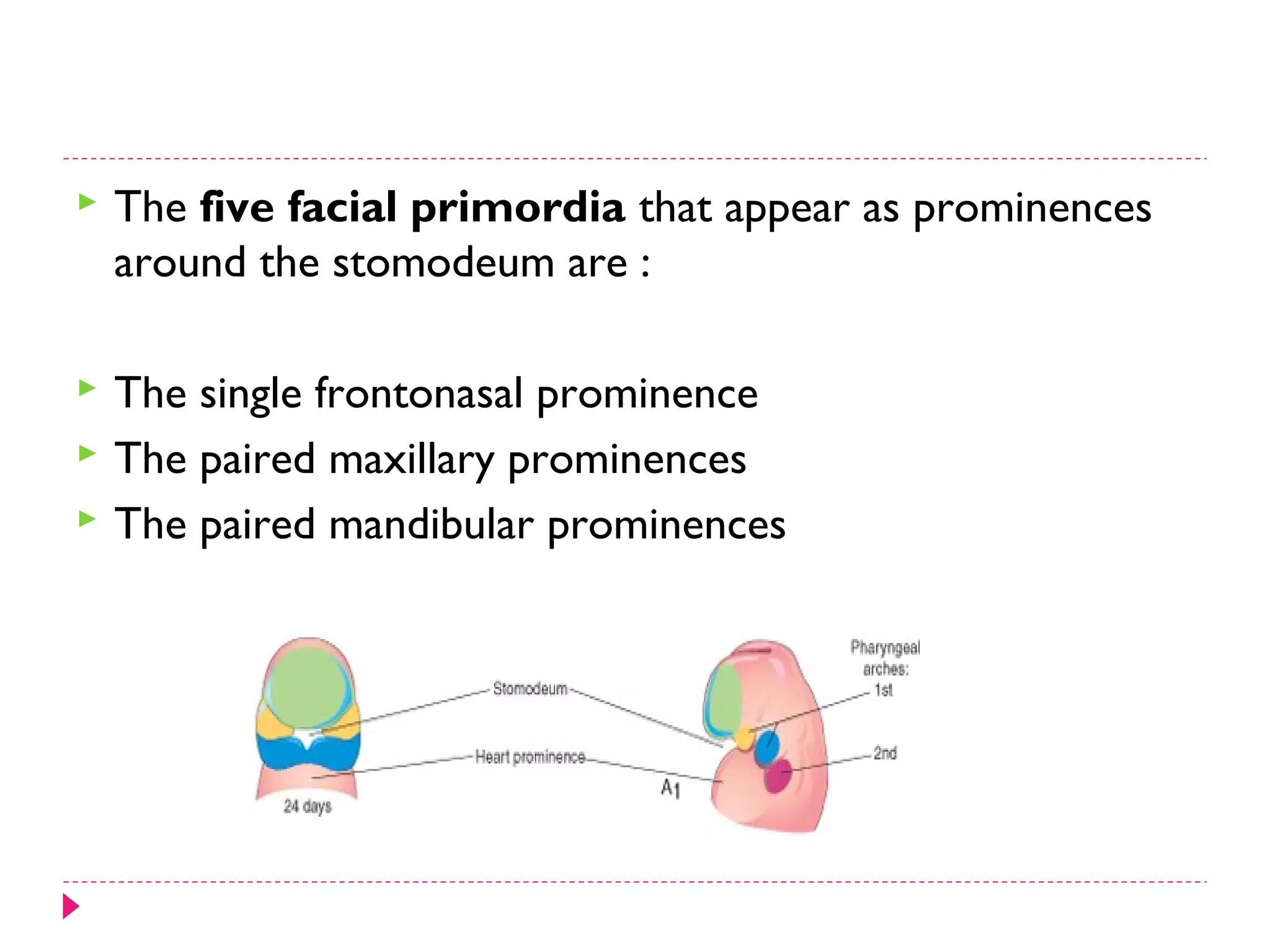
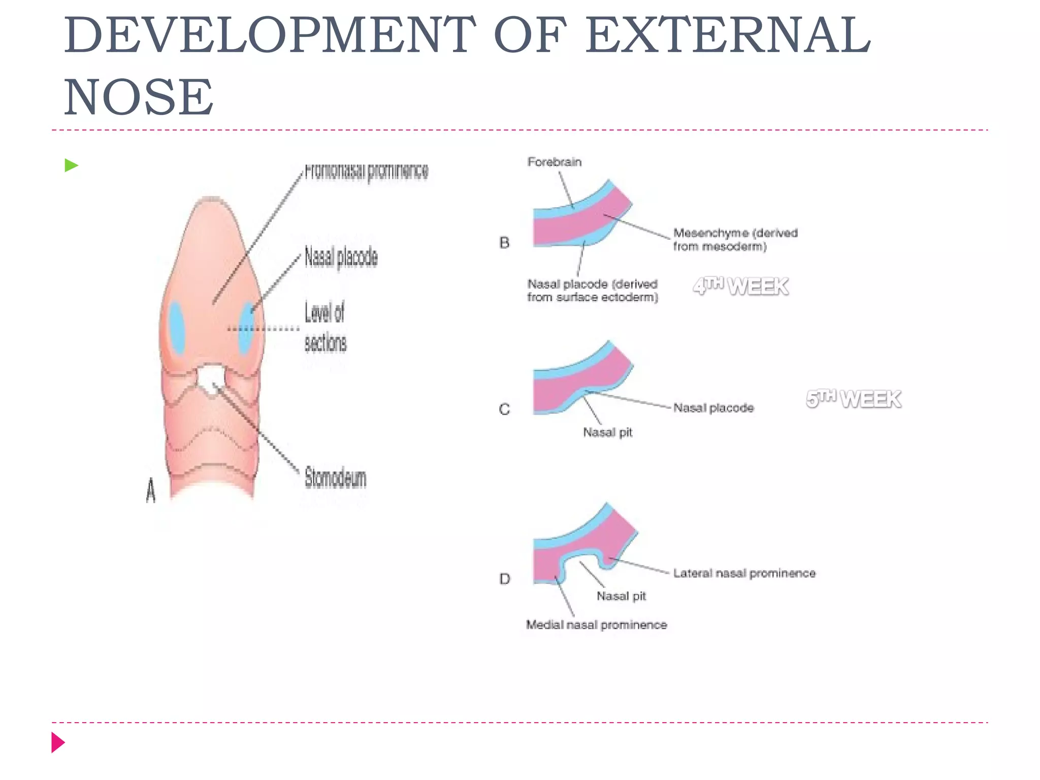
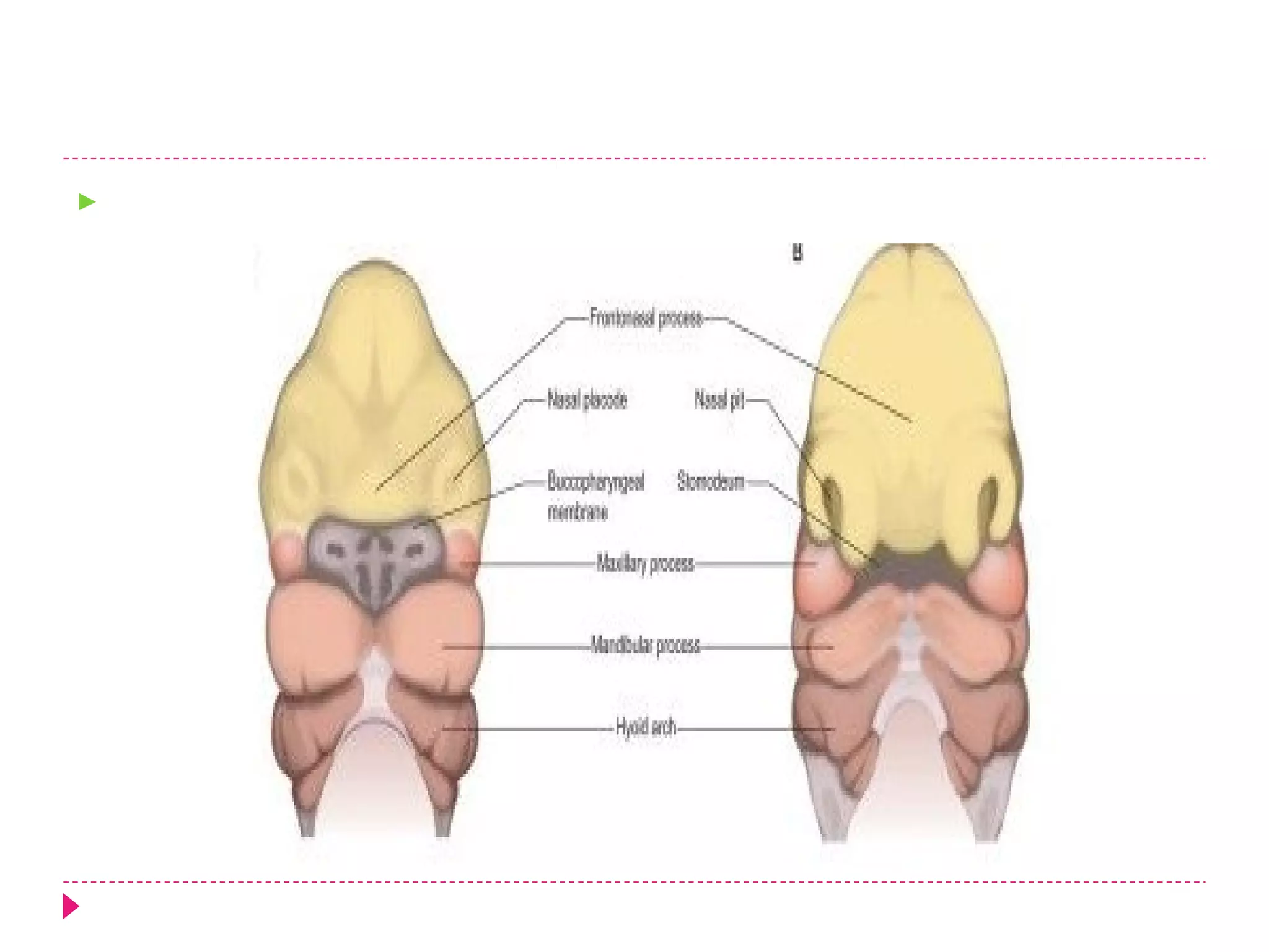
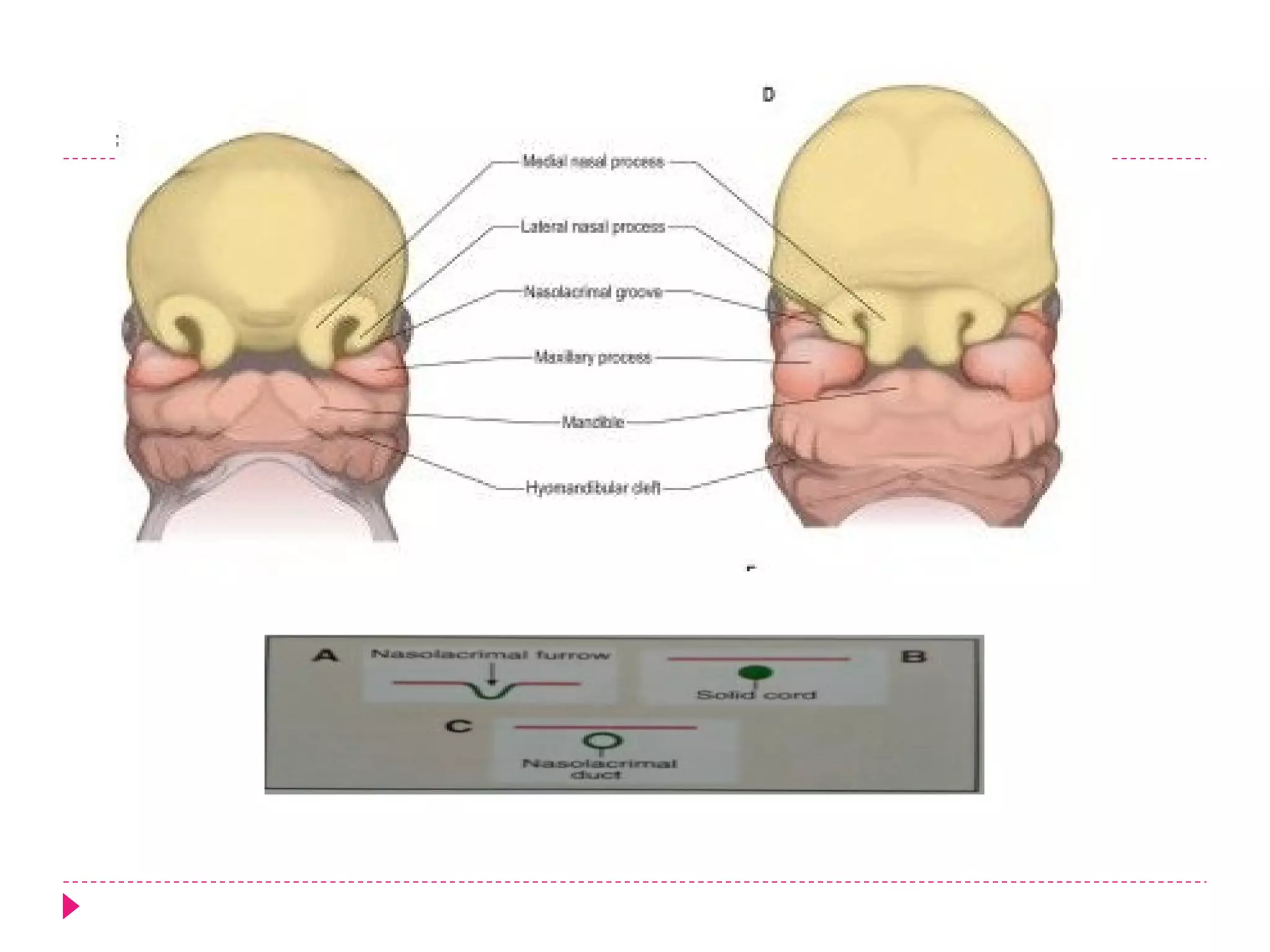
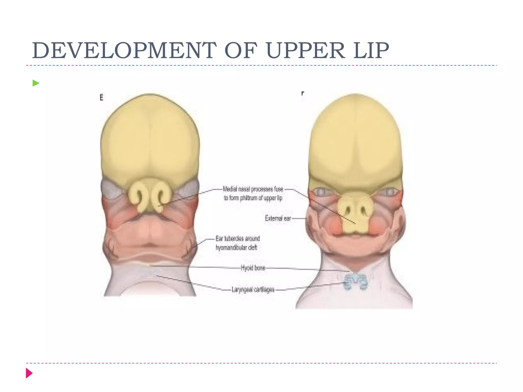
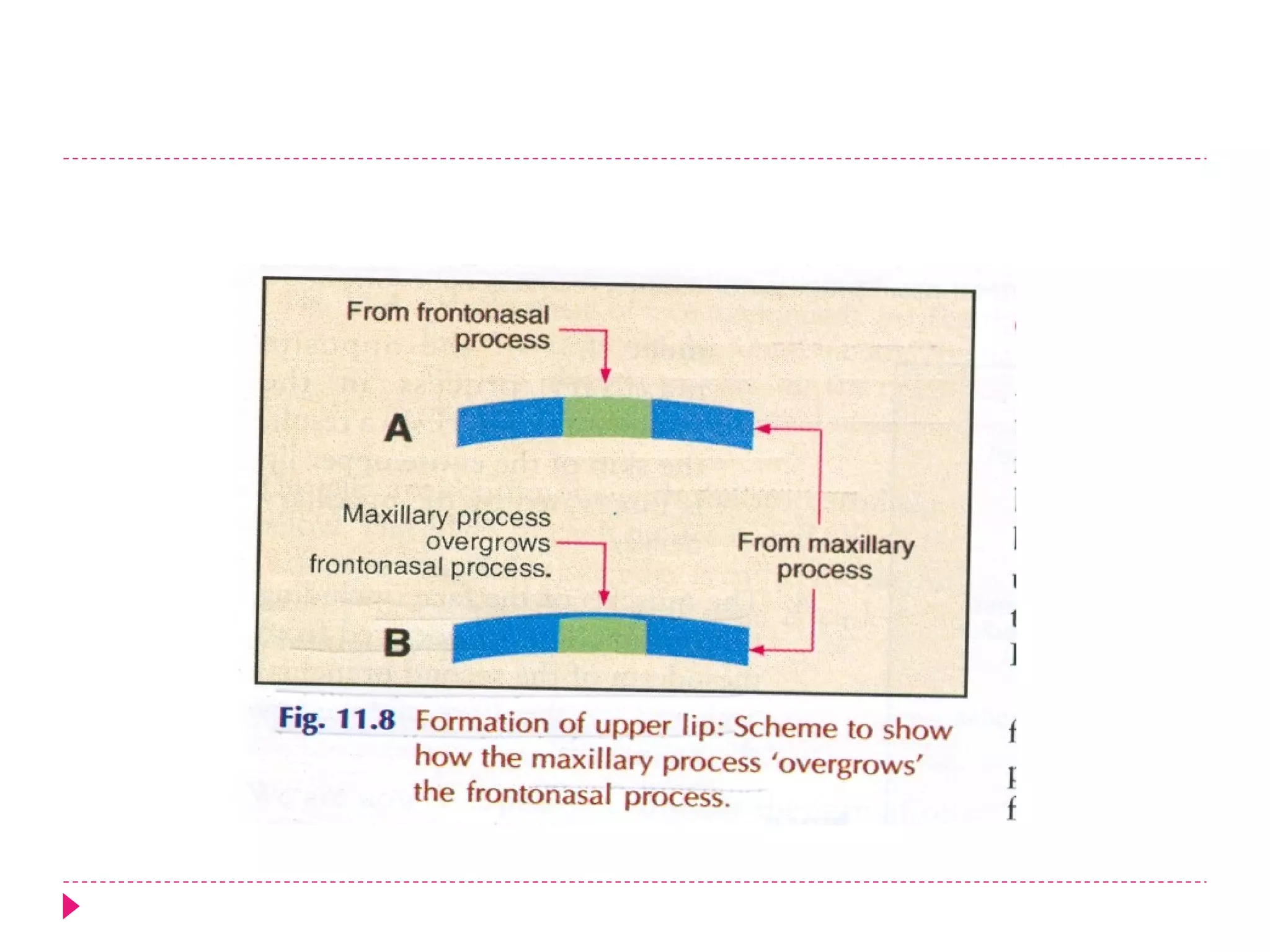
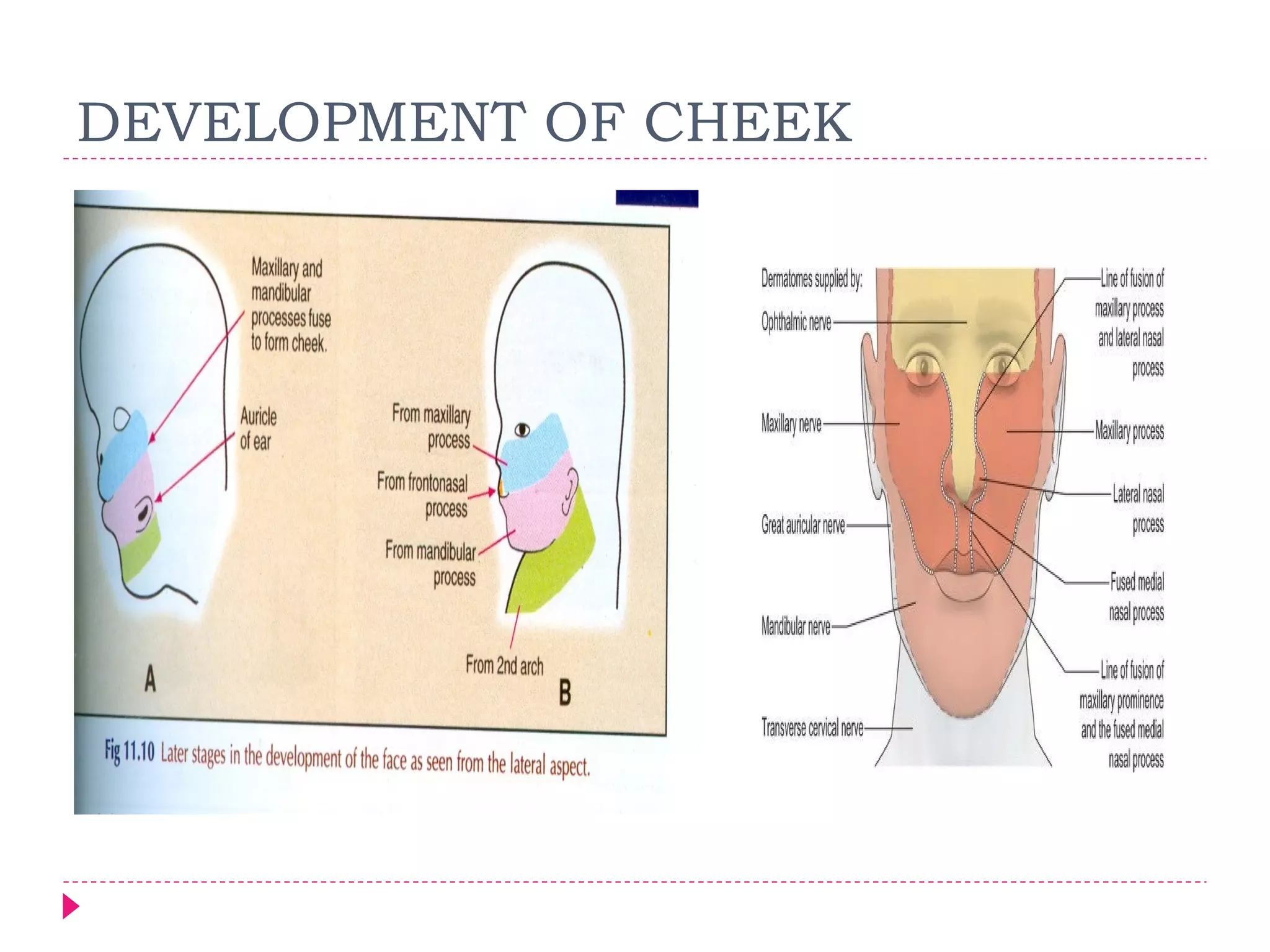
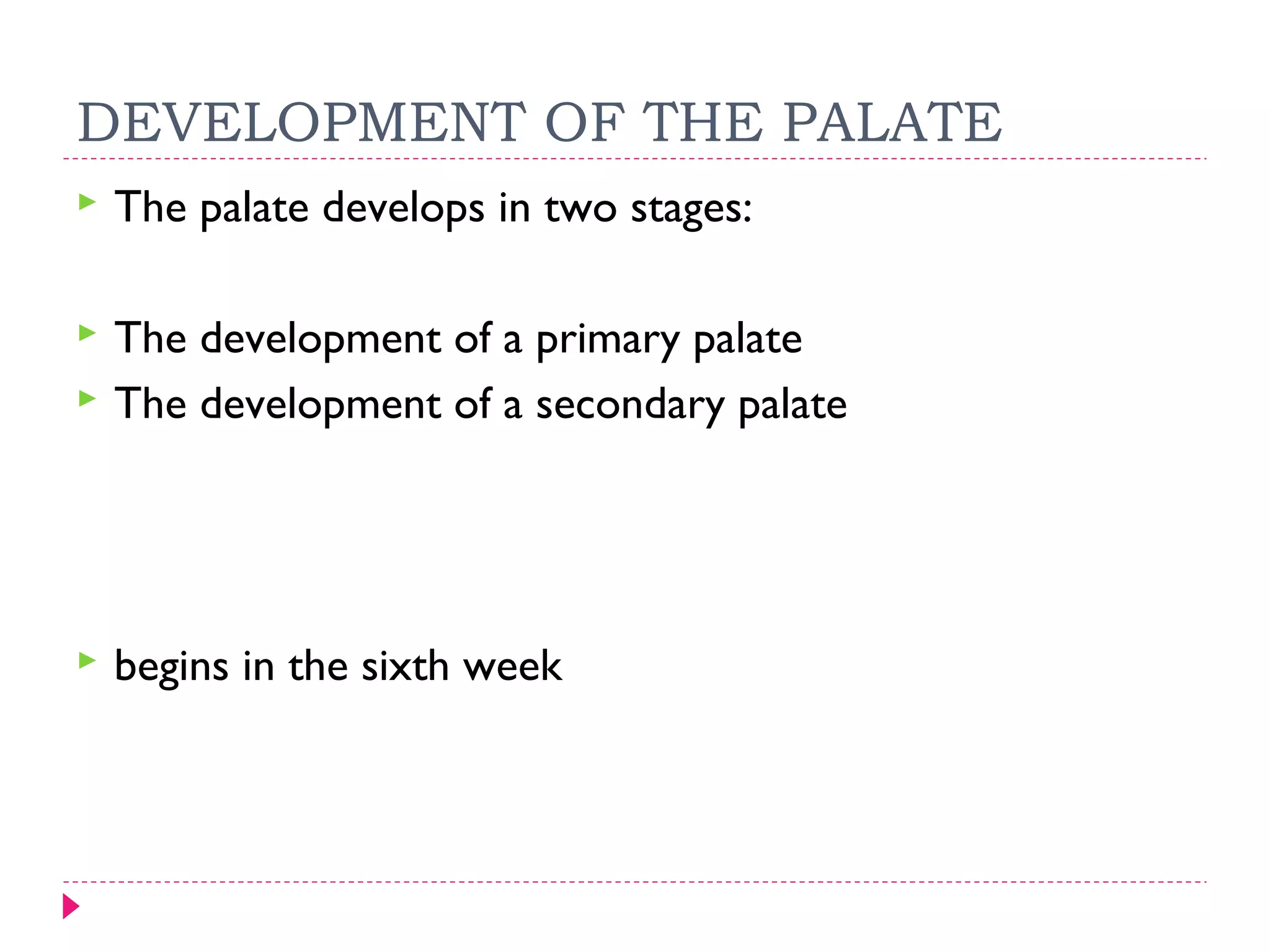
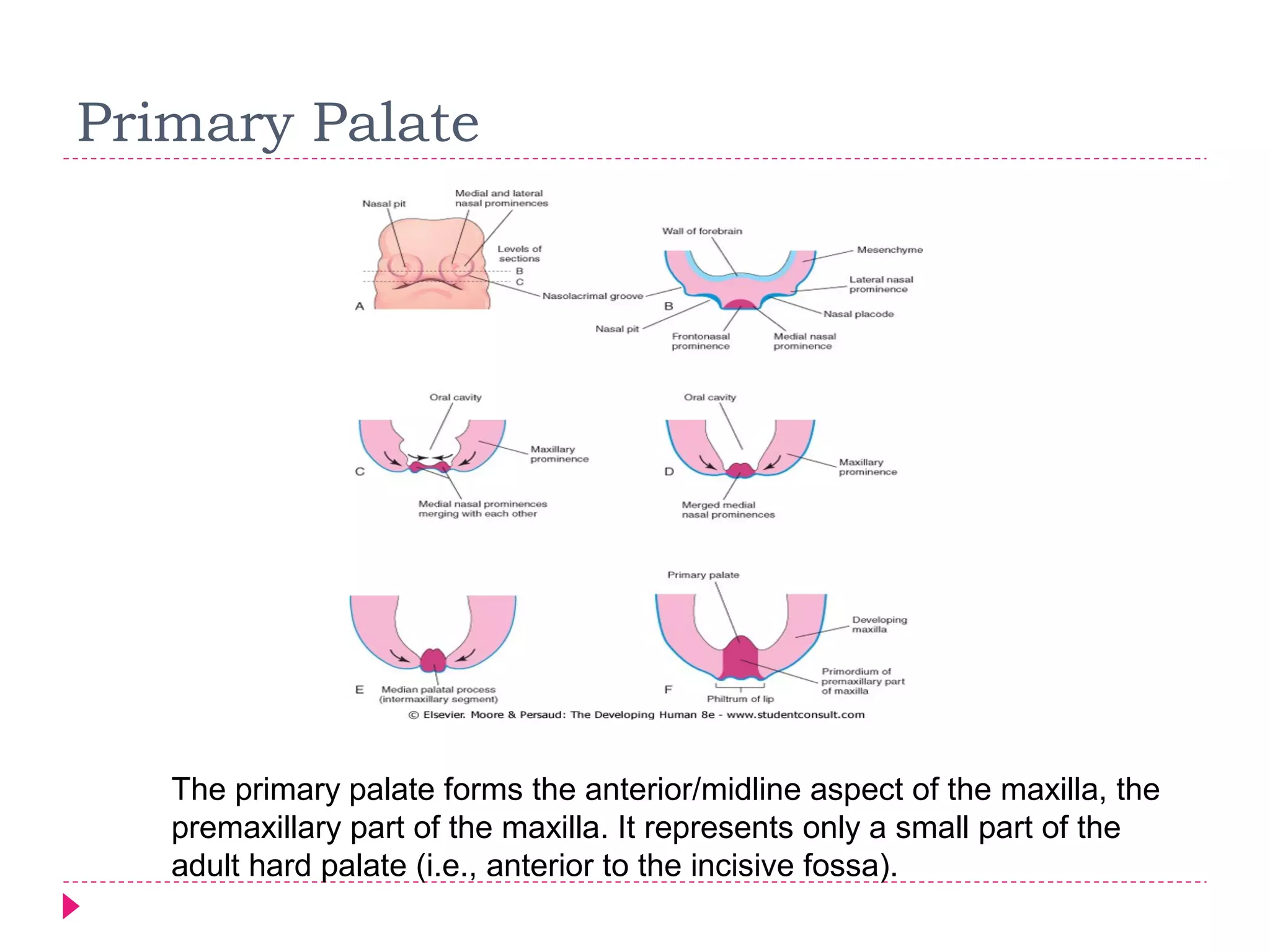
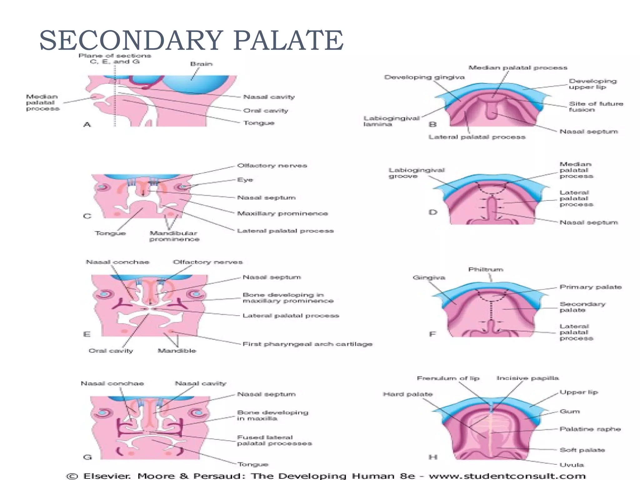
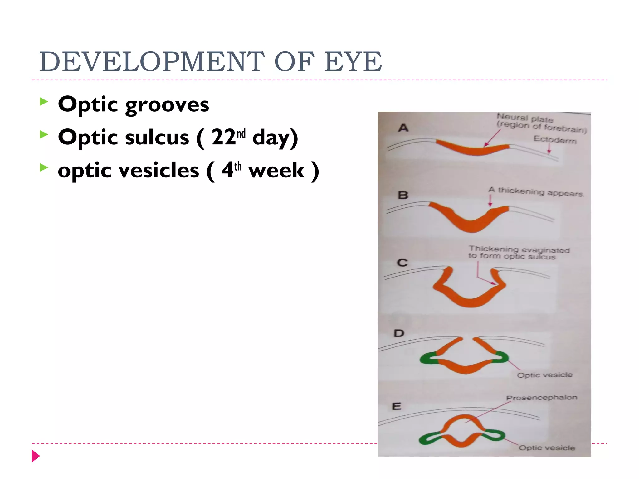
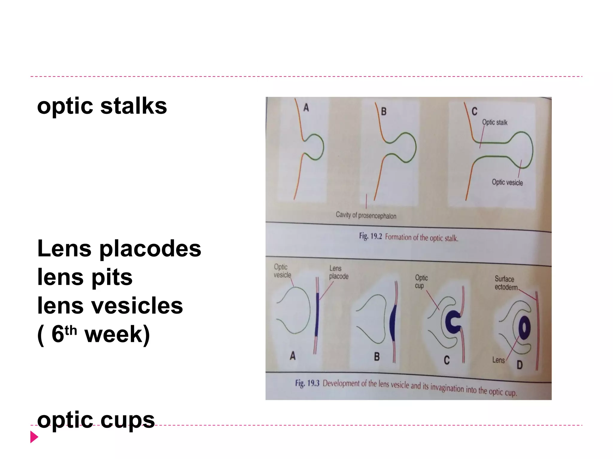
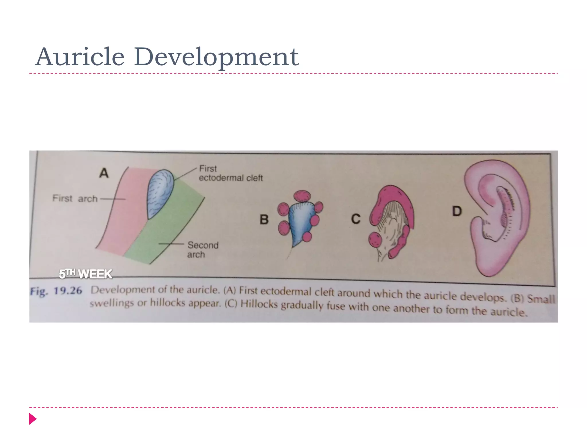
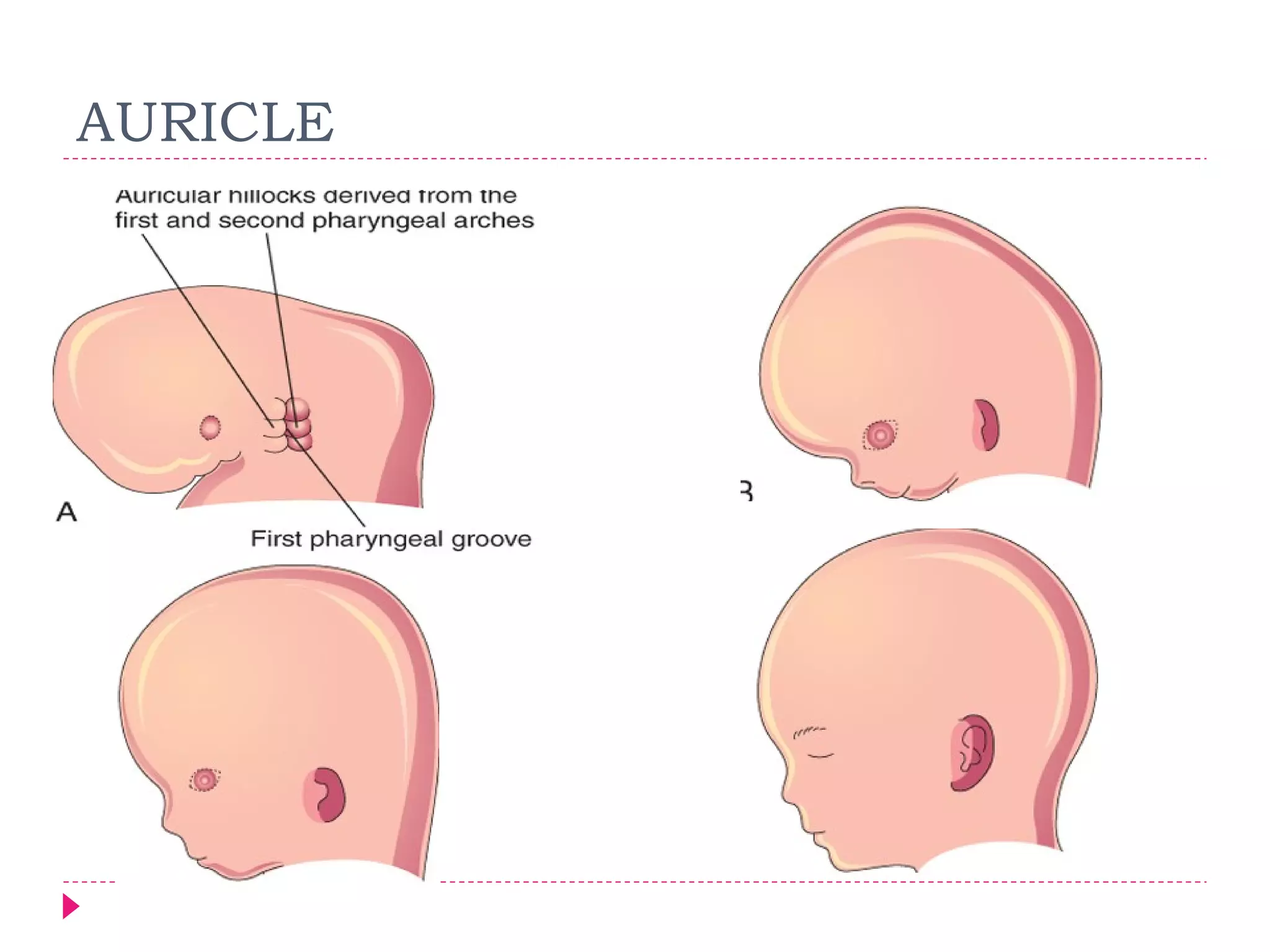
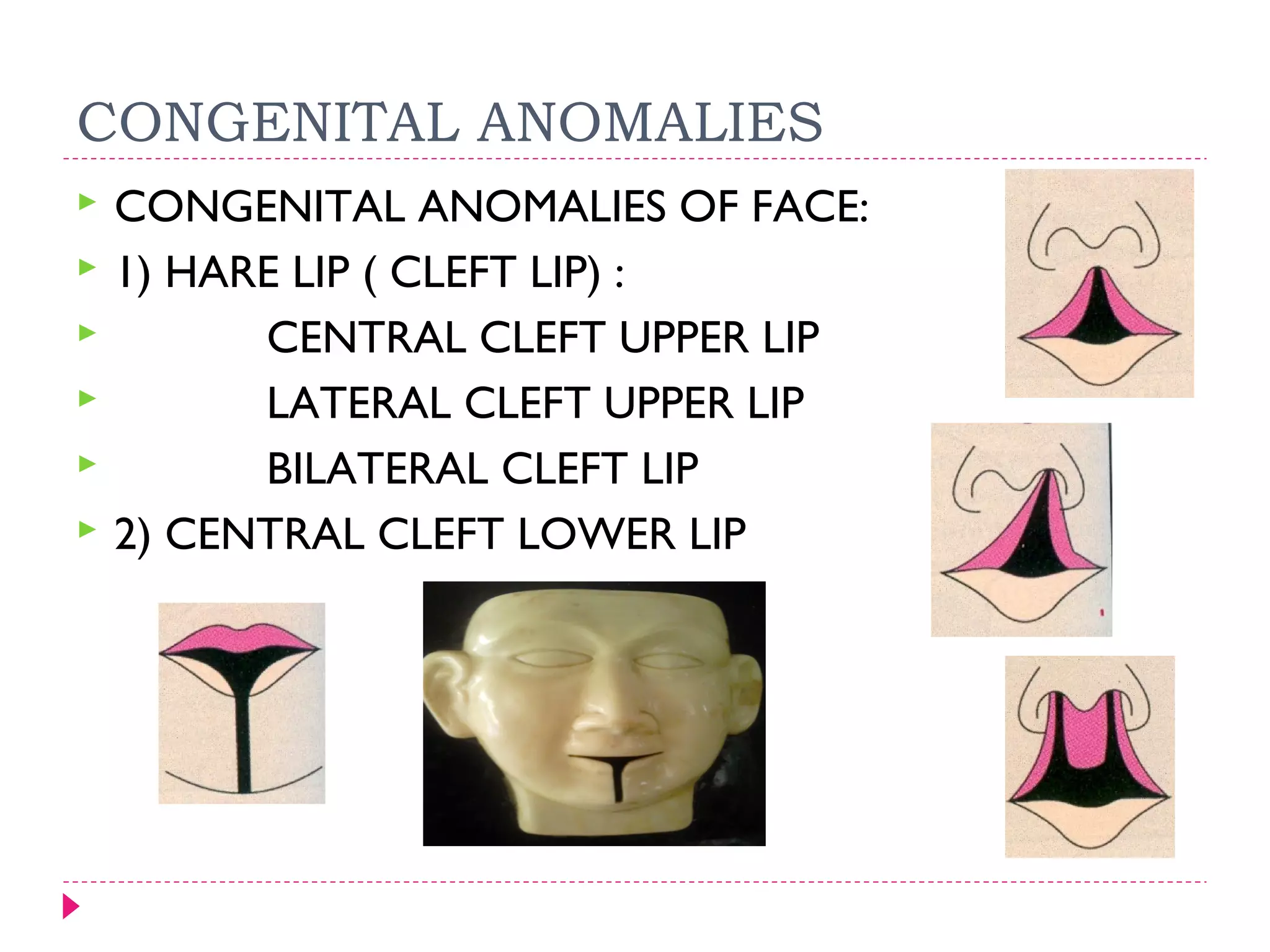
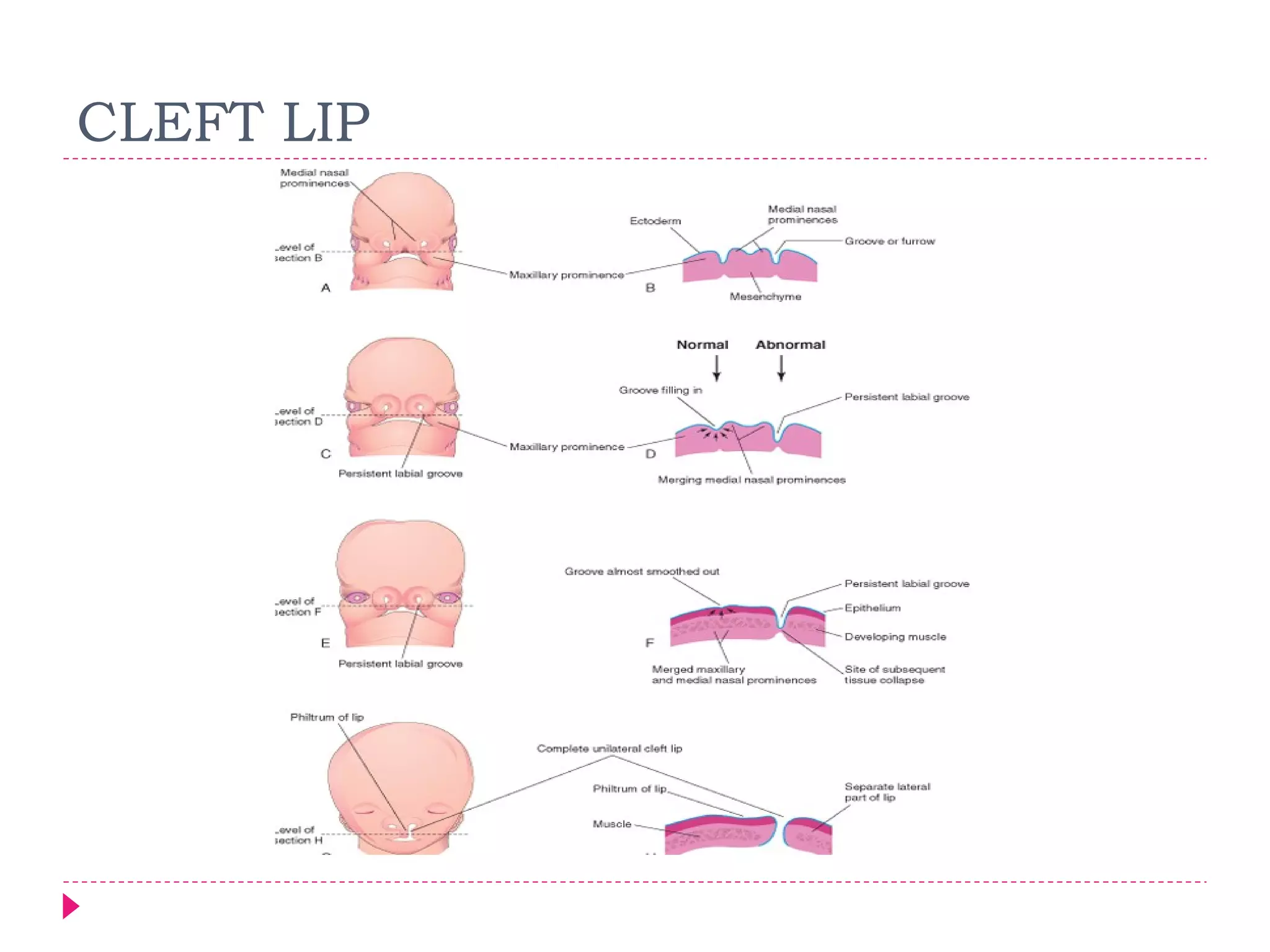
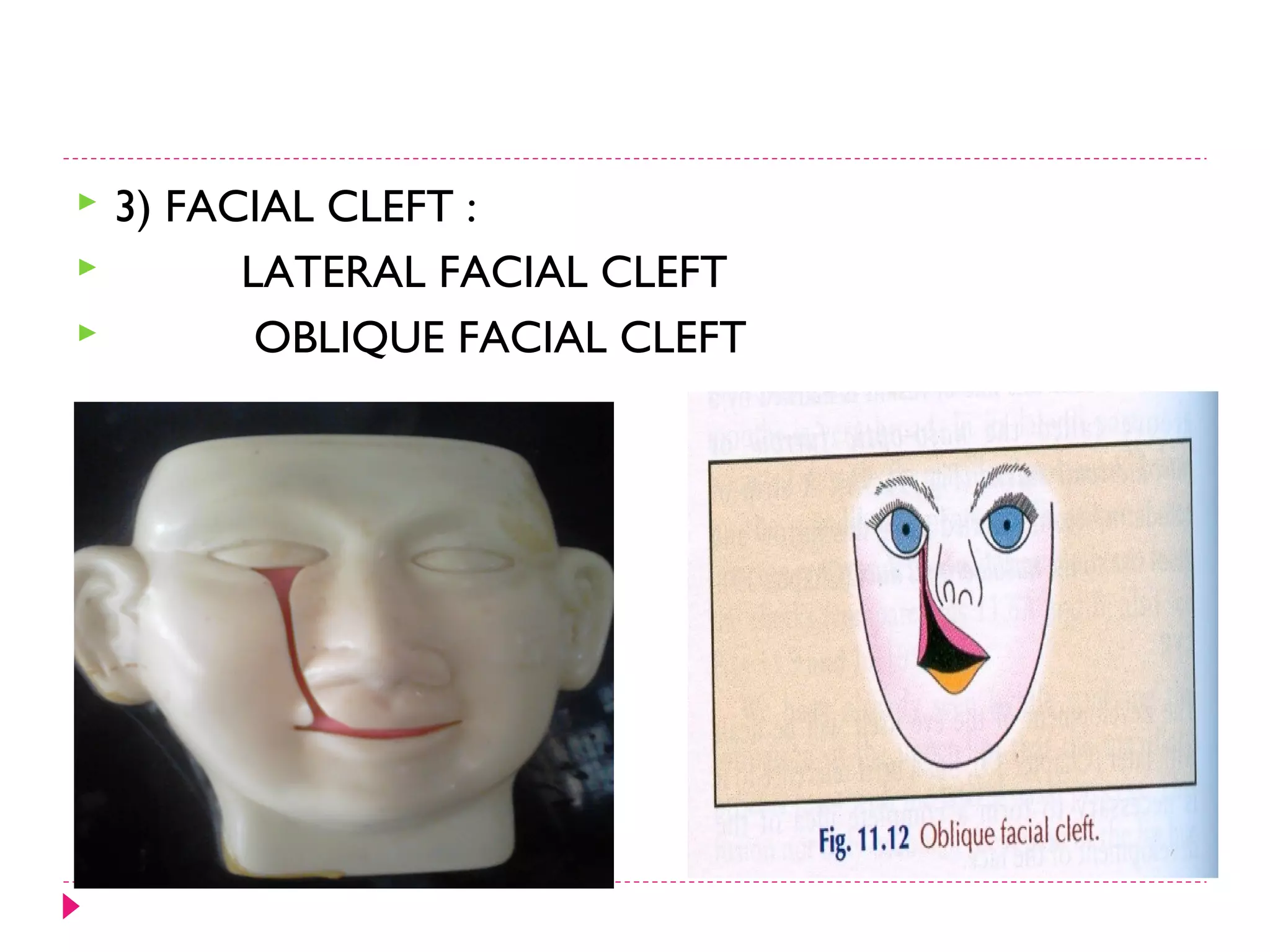
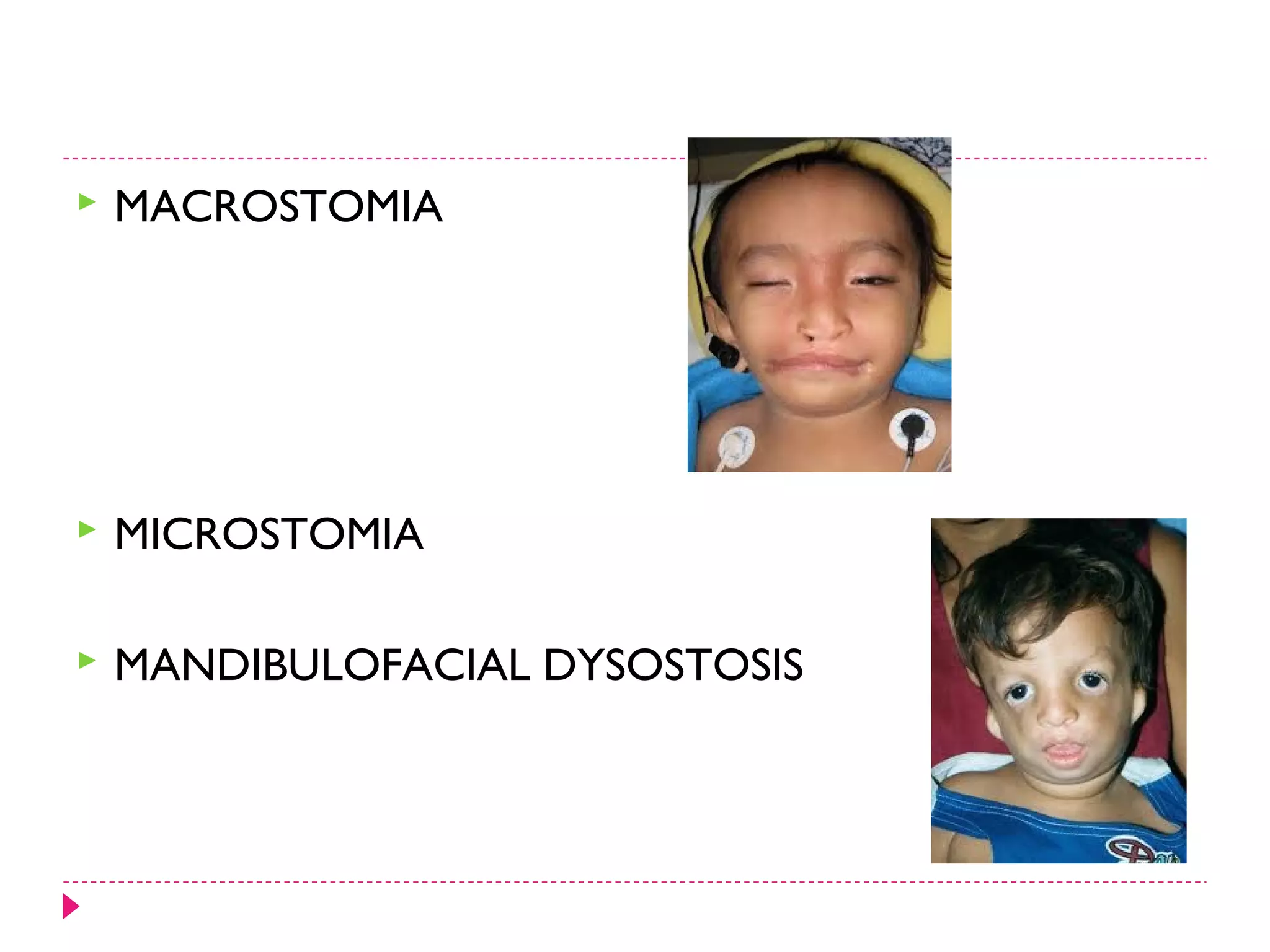
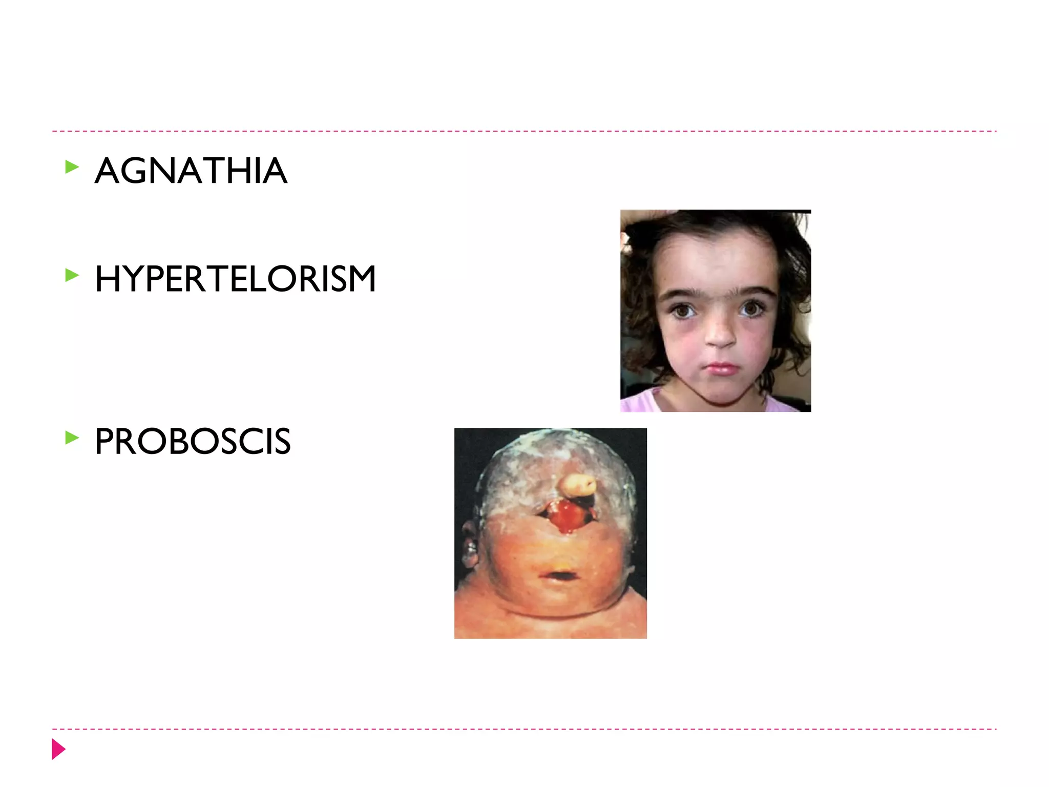
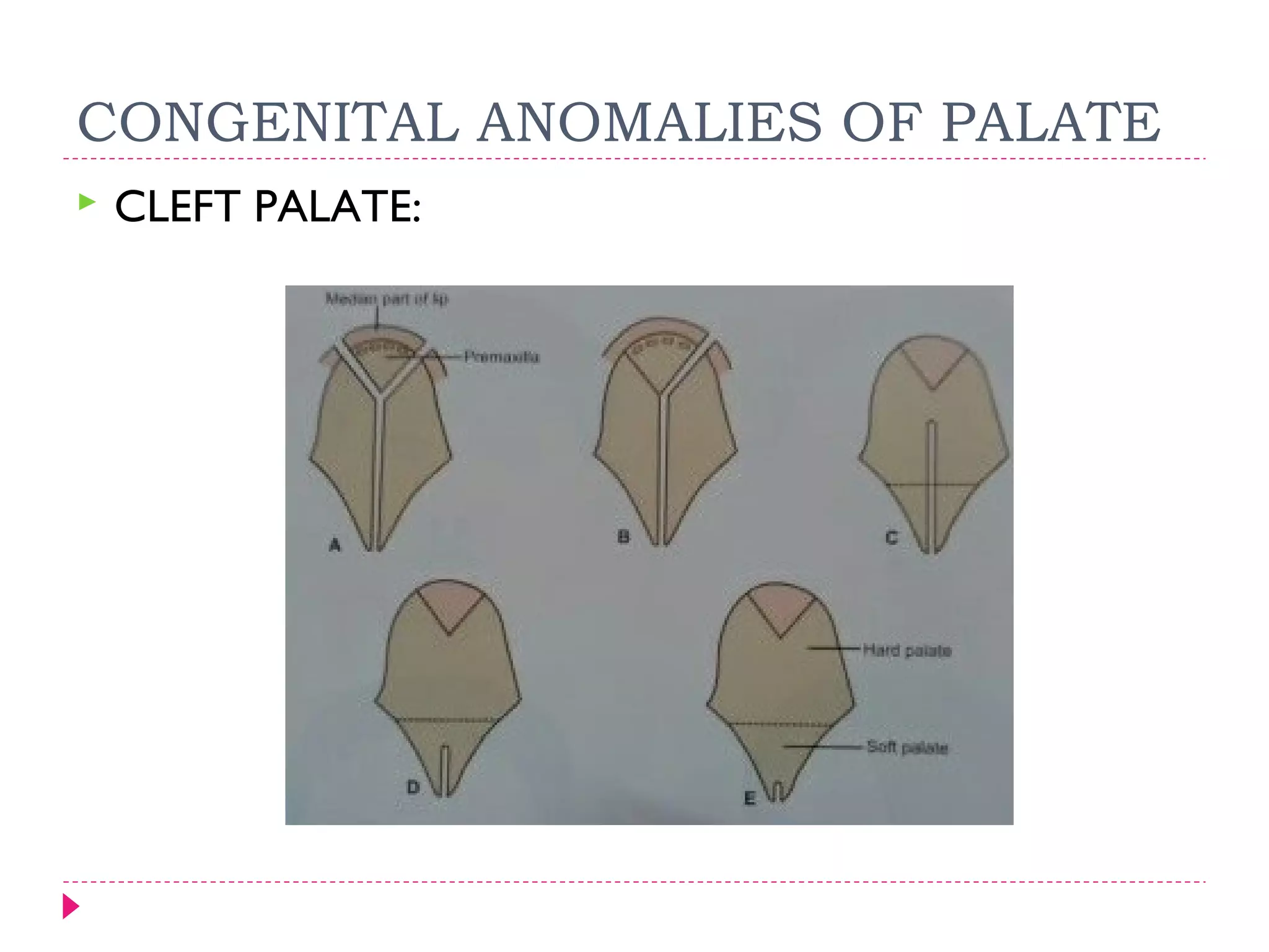
DEVELOPMENT OF FACE

PRESENTER- DR KAPIL AMGAIN
SCHEME OF PRESENTATION:
INTRODUCTION
 DEVELOPMENT OF FACE:
EXTERNAL NOSE
LOWER LIP
UPPER LIP
CHEEKS
PALATE
NASAL CAVITY
EYES IN BRIEF
AURICLE
 ANOMALIES

INTRODUCTION


The paired maxillary and mandibular prominences are produced mainly by
the expansion of neural crest populations that originate from the
mesencephalic and rostral rhombencephalic neural folds during the fourth
week.
These cells are the major source of connective tissue components, including
cartilage, bone, and ligaments in the facial and oral regions


The five facial primordia that appear as prominences
around the stomodeum are :



The single frontonasal prominence
The paired maxillary prominences
The paired mandibular prominences











The frontal part of the FNP forms the forehead; the nasal
part of the FNP forms the rostral boundary of the
stomodeum and nose .
Most of the upper lip, maxilla, and secondary palate form
from the maxillary prominences .
The primordial lips and cheeks are invaded by
mesenchyme from the second pair of pharyngeal arches,
which differentiates into the facial muscles.
The intermaxillary segment gives rise to: The middle part
(philtrum) of the upper lip,the premaxillary part of the
maxilla and its associated gingiva (gum), primary palate.
DEVELOPMENT OF EXTERNAL
NOSE


DEVELOPMENT OF UPPER LIP

DEVELOPMENT OF CHEEK
DEVELOPMENT OF THE PALATE


The palate develops in two stages:




The development of a primary palate
The development of a secondary palate



begins in the sixth week
Primary Palate

The primary palate forms the anterior/midline aspect of the maxilla, the
premaxillary part of the maxilla. It represents only a small part of the
adult hard palate (i.e., anterior to the incisive fossa).
SECONDARY PALATE
DEVELOPMENT OF NASAL CAVITY
DEVELOPMENT OF EYE




Optic grooves
Optic sulcus ( 22nd day)
optic vesicles ( 4th week )
optic stalks

Lens placodes
lens pits
lens vesicles
( 6th week)
optic cups
Auricle Development
AURICLE
CONGENITAL ANOMALIES







CONGENITAL ANOMALIES OF FACE:
1) HARE LIP ( CLEFT LIP) :
CENTRAL CLEFT UPPER LIP
LATERAL CLEFT UPPER LIP
BILATERAL CLEFT LIP
2) CENTRAL CLEFT LOWER LIP
CLEFT LIP




3) FACIAL CLEFT :
LATERAL FACIAL CLEFT
OBLIQUE FACIAL CLEFT


MACROSTOMIA



MICROSTOMIA



MANDIBULOFACIAL DYSOSTOSIS


AGNATHIA



HYPERTELORISM



PROBOSCIS
CONGENITAL ANOMALIES OF PALATE


CLEFT PALATE:
ANOMALIES OF AURICLE
AURICLE –
 TOTALLY ABSENT
 PARTIALLY ABSENT
 ISOLATED NODULES
 FAILURE OF MIGRATION
 AURICULAR SINUSES
DEVELOPMENT OF EYE
The eyes are derived from four sources:





The neuroectoderm of the forebrain
The surface ectoderm of the head
The mesoderm between the above layers
Neural crest cells
The neuroectoderm
 of the forebrain differentiates into the retina,
 posterior layers of the iris,
 optic nerve.
The surface ectoderm
 forms the lens of the eye and
 the corneal epithelium.
The mesoderm
 fibrous and vascular coats of the eye.


Mesenchyme is derived from mesoderm.

Neural crest cells
 differentiate into the choroid
 sclera
 corneal endothelium.
Development of face

Development of face