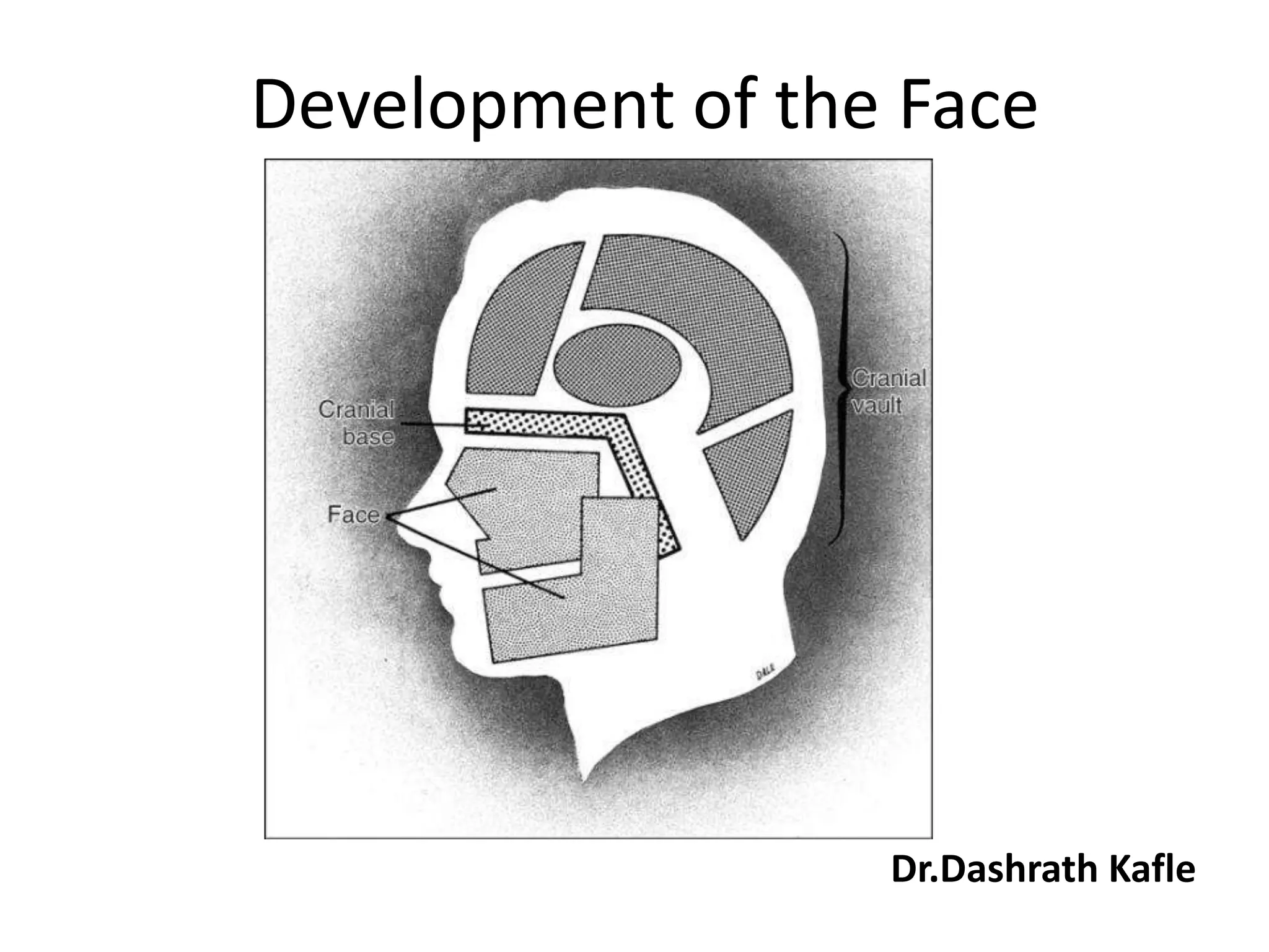
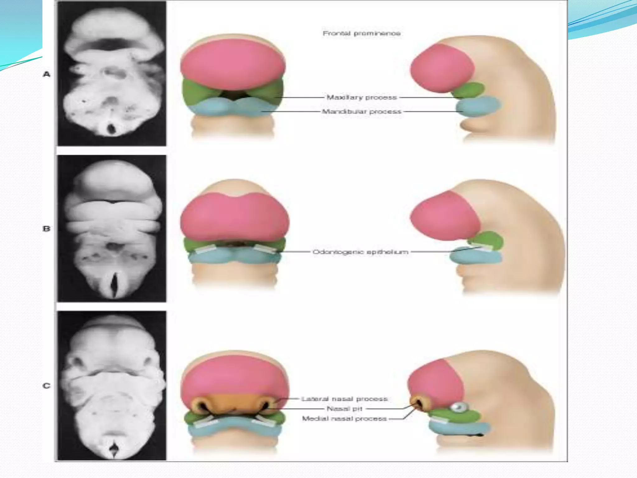
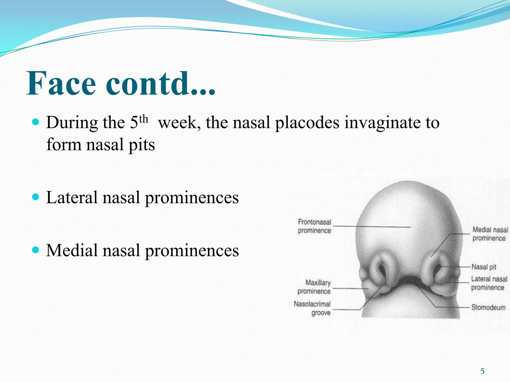
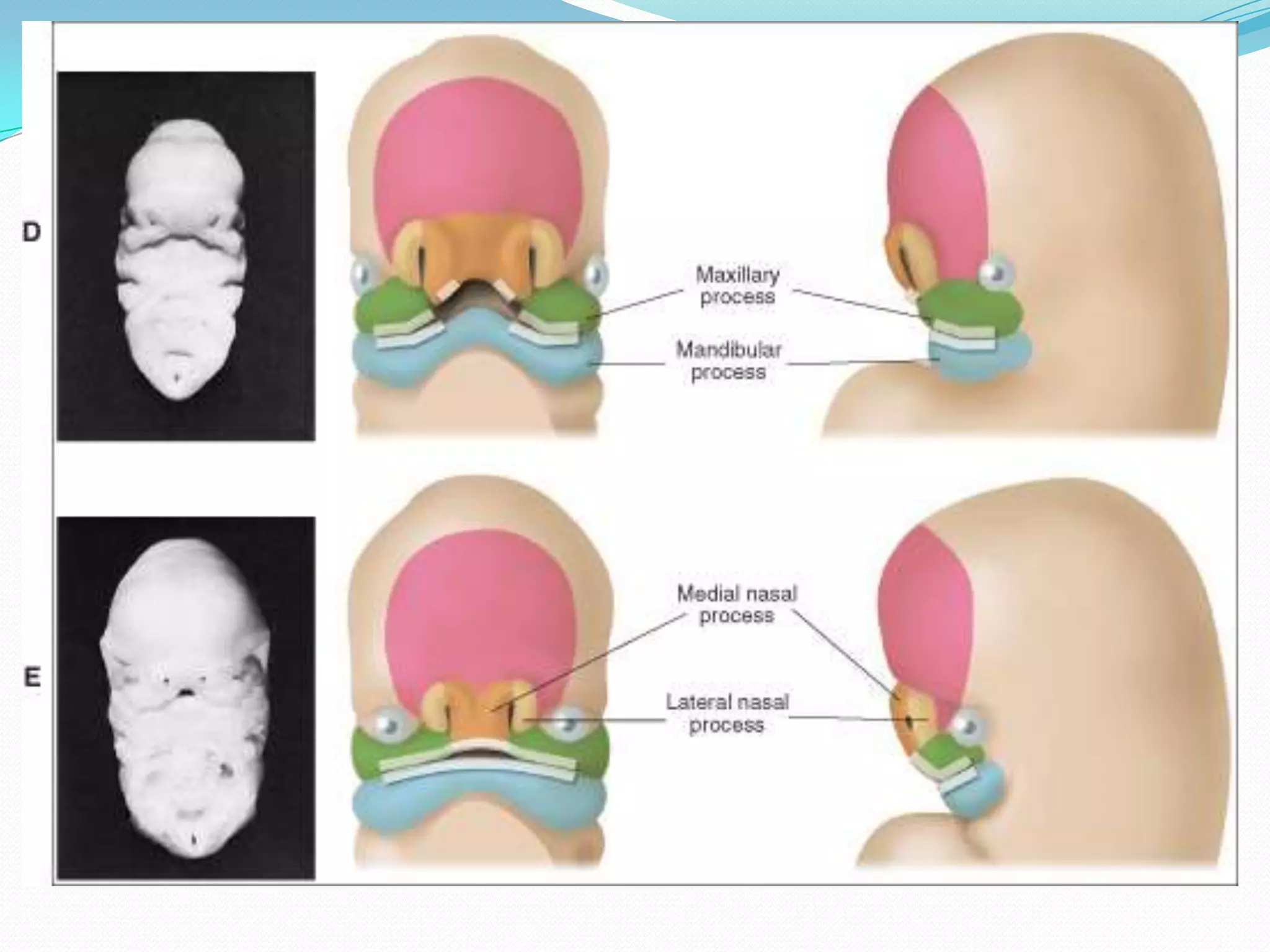
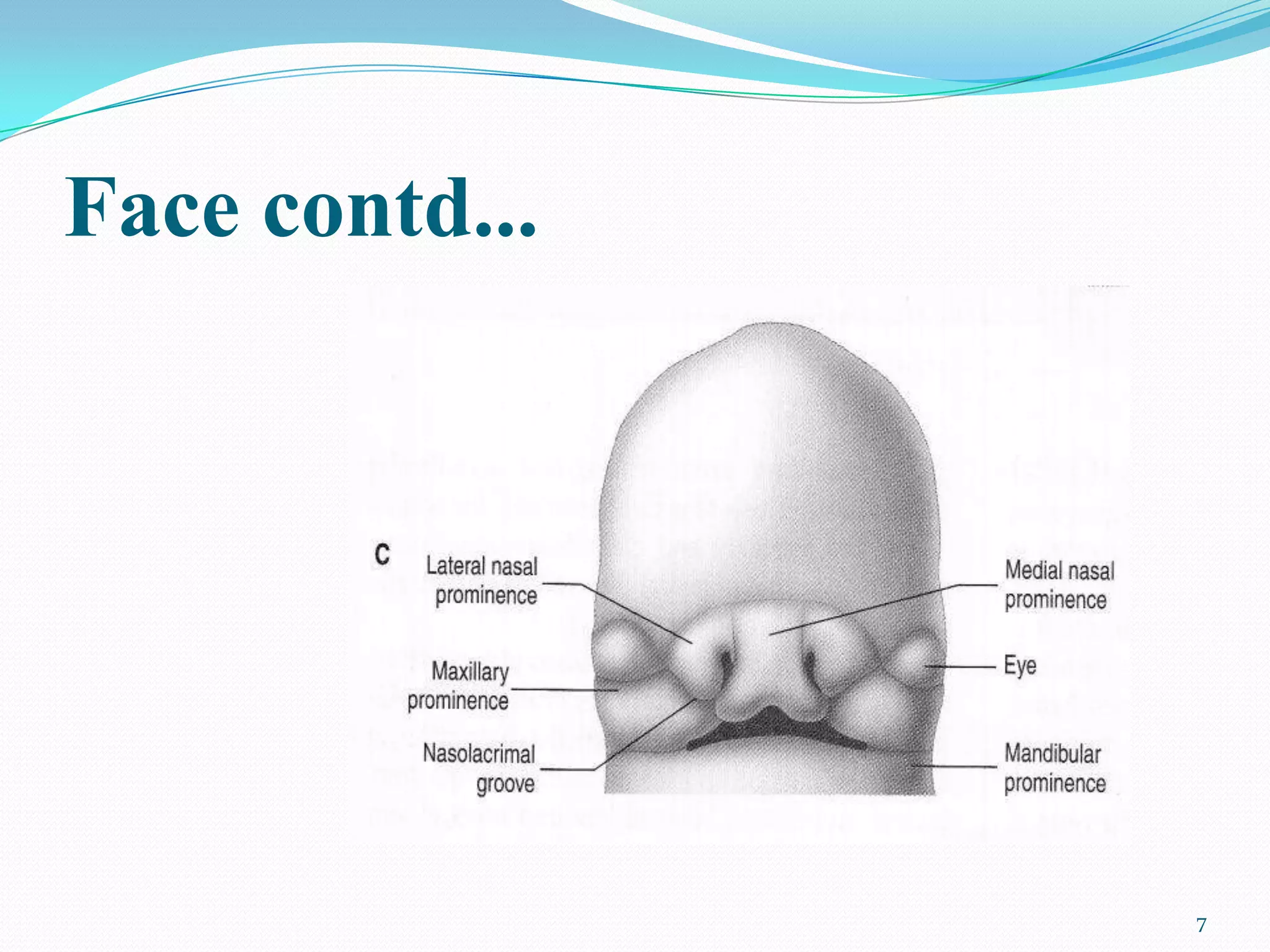
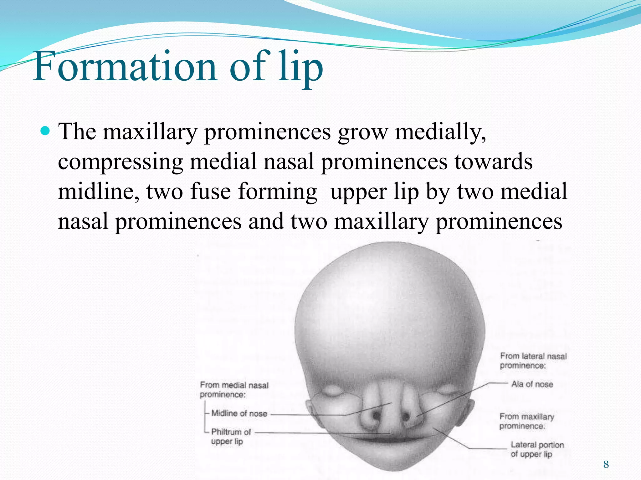
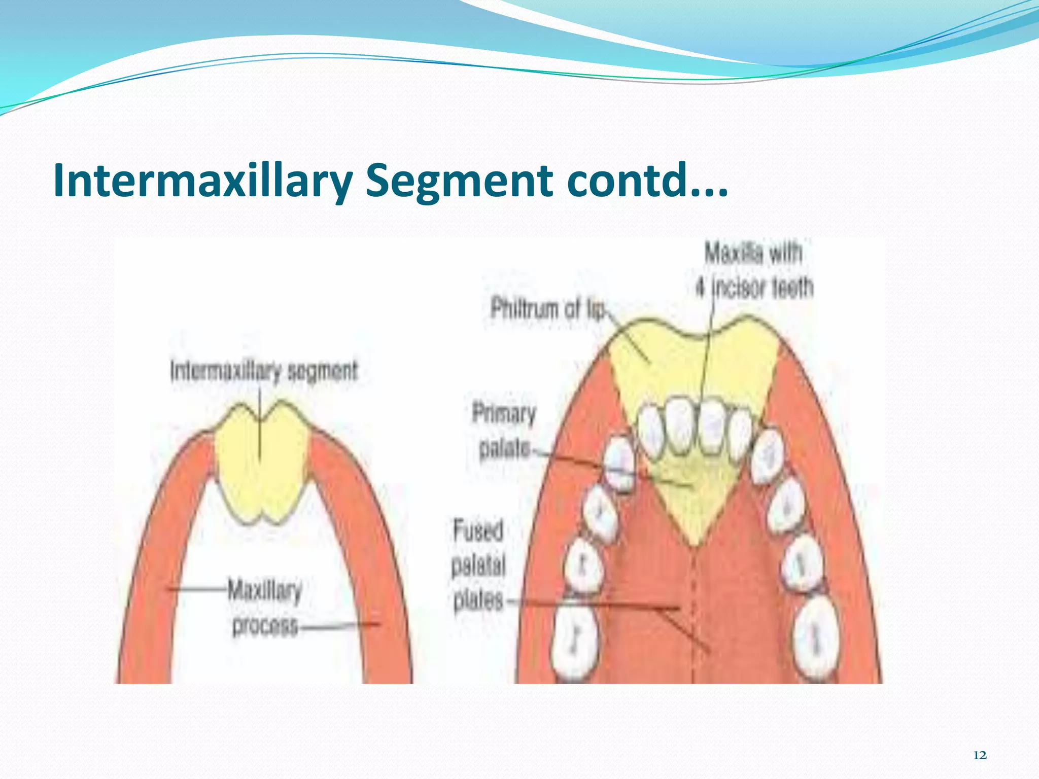
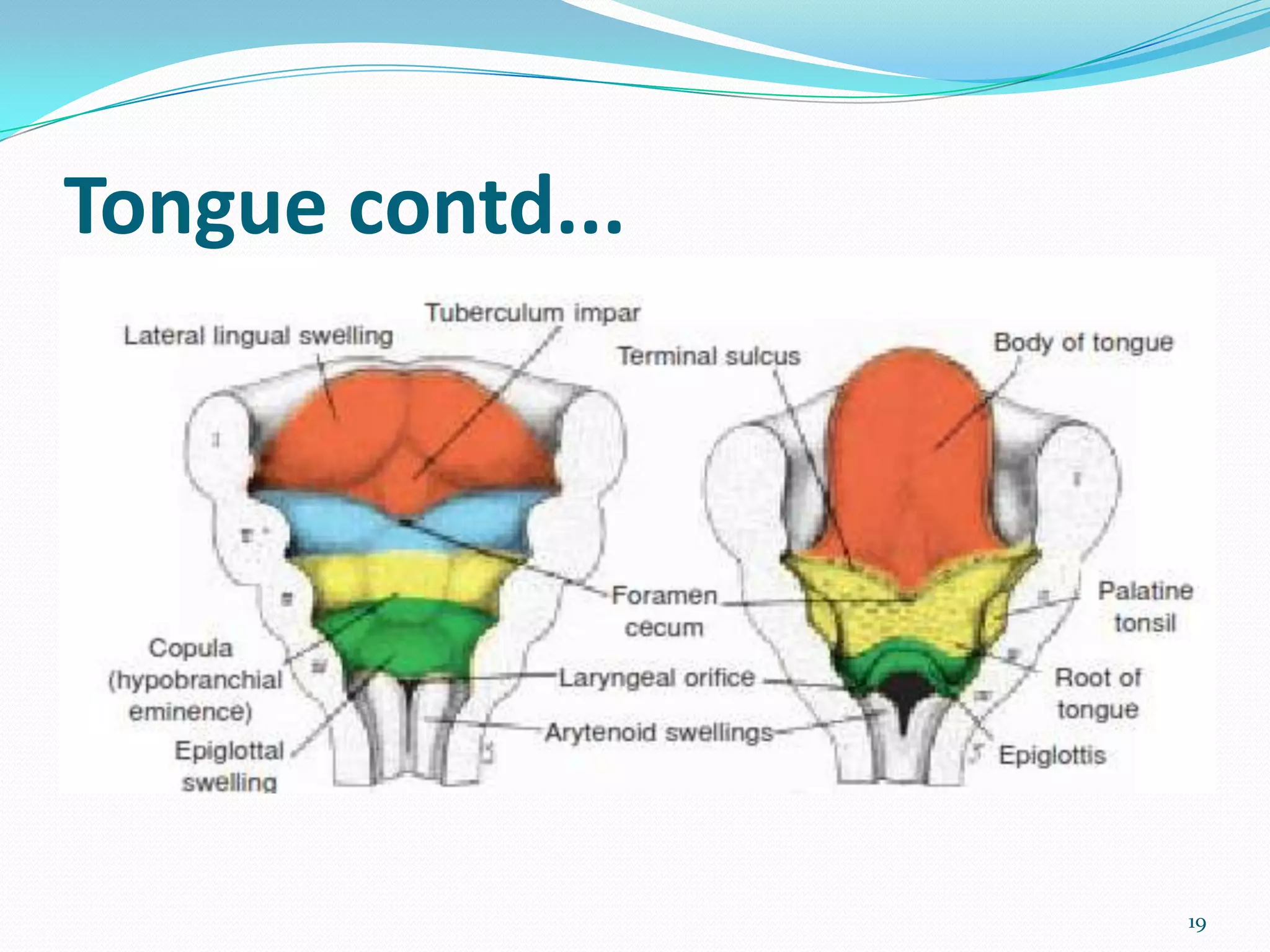
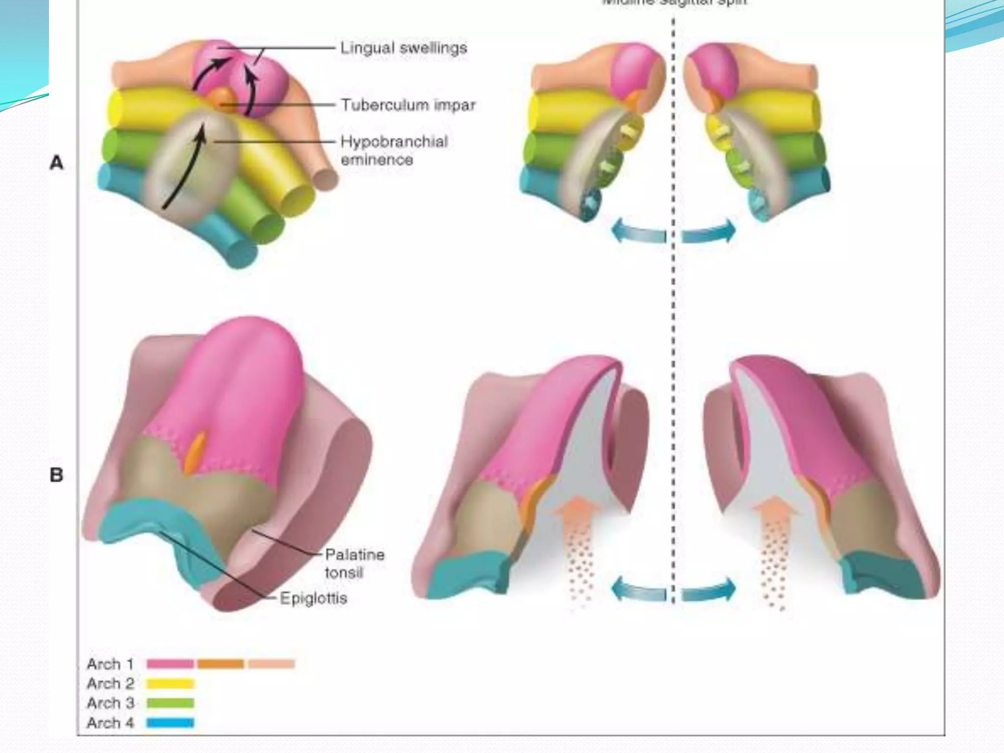
The document summarizes the development of the face and its structures from the 4th to 7th weeks of gestation. It describes how facial prominences like the maxillary and mandibular prominences form and contribute to structures like the upper lip, nose, and palate. It also discusses the development of the tongue from lingual swellings and how various nerves innervate parts of the developing face and tongue.
Face develops in humans between 4th – 10th week of intrauterine life.
prenatal growth of the maxilla
DEVELOPMENT OF UPPER LIP
Development of lower lip
Development of nose
hare lip
OBLIQUE FACIAL CLEFT
macrostomia
lateral facial cleft
microstomia
Face develops in humans between 4th – 10th week of intrauterine life.
prenatal growth of the maxilla
DEVELOPMENT OF UPPER LIP
Development of lower lip
Development of nose
hare lip
OBLIQUE FACIAL CLEFT
macrostomia
lateral facial cleft
microstomia
Central face begins to develop by 4th week, when olfactory placodes appear on both sides of the frontonasal process.
Gradually both placodes develop to form the median and lateral nasal process.
Upper lip is formed by 6th week by fusion of two median nasal processes in midline and the maxilllary process of the 1st branchial arch.
PRE-NATAL GROWTH AND DEVELOPMENT OF PALATEFormation of primary and secondary palate
Elevation of palatal shelves
Fusion of palatal shelves
2. Face
At the end of the 4th week, facial prominences consisting
primarily of neural crest-derived mesenchyme appear
Maxillary prominences- lateral to the stomodeum
mandibular prominences- caudal to stomodeum
2
3. Face contd...
Frontonasal prominence - proliferation of mesenchyme
ventral to the brain vesicles
On both sides, local thickenings of the surface ectoderm,
the nasal placodes originate
3
4.
5. Face contd...
During the 5th week, the nasal placodes invaginate to
form nasal pits
Lateral nasal prominences
Medial nasal prominences
5
8. Formation of lip
The maxillary prominences grow medially,
compressing medial nasal prominences towards
midline, two fuse forming upper lip by two medial
nasal prominences and two maxillary prominences
8
9.
10. Development of nose
Maxillary prominences enlarge to form the cheeks and
maxilla
Nose is formed from five facial prominences:
- frontal prominence gives rise to bridge
- merged medial nasal prominences provide the
crest and tip
- lateral nasal prominences form the alae
10
11. Intermaxillary Segment
Two medial nasal prominences merge at a deeper level
forming the intermaxillary segment
Composed of
(a) philtrum of the upper lip
(b) upper jaw component- carries the four incisor teeth
(c) primary palate
11
13. Secondary Palate
Formed by two palatine shelves (6th week of
development) –maxillary process
Directed obliquely downward on each side of the tongue
In the 7th week, the palatine shelves ascend to attain a
horizontal position above the tongue and fuse, forming the
secondary palate
13
18. Development of Tongue
Appear at 4 weeks
As two lateral lingual swellings and one medial swelling,
the tuberculum impar (all originate from the first
pharyngeal arch)
A second median swelling, the copula, formed by
mesoderm of the second, third, and part of the fourth arch
18
20. Tongue contd...
Lateral lingual swellings increase in size, overgrow the
tuberculum impar and merge, forming the anterior two-
thirds, or body, of the tongue
The body of the tongue is separated from the posterior
third by a V-shaped groove, the terminal sulcus
20
21.
22. Tongue contd...
A third median swelling, formed by the posterior part of
the fourth arch, marks development of the epiglottis
22
24. Tongue contd...
The mucosa covering the body of the tongue originates
from the first pharyngeal arch
Sensory innervation to the posterior part, or root part of
the tongue is supplied by the glossopharyngeal nerve
24
25. Tongue contd...
The epiglottis and the extreme posterior part of the tongue
are innervated by the superior laryngeal nerve, reflecting
their development from the fourth arch
Most tongue muscles are derived from myoblasts
originating in occipital somites. So innervated by the
hypoglossal nerve
25
26. Tongue contd...
Special sensory innervation (taste) to the anterior two
thirds of the tongue is provided by the chorda tympani
branch of the facial nerve, while the posterior third is
supplied by the glossopharyngeal nerve
26
On both sides, local thickenings of the surface ectoderm, the nasal (olfactory) placodes, originate under inductive influence of the ventral portion of the forebrain
In so doing, they create a ridge of tissue that surrounds each pit and forms the nasal prominences
medial growth of the maxillary prominences helps two medial nasal prominences merge at a deeper level forming the intermaxillary segment
sensory innervation to the posterior part, or root part of the tongue is supplied by the glossopharyngeal nerveindicates thattissue of the third arch overgrows that of the second