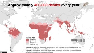
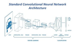
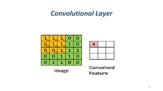
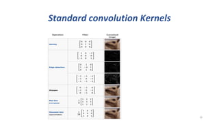
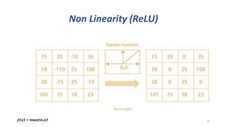
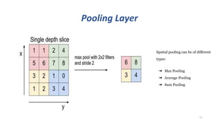
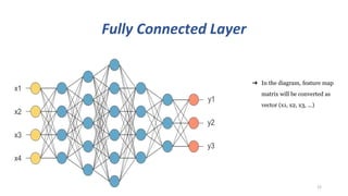
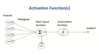
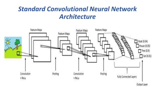
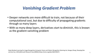
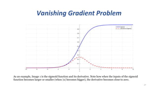
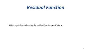
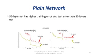
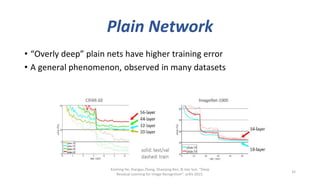
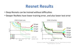
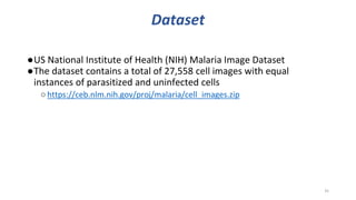
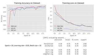
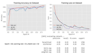
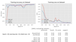
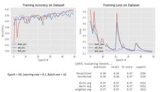
Detecting malaria using a Deep Residual
Convolutional Neural Network (ResNet)
Presented by
Yusuf Brima
MSc. Computer Science and Engineering
yusufbrima@cse.du.ac.bd
Jargis Ahmed,
MSc. Computer Science and Engineering
jargisahmed@gmail.com
2
3
Current Diagnostic Methods:
Microscopy
• Gold standard of malaria diagnosis (Murphy et al, 2013)
• High positive and negative predictive powers
• Uses microscopic examination giemsa stained in blood
film
✔ painstaking counting of bacteria cells!
• Long patient waiting period
• Invasive
• Requires expertise (competency)
4
Current Diagnostic Methods:
Rapid Diagnostic Test (RDT)
• Introduced as a front-line defense mechanism for malaria
diagnosis
• Malaria RDT or “dipstick RDT detects specific antigens or
proteins developed by malaria parasites
• Uses Principle-lateral flow or immunochromatographic
stick method
• Presence of antigen is detected by variational colour
change on absorbing nitrocellulose strip
• Has a lower sensitivity and specificity relative to
Microscopy approach
5
Problem Context
• How can we reduce the wait time and
improve the sensitivity and specificity of
malaria diagnostic process?
•Deep Learning!
6
Related Literature
•A simple and efficient method for parasite and
erythrocyte detection in thin blood images was proposed;
the approach is based on a classification process that finds
boundaries that optimally separate a given color space in
three classes, namely, background, erythrocyte and
parasite.(DíazFabio & Romero, 2007).
7
Related Literature
•The assessment was also carried out in terms of precision and
recall and combined in the F-measure providing results
generally in the range of 92% to 97% for a variety of smears. In
this context the observed trade-off relation between precision
and recall guaranteed stable results. Finally, relating the F-
measure with the degree of cell overlaps, showed that up to
50% total cell overlap can be tolerated if the smear image is
well-focused and the smear itself adequately stained(Le, et al.,
2008).
8
Related Literature
•Two back propagation Artificial Neural Network models (3
layers and 4 layers) was employed together with image analysis
techniques to evaluate the accuracy of the classification in the
recognition of medical image patterns associated with
morphological features of erythrocytes in the blood. The three
layers Artificial Neural Network (ANN) architecture had the best
performance with an error of 2.74545e-005 and 86.54% correct
recognition rate (Hirimutugoda & Wijayarathna, 2010).
9
Related Literature
•This programme has been validated for use in estimation of
parasitemia in mouse infection by Plasmodium yoelii and used
to monitor parasitaemia on a daily basis for an entire challenge
infection(Ma, et al., 2010).
10
Related Literature
•The image-based method is tested over more than 500 images
from two independent laboratories; overall sensitivity to
capture cases of malaria is 100% and specificity ranges from 50-
88% for all species of malaria parasites. (Purwar, et al., 2011).
11
Related Literature
•The researchers proposed an identification system of
plasmodium falciparum development phase in preparations of
red blood cells infected with malaria has been designed the
study. The results accuracy of the system in identifying the
plasmodium falciparum development phase is 87.67%
(Pamungkas, et al., 2015).
12
Related Literature
•The training and evaluation have been carried out on image
dataset with respect to ground truth data, determining the
degree of infection with the sensitivity of 98 % and specificity of
97 %. The accuracy and efficiency of the proposed scheme in
the context of being automatic were proved experimentally,
surpassing other state-of-the-art schemes (Abbas, et al., 2018).
13
Standard Convolutional Neural Network
Architecture
14
Image Data
15
Array of RGB Matrix
Convolutional Layer
16
Convolutional Layer
17
Convolutional Layer
18
Standard convolution Kernels
19
Non Linearity (ReLU)
20ƒ(x) = max(0,x)
Pooling Layer
21
Spatial pooling can be of different
types:
➔ Max Pooling
➔ Average Pooling
➔ Sum Pooling
Fully Connected Layer
22
➔ In the diagram, feature map
matrix will be converted as
vector (x1, x2, x3, …)
Activation Function(s)
23
Standard Convolutional Neural Network
Architecture
24
Proposed Model
• Deep Residual Learning for Image Recognition (He et al, 2015)1
✔Best paper of the year (CVPR)
• Deep Learning fallacy: The deeper network can cover more complex
problems!
• However, training the deeper network is more difficult because of
vanishing/exploding gradients problem
25
Deep Residual Learning for Image Recognition,Computer Vision and Pattern Recognition (Kaiming He, Xiangyu Zhang, Shaoqing Ren
and Jian Sun, 2015),Microsoft Research Asia (MSRA) (https://arxiv.org/abs/1603.05027)
Vanishing Gradient Problem
• Deeper networks are more difficult to train, not because of their
computational cost, but due to difficulty of propagating gradients
through so many layers
• With so many deep layers, derivatives start to diminish, this is known
as the gradient vanishing problem
26
Deep Residual Learning for Image Recognition,Computer Vision and Pattern Recognition (Kaiming He, Xiangyu Zhang, Shaoqing Ren
and Jian Sun, 2015),Microsoft Research Asia (MSRA) (https://arxiv.org/abs/1603.05027)
Vanishing Gradient Problem
27
As an example, Image 1 is the sigmoid function and its derivative. Note how when the inputs of the sigmoid
function becomes larger or smaller (when |x| becomes bigger), the derivative becomes close to zero.
Rectified Linear Units (ReLU) function: a
possible solution to the Vanishing Gradient
Problem
28
Residual Function
29
Residual connections are simply connections between a layer and layers after the next. The residual connections on
the other hand takes the feature map from layer t and adds it to the output of layer t + 2.
Residual Function
30
This is equivalent to learning the residual function y = f(x) + x
Plain Network
• 56-layer net has higher training error and test error than 20-layers
net
Kaiming He, Xiangyu Zhang, Shaoqing Ren, & Jian Sun. “Deep
Residual Learning for Image Recognition”. arXiv 2015.
31
Plain Network
• “Overly deep” plain nets have higher training error
• A general phenomenon, observed in many datasets
Kaiming He, Xiangyu Zhang, Shaoqing Ren, & Jian Sun. “Deep
Residual Learning for Image Recognition”. arXiv 2015.
32
Resnet Results
• Deep Resnets can be trained without difficulties
• Deeper ResNets have lower training error, and also lower test error
Kaiming He, Xiangyu Zhang, Shaoqing Ren, & Jian Sun. “Deep Residual Learning for
Image Recognition”. arXiv 2015.
ResNets Results cont’d
• Deep Resnets can be trained without difficulties
• Deeper ResNets have lower training error, and also lower test error
Kaiming He, Xiangyu Zhang, Shaoqing Ren, & Jian Sun. “Deep Residual Learning for
Image Recognition”. arXiv 2015.
Dataset
●US National Institute of Health (NIH) Malaria Image Dataset
●The dataset contains a total of 27,558 cell images with equal
instances of parasitized and uninfected cells
○https://ceb.nlm.nih.gov/proj/malaria/cell_images.zip
35
Our findings
36
Epoch = 20, Learning rate = 0.05, Batch size = 32
Epoch = 20, Learning rate = 0.1, Batch size = 32
Epoch = 20, Learning rate = 0.2, Batch size = 32
Epoch = 50, Learning rate = 0.1, Batch size = 32
Conclusion
• One of the barriers toward a successful mortality reduction has been
inadequate malaria diagnosis in particular
• To improve diagnosis, image analysis software and machine learning
methods have been used to quantify parasitemia in microscopic
blood slides
41
References
• Abbas, N. et al., 2018. Machine aided malaria parasitemia detection in Giemsa-stained thin blood smears. Neural Computing and
Applications, 29(3), p. 803–818.
• DíazFabio, G. & Romero, G. E., 2007. Infected Cell Identification in Thin Blood Images Based on Color Pixel Classification: Comparison
and Analysis. Iberoamerican Congress on Pattern Recognition, pp. 812-821.
• Hirimutugoda, Y. M. & Wijayarathna, G., 2010. Image Analysis System for Detection of Red Cell Disorders Using Artificial. Sri Lanka
Journal of Bio-Medical Informatics, 1(1), pp. 35-42.
• Le, M.-T., Bretschneider, T. R., Kuss, C. & Preiser, P. R., 2008. A novel semi-automatic image processing approach to determine
Plasmodium falciparum parasitemia in Giemsa-stained thin blood smears. BMC Molecular and Cell Biology, 9(15).
• Ma, C., Harrison, P., Wang, L. & Coppel, R. L., 2010. Automated estimation of parasitaemia of Plasmodium yoelii-infected mice by digital
image analysis of Giemsa-stained thin blood smears. Malaria Journal, 9(348).
• Pamungkas, A., Adi, K. & Gernowo, R., 2015. Identification of Plasmodium Falciparum Development Phase in Malaria Infected Red Blood
Cells using Adaptive Color Segmentation and Decision Tree based Classification. International Journal of Applied Engineering Research,
10(2), pp. 4043-4055.
• Prasad, K. et al., 2012. Image Analysis Approach for Development of a Decision Support System for Detection of Malaria Parasites in
Thin Blood Smear Images. Journal of Digital Imaging, 25(4), p. 542–549.
• Purwar, Y. et al., 2011. Automated and unsupervised detection of malarial parasites in microscopic images. Malaria Journal, 110(364).
42
Thank you
for your attention!
43

Detecting malaria using a deep convolutional neural network

  • 1.
    Detecting malaria usinga Deep Residual Convolutional Neural Network (ResNet) Presented by Yusuf Brima MSc. Computer Science and Engineering yusufbrima@cse.du.ac.bd Jargis Ahmed, MSc. Computer Science and Engineering jargisahmed@gmail.com
  • 2.
  • 3.
  • 4.
    Current Diagnostic Methods: Microscopy •Gold standard of malaria diagnosis (Murphy et al, 2013) • High positive and negative predictive powers • Uses microscopic examination giemsa stained in blood film ✔ painstaking counting of bacteria cells! • Long patient waiting period • Invasive • Requires expertise (competency) 4
  • 5.
    Current Diagnostic Methods: RapidDiagnostic Test (RDT) • Introduced as a front-line defense mechanism for malaria diagnosis • Malaria RDT or “dipstick RDT detects specific antigens or proteins developed by malaria parasites • Uses Principle-lateral flow or immunochromatographic stick method • Presence of antigen is detected by variational colour change on absorbing nitrocellulose strip • Has a lower sensitivity and specificity relative to Microscopy approach 5
  • 6.
    Problem Context • Howcan we reduce the wait time and improve the sensitivity and specificity of malaria diagnostic process? •Deep Learning! 6
  • 7.
    Related Literature •A simpleand efficient method for parasite and erythrocyte detection in thin blood images was proposed; the approach is based on a classification process that finds boundaries that optimally separate a given color space in three classes, namely, background, erythrocyte and parasite.(DíazFabio & Romero, 2007). 7
  • 8.
    Related Literature •The assessmentwas also carried out in terms of precision and recall and combined in the F-measure providing results generally in the range of 92% to 97% for a variety of smears. In this context the observed trade-off relation between precision and recall guaranteed stable results. Finally, relating the F- measure with the degree of cell overlaps, showed that up to 50% total cell overlap can be tolerated if the smear image is well-focused and the smear itself adequately stained(Le, et al., 2008). 8
  • 9.
    Related Literature •Two backpropagation Artificial Neural Network models (3 layers and 4 layers) was employed together with image analysis techniques to evaluate the accuracy of the classification in the recognition of medical image patterns associated with morphological features of erythrocytes in the blood. The three layers Artificial Neural Network (ANN) architecture had the best performance with an error of 2.74545e-005 and 86.54% correct recognition rate (Hirimutugoda & Wijayarathna, 2010). 9
  • 10.
    Related Literature •This programmehas been validated for use in estimation of parasitemia in mouse infection by Plasmodium yoelii and used to monitor parasitaemia on a daily basis for an entire challenge infection(Ma, et al., 2010). 10
  • 11.
    Related Literature •The image-basedmethod is tested over more than 500 images from two independent laboratories; overall sensitivity to capture cases of malaria is 100% and specificity ranges from 50- 88% for all species of malaria parasites. (Purwar, et al., 2011). 11
  • 12.
    Related Literature •The researchersproposed an identification system of plasmodium falciparum development phase in preparations of red blood cells infected with malaria has been designed the study. The results accuracy of the system in identifying the plasmodium falciparum development phase is 87.67% (Pamungkas, et al., 2015). 12
  • 13.
    Related Literature •The trainingand evaluation have been carried out on image dataset with respect to ground truth data, determining the degree of infection with the sensitivity of 98 % and specificity of 97 %. The accuracy and efficiency of the proposed scheme in the context of being automatic were proved experimentally, surpassing other state-of-the-art schemes (Abbas, et al., 2018). 13
  • 14.
    Standard Convolutional NeuralNetwork Architecture 14
  • 15.
  • 16.
  • 17.
  • 18.
  • 19.
  • 20.
  • 21.
    Pooling Layer 21 Spatial poolingcan be of different types: ➔ Max Pooling ➔ Average Pooling ➔ Sum Pooling
  • 22.
    Fully Connected Layer 22 ➔In the diagram, feature map matrix will be converted as vector (x1, x2, x3, …)
  • 23.
  • 24.
    Standard Convolutional NeuralNetwork Architecture 24
  • 25.
    Proposed Model • DeepResidual Learning for Image Recognition (He et al, 2015)1 ✔Best paper of the year (CVPR) • Deep Learning fallacy: The deeper network can cover more complex problems! • However, training the deeper network is more difficult because of vanishing/exploding gradients problem 25 Deep Residual Learning for Image Recognition,Computer Vision and Pattern Recognition (Kaiming He, Xiangyu Zhang, Shaoqing Ren and Jian Sun, 2015),Microsoft Research Asia (MSRA) (https://arxiv.org/abs/1603.05027)
  • 26.
    Vanishing Gradient Problem •Deeper networks are more difficult to train, not because of their computational cost, but due to difficulty of propagating gradients through so many layers • With so many deep layers, derivatives start to diminish, this is known as the gradient vanishing problem 26 Deep Residual Learning for Image Recognition,Computer Vision and Pattern Recognition (Kaiming He, Xiangyu Zhang, Shaoqing Ren and Jian Sun, 2015),Microsoft Research Asia (MSRA) (https://arxiv.org/abs/1603.05027)
  • 27.
    Vanishing Gradient Problem 27 Asan example, Image 1 is the sigmoid function and its derivative. Note how when the inputs of the sigmoid function becomes larger or smaller (when |x| becomes bigger), the derivative becomes close to zero.
  • 28.
    Rectified Linear Units(ReLU) function: a possible solution to the Vanishing Gradient Problem 28
  • 29.
    Residual Function 29 Residual connectionsare simply connections between a layer and layers after the next. The residual connections on the other hand takes the feature map from layer t and adds it to the output of layer t + 2.
  • 30.
    Residual Function 30 This isequivalent to learning the residual function y = f(x) + x
  • 31.
    Plain Network • 56-layernet has higher training error and test error than 20-layers net Kaiming He, Xiangyu Zhang, Shaoqing Ren, & Jian Sun. “Deep Residual Learning for Image Recognition”. arXiv 2015. 31
  • 32.
    Plain Network • “Overlydeep” plain nets have higher training error • A general phenomenon, observed in many datasets Kaiming He, Xiangyu Zhang, Shaoqing Ren, & Jian Sun. “Deep Residual Learning for Image Recognition”. arXiv 2015. 32
  • 33.
    Resnet Results • DeepResnets can be trained without difficulties • Deeper ResNets have lower training error, and also lower test error Kaiming He, Xiangyu Zhang, Shaoqing Ren, & Jian Sun. “Deep Residual Learning for Image Recognition”. arXiv 2015.
  • 34.
    ResNets Results cont’d •Deep Resnets can be trained without difficulties • Deeper ResNets have lower training error, and also lower test error Kaiming He, Xiangyu Zhang, Shaoqing Ren, & Jian Sun. “Deep Residual Learning for Image Recognition”. arXiv 2015.
  • 35.
    Dataset ●US National Instituteof Health (NIH) Malaria Image Dataset ●The dataset contains a total of 27,558 cell images with equal instances of parasitized and uninfected cells ○https://ceb.nlm.nih.gov/proj/malaria/cell_images.zip 35
  • 36.
  • 37.
    Epoch = 20,Learning rate = 0.05, Batch size = 32
  • 38.
    Epoch = 20,Learning rate = 0.1, Batch size = 32
  • 39.
    Epoch = 20,Learning rate = 0.2, Batch size = 32
  • 40.
    Epoch = 50,Learning rate = 0.1, Batch size = 32
  • 41.
    Conclusion • One ofthe barriers toward a successful mortality reduction has been inadequate malaria diagnosis in particular • To improve diagnosis, image analysis software and machine learning methods have been used to quantify parasitemia in microscopic blood slides 41
  • 42.
    References • Abbas, N.et al., 2018. Machine aided malaria parasitemia detection in Giemsa-stained thin blood smears. Neural Computing and Applications, 29(3), p. 803–818. • DíazFabio, G. & Romero, G. E., 2007. Infected Cell Identification in Thin Blood Images Based on Color Pixel Classification: Comparison and Analysis. Iberoamerican Congress on Pattern Recognition, pp. 812-821. • Hirimutugoda, Y. M. & Wijayarathna, G., 2010. Image Analysis System for Detection of Red Cell Disorders Using Artificial. Sri Lanka Journal of Bio-Medical Informatics, 1(1), pp. 35-42. • Le, M.-T., Bretschneider, T. R., Kuss, C. & Preiser, P. R., 2008. A novel semi-automatic image processing approach to determine Plasmodium falciparum parasitemia in Giemsa-stained thin blood smears. BMC Molecular and Cell Biology, 9(15). • Ma, C., Harrison, P., Wang, L. & Coppel, R. L., 2010. Automated estimation of parasitaemia of Plasmodium yoelii-infected mice by digital image analysis of Giemsa-stained thin blood smears. Malaria Journal, 9(348). • Pamungkas, A., Adi, K. & Gernowo, R., 2015. Identification of Plasmodium Falciparum Development Phase in Malaria Infected Red Blood Cells using Adaptive Color Segmentation and Decision Tree based Classification. International Journal of Applied Engineering Research, 10(2), pp. 4043-4055. • Prasad, K. et al., 2012. Image Analysis Approach for Development of a Decision Support System for Detection of Malaria Parasites in Thin Blood Smear Images. Journal of Digital Imaging, 25(4), p. 542–549. • Purwar, Y. et al., 2011. Automated and unsupervised detection of malarial parasites in microscopic images. Malaria Journal, 110(364). 42
  • 43.
    Thank you for yourattention! 43