Download as PDF, PPTX

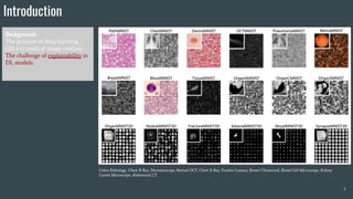
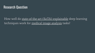
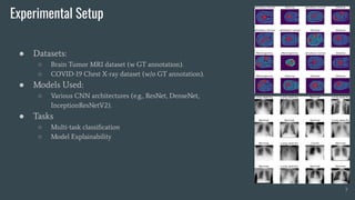
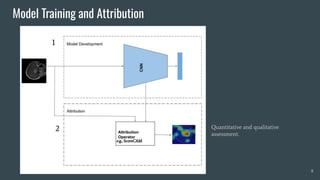
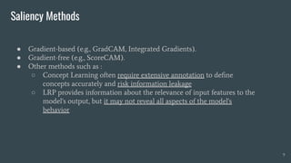
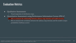
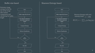
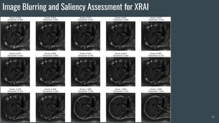
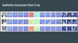
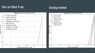


The document discusses the evaluation of explainable deep learning techniques in medical image analysis, focusing on saliency methods applied to datasets like brain tumor MRI and COVID-19 chest X-rays. It highlights the significance of explainability for clinical adoption and the lack of quantitative analysis in existing methods. Key findings include the effectiveness of specific saliency methods and the proposal of a framework combining qualitative and quantitative assessments to enhance model explainability.


![AIMS Block Presentation]{Deep Transfer Learning for Magnetic Resonance Image ...](https://cdn.slidesharecdn.com/ss_thumbnails/aimspresentation-210209214939-thumbnail.jpg?width=640&height=640&fit=bounds)


![PERI-PROSTHETIC FRACTURE NAIL-PLATE CONSTRUCT [NPC].pptx](https://cdn.slidesharecdn.com/ss_thumbnails/drarunkumardrmohamedashrafperiprostheticfrasturenail-plateconstructnpc-260209164459-7e9d15a1-thumbnail.jpg?width=640&height=640&fit=bounds)


