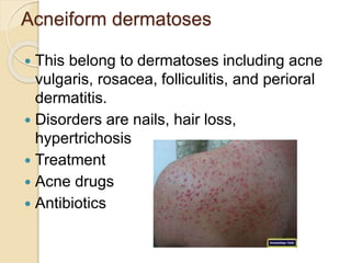
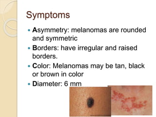
• Dermotoxicants, their toxico kinetics, mechanisms
of toxicity and treatment
•
Contents
• Introduction
• Anatomy of skin
• Percutaneous absorption
• Factors effecting stratum corneum
• Manifestation
• Contact dermatitis
• Ulcers
 Utricaria
 Toxic epidermal necrolysis
 Acneiform dermatoses
 Pigment disturbances
 Skin cancer
Introduction
 Dermal toxicology
 Also known as cutaneous toxicity is the ability of
substance to poison people or animals by contact with
skin. Toxic materials absorb through the skin to various
degrees depending on their chemical composition and
whether they are dissolved in solvent.
Anatomy of skin
 Skin is composed of three primary layers
1. The epidermis
2. The dermis
3. The hypodermis
Percutaneous Absorption
• To be absorbed through the skin, a toxicant
must pass through the epidermis or the
appendages (sweat and sebaceous glands
and hair follicles).
Once absorbed through the skin, toxicants
must pass through several tissue layers
before entering the small blood and lymph
capillaries in the dermis
 The rate-determining barrier in the
dermal absorption of chemicals is the
epidermis—especially the stratum
corneum (horny layer), the upper most
layer of the epidermis.
• Once a toxicant is absorbed through
the stratum corneum, absorption
through the other epidermal layers is
rapid.
All toxicants move across the stratum
corneum by passive diffusion
• Polar substances diffuse through the outer surface
of protein filaments of the hydrated stratum
corneum.
• Non-polar molecules dissolve and diffuse through
the lipid matrix between protein filaments.
• The rate of diffusion is proportional to lipid solubility
and inversely proportional to molecular weight.
 Once absorbed, the toxicant enters the
systemic circulation by-passing first-pass
metabolism
Factors that Affect Stratum Corneum
Absorption of Toxicants
1. Hydration of the stratum corneum
• The stratum corneum is normally 7% hydrated which greatly
increases permeability of toxicants. (10-fold better than
completely dry skin)
• On additional contact with water, toxicant absorption can
increase by 2- to 3-fold.
 2. Damage to the stratum corneum
• Acids, alkalis and mustard gases injure the epidermis and
increase absorption of toxicants.
• Burns and skin diseases can increase permeability to
toxicants.
 3. Solvent Administration
• Carrier solvents and creams can aid in increased absorption
of toxicants and drugs (e.g. dimethylsulfoxide (DMSO)).
Special Routes of Exposure
 Toxicants usually enter the bloodstream after
absorption through the skin, lungs or GI tract.
Special routes include:
 1. Subcutaneous injection (SC) (under the skin)
◦ -by-passes the epidermal barrier, slow absorption but directly into
systemic circulation; affected by blood flow
◦ 2. Intramuscular injection (IM) (into muscle)
◦ -slower absorption than IP but steady and directly into systemic
circulation; affected by blood flow
◦ 3. Intraperitoneal injection (IP) (into the peritoneal cavity)
◦ -quick absorption due to high vascularization and large surface area
◦ -absorbed primarily into the portal circulation (to liver—first-pass
metabolism) as well as directly into the systemic circulation.
 4. Intravenous injection (IV) (into blood stream) -
directly into systemic circulation
Manifestation
 Contact dermatitis
 Ulcers
 Utricaria
 Toxic epidermal necrolysis
 Acneiform dermatoses
 Pigment disturbances
 Skin cancer
Contact dermatitis
 Most common occupational disease
 Symptoms
 Hives, reddening of the skin (erythema),
rashes, hyperkeratosis (thickening of the
skin), dryness and roughness of skin
 Treatment
 Avoiding the amount of exposure to the
irritant
 Wearing gloves
 Hand washing
 Avoiding from chemicals
Ulcers
 Some chemicals cause ulceration of the
skin
 It involves sloughing of the epidermis and
damage to the exposed dermis
 It is cause by
 Acids, burns, trauma and can occur on
mucous membranes and skin
 Plants and trees,
 rubber products, leather
Treatment
 Treatment
 remove any excess discharge
 maintain a moist wound environment
 Using of antibiotics
 Change diet
 Recommended exercise
 Stop smoking and loose wieght
Utricaria
 It is a kind of skin rash
 They are frequently caused by allergic
reactions
 Chronic urticaria (hives lasting longer than
six weeks) is rarely due to an allergy.
 Symptoms
 Wheals appear on the
surface of skin
 Edema of the upper dermis occur
Treatment
 Don’t eat foods that have been
identified to cause your symptoms.
 Avoid harsh soaps. Frequent baths
may reduce itching and scratching
 Avoid tight clothing
 Wear protective clothing; apply
sunblock.
Toxic epidermal necrolysis
 also known as Lyell's syndrome, is a rare,
life-threatening skin condition that is usually
caused by a reaction to drugs.
 Symptoms
 Prodrome
 Skin Findings
 Mucosal Findings
 Treatment
 intravenous immunoglobulins (IVIG)
treatment is necesaary
Acneiform dermatoses
 This belong to dermatoses including acne
vulgaris, rosacea, folliculitis, and perioral
dermatitis.
 Disorders are nails, hair loss,
hypertrichosis
 Treatment
 Acne drugs
 Antibiotics
Pigment disturbances
 it includes
 hyperpigmentary disorders (darkening of
the skin) and
 hypopigmentary disorders (decrease in
the normal skin color)
 Disorders
 Albinism
 Melasma
 Pigment loss after skin
 vitligo
Tratment
 Avoid sunlight
 Applying of cream
 Cosmetics
 Light sensitive drugs
Skin cancer
 Skin cancers are cancers that arise
from the skin. They are due to the
development of abnormal cells that
have the ability to invade or spread to
other parts of the body.[
 There are three main types:
 basal-cell cancer (BCC)
 squamous-cell cancer (SCC)
 and melanoma
Symptoms
 Asymmetry: melanomas are rounded
and symmetric
 Borders: have irregular and raised
borders.
 Color: Melanomas may be tan, black
or brown in color
 Diameter: 6 mm
Treatment
 Surgery. Most basal cell and
squamous cell skin cancers can be
successfully treated with surgery. ...
 Radiotherapy. ...
 Chemotherapy. ...
 Immunotherapy. ...
 Photodynamic therapy (PDT)
Toxicant present in foods which
cause dermal disease
 Bleached starch: Can be used in many
dairy products. Thought to be related to
asthma and skin irritations.
 BHT
 It cause liver cancer
 Potassium bromate: Added to breads to
increase volume. Linked to cancer in
humans.
 Carrageenan: Stabilizer and thickening
agent used in many prepared foods. Can
cause ulcers and cancer
 Tert butylhydroquinone: Used to
preserve fish products. Could cause
stomach tumors at high doses.
 Aluminum: A preservative in some
packaged foods that can cause
cancer.
 Agave nectar: Sweetener derived
from a cactus. Contains high levels of
fructose, which causes insulin
resistance, liver disease and
Dermo toxicology

Dermo toxicology

  • 1.
    • Dermotoxicants, theirtoxico kinetics, mechanisms of toxicity and treatment •
  • 2.
    Contents • Introduction • Anatomyof skin • Percutaneous absorption • Factors effecting stratum corneum • Manifestation • Contact dermatitis • Ulcers  Utricaria  Toxic epidermal necrolysis  Acneiform dermatoses  Pigment disturbances  Skin cancer
  • 3.
    Introduction  Dermal toxicology Also known as cutaneous toxicity is the ability of substance to poison people or animals by contact with skin. Toxic materials absorb through the skin to various degrees depending on their chemical composition and whether they are dissolved in solvent.
  • 4.
    Anatomy of skin Skin is composed of three primary layers 1. The epidermis 2. The dermis 3. The hypodermis
  • 5.
    Percutaneous Absorption • Tobe absorbed through the skin, a toxicant must pass through the epidermis or the appendages (sweat and sebaceous glands and hair follicles). Once absorbed through the skin, toxicants must pass through several tissue layers before entering the small blood and lymph capillaries in the dermis
  • 6.
     The rate-determiningbarrier in the dermal absorption of chemicals is the epidermis—especially the stratum corneum (horny layer), the upper most layer of the epidermis. • Once a toxicant is absorbed through the stratum corneum, absorption through the other epidermal layers is rapid.
  • 7.
    All toxicants moveacross the stratum corneum by passive diffusion • Polar substances diffuse through the outer surface of protein filaments of the hydrated stratum corneum. • Non-polar molecules dissolve and diffuse through the lipid matrix between protein filaments. • The rate of diffusion is proportional to lipid solubility and inversely proportional to molecular weight.  Once absorbed, the toxicant enters the systemic circulation by-passing first-pass metabolism
  • 8.
    Factors that AffectStratum Corneum Absorption of Toxicants 1. Hydration of the stratum corneum • The stratum corneum is normally 7% hydrated which greatly increases permeability of toxicants. (10-fold better than completely dry skin) • On additional contact with water, toxicant absorption can increase by 2- to 3-fold.  2. Damage to the stratum corneum • Acids, alkalis and mustard gases injure the epidermis and increase absorption of toxicants. • Burns and skin diseases can increase permeability to toxicants.  3. Solvent Administration • Carrier solvents and creams can aid in increased absorption of toxicants and drugs (e.g. dimethylsulfoxide (DMSO)).
  • 9.
    Special Routes ofExposure  Toxicants usually enter the bloodstream after absorption through the skin, lungs or GI tract. Special routes include:  1. Subcutaneous injection (SC) (under the skin) ◦ -by-passes the epidermal barrier, slow absorption but directly into systemic circulation; affected by blood flow ◦ 2. Intramuscular injection (IM) (into muscle) ◦ -slower absorption than IP but steady and directly into systemic circulation; affected by blood flow ◦ 3. Intraperitoneal injection (IP) (into the peritoneal cavity) ◦ -quick absorption due to high vascularization and large surface area ◦ -absorbed primarily into the portal circulation (to liver—first-pass metabolism) as well as directly into the systemic circulation.  4. Intravenous injection (IV) (into blood stream) - directly into systemic circulation
  • 10.
    Manifestation  Contact dermatitis Ulcers  Utricaria  Toxic epidermal necrolysis  Acneiform dermatoses  Pigment disturbances  Skin cancer
  • 11.
    Contact dermatitis  Mostcommon occupational disease  Symptoms  Hives, reddening of the skin (erythema), rashes, hyperkeratosis (thickening of the skin), dryness and roughness of skin  Treatment  Avoiding the amount of exposure to the irritant  Wearing gloves  Hand washing  Avoiding from chemicals
  • 12.
    Ulcers  Some chemicalscause ulceration of the skin  It involves sloughing of the epidermis and damage to the exposed dermis  It is cause by  Acids, burns, trauma and can occur on mucous membranes and skin  Plants and trees,  rubber products, leather
  • 13.
    Treatment  Treatment  removeany excess discharge  maintain a moist wound environment  Using of antibiotics  Change diet  Recommended exercise  Stop smoking and loose wieght
  • 14.
    Utricaria  It isa kind of skin rash  They are frequently caused by allergic reactions  Chronic urticaria (hives lasting longer than six weeks) is rarely due to an allergy.  Symptoms  Wheals appear on the surface of skin  Edema of the upper dermis occur
  • 15.
    Treatment  Don’t eatfoods that have been identified to cause your symptoms.  Avoid harsh soaps. Frequent baths may reduce itching and scratching  Avoid tight clothing  Wear protective clothing; apply sunblock.
  • 16.
    Toxic epidermal necrolysis also known as Lyell's syndrome, is a rare, life-threatening skin condition that is usually caused by a reaction to drugs.  Symptoms  Prodrome  Skin Findings  Mucosal Findings  Treatment  intravenous immunoglobulins (IVIG) treatment is necesaary
  • 17.
    Acneiform dermatoses  Thisbelong to dermatoses including acne vulgaris, rosacea, folliculitis, and perioral dermatitis.  Disorders are nails, hair loss, hypertrichosis  Treatment  Acne drugs  Antibiotics
  • 18.
    Pigment disturbances  itincludes  hyperpigmentary disorders (darkening of the skin) and  hypopigmentary disorders (decrease in the normal skin color)  Disorders  Albinism  Melasma  Pigment loss after skin  vitligo
  • 19.
    Tratment  Avoid sunlight Applying of cream  Cosmetics  Light sensitive drugs
  • 20.
    Skin cancer  Skincancers are cancers that arise from the skin. They are due to the development of abnormal cells that have the ability to invade or spread to other parts of the body.[  There are three main types:  basal-cell cancer (BCC)  squamous-cell cancer (SCC)  and melanoma
  • 21.
    Symptoms  Asymmetry: melanomasare rounded and symmetric  Borders: have irregular and raised borders.  Color: Melanomas may be tan, black or brown in color  Diameter: 6 mm
  • 22.
    Treatment  Surgery. Mostbasal cell and squamous cell skin cancers can be successfully treated with surgery. ...  Radiotherapy. ...  Chemotherapy. ...  Immunotherapy. ...  Photodynamic therapy (PDT)
  • 23.
    Toxicant present infoods which cause dermal disease  Bleached starch: Can be used in many dairy products. Thought to be related to asthma and skin irritations.  BHT  It cause liver cancer  Potassium bromate: Added to breads to increase volume. Linked to cancer in humans.  Carrageenan: Stabilizer and thickening agent used in many prepared foods. Can cause ulcers and cancer
  • 24.
     Tert butylhydroquinone:Used to preserve fish products. Could cause stomach tumors at high doses.  Aluminum: A preservative in some packaged foods that can cause cancer.  Agave nectar: Sweetener derived from a cactus. Contains high levels of fructose, which causes insulin resistance, liver disease and