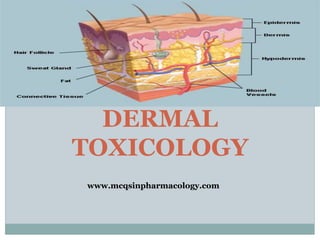
2. SKIN STRUCTURE
Skin is a water resistant covering of multiple cell layers.
It has three major divisions
Epidermis
Dermis
Hypodermis
www.mcqsinpharmacology.com
3. Epidermis has five layers(from superficial to deep)
Stratum corneum
Stratum lucidum
Stratum granuolosum
Stratum spinosum
Stratum basale (include melanocytes)
or stratum germinativum
Dermis
Supported by network of loose
connective tissue containing
collagen and elastin.
www.mcqsinpharmacology.com
4. Supplied by extensive blood vessels that are under active
neurogenic control.
Hypodermis(fat storage)
OTHER COMPONENETS OF SKIN:
Hair follicles
Sebaceous glands
Sweat glands
www.mcqsinpharmacology.com
5. BIOTRANSFORMATION OF
XENOBIOTICS
The deep layers of the epidermis have significant capability
to metabolize foreign compounds. The dermis has no
significant xenobiotic metabolizing ability.
Compounds that are biotransformed in other organs are
delivered to the skin, where they exert toxic or
photodynamic activity that damages the skin.
www.mcqsinpharmacology.com
6. EXPOSURE TO TOXICANTS
Skin especially the stratum corneum is relatively
impermeable to water soluble substances.
Small non polar lipophilic toxicants readily penetrate the
epidermis and are absorbed by dermal vasculature.
Axillary, inguinal, mammary and scrotal skin is highly
permeable compared with the skin in the dorsal and lateral
regions of most of the mammals.
www.mcqsinpharmacology.com
7. DERMAL HYPERMIA
Increases in environmental temperature may enhances
absorption e.g in hot climates, dermal hyperemia may
increase the toxicity of organophosphate insecticide to
mammals.
Highly lipophilic substances that contain surfactants or
detergents.
www.mcqsinpharmacology.com
8. RESPONSE TO TOXICANTS
Irritation, degeneration and necrosis
Direct irritation of the skin by corrosive, caustic and
necrotizing chemicals elicits an inflammatory response.
Generally materials with less than pH 2 and Ph 12 are
strong irritants. e.g acids, alkalis and metal salts.
a) Agents that damage cell membrane results in irritation
followed by the cardinal signs of inflammation(i.e.
erythema, swelling , heat and pain) leucocytosis and
increased vascular permeability.
Eschar formation, ulceration and necrosis may
permanently damage the skin.
www.mcqsinpharmacology.com
10. Allergic Contact
Dermatitis
Some chemicals act as antigens by combining with a carrier
proteins, eliciting a response from cellular components of
the immune system.
Langerhans’ cells in the epidermis process the antigen and
interact with appropriate T lymphocytes to form sensitized
T lymphocytes.
Sensitized T lymphocytes react to later exposure to the
antigen by producing a variety of cytokines. The cytokines
initiate a series of changes that characterize the allergic
response(i.e. erythema, itching , edema)
12. Photosensitization
Is an increase in susceptibility to ultraviolet light
Free radicals produced by photodynamic reactions damage
cells and lysosomal membranes.
Conditions lead to photosensitization
Photosensitization depends on the absorption of UV light
within the specific range of wavelengths (280-790 nm) and
the presence of photodynamic agent in the skin.
Released energy of the photodynamic agents damages
epidermal cell membrane and forms free radicals that can
initiate a chain reaction of membrane damage.
www.mcqsinpharmacology.com
13. photosensitization is most prominent on areas of the body
where protection from sunlight is least effective.
dorsal and lateral areas of the body
thin and unpigmented skin of the body
photosensitization is most likely to occur in sunny climates
and during the spring and summer when sunlight is more
intense or of longer duration each day.
www.mcqsinpharmacology.com
14. Clinical effects of photosensitization
early signs of erythema and edema
pruritis, photophobia and hyperesthesia follow
serious signs that occur later in the course of the disease
include exudation of serum, formation of vesicles ,
ulceration, exfoliation of damaged epidermis, and,
possibly, blindness.
www.mcqsinpharmacology.com
15. Types of photosensitization
primary photosensitization occurs when a
photodynamic agent is directly ingested, absorbed through
the skin, or injected, or when a chemical is biotransformed
to a photodynamic metabolite.
the major effects of primary photosensitizers occur in the
skin; other organs are usually spared.
prompt removal of the photosensitizer and supportive
treatment often results in recovery with new sequelae.
secondary photosensitization occur as a result of
compromised liver function, which reduces the excretion of
plant pigment metabolites from the body.
www.mcqsinpharmacology.com
16. several toxic plants are known to cause hepatogenous
photosensitization.
normally chlorophyll is metabolized to phylloerythrin by
intestinal and colonic bacteria. Phylloerythrin reabsorbed
from the gut is conjugated by the liver and excreted in the
bile.
failure of the liver to conjugate or excrete phylloerythrin
allows it to accumulate in the dermal vasculature, where it
is activated to a photodynamic state by ultraviolet light.
Liver damage and involvement of other organ system may
accompany the expected skin related signs of
photosensitization.
www.mcqsinpharmacology.com
17. Cutaneous Porphyrias(Vampires disease)
porphyria, which can cause photosensitization, is the
presence of abnormal levels of porphyrins in the blood as a
result of abnormal heme synthesis.
www.mcqsinpharmacology.com
18.
19. Hyperkeratosis
is the abnormal proliferation or keratinization of the
superficial epidermis .Toxicants include highly chlorinated
naphthalenes.
20. Alopecia
associated with thallium, arsenic, and selenium toxicosis .
chemotherapy with cytostatic drugs.
21. Skin cancer
Skin cancers are named after the type of skin cell from
which they arise.
Basal cell carcinoma originates from the lowest layer of the
epidermis, and is the most common but least dangerous
skin cancer.
Squamous cell carcinoma originates from the middle layer,
and is less common but more likely to spread and, if
untreated, become fatal.
Melanoma, which originates in the pigment-producing cells
(melanocytes), is the least common, but most aggressive,
most likely to spread and, if untreated, become fatal.
www.mcqsinpharmacology.com
22. Basal cell carcinoma
Note the pearly translucency to
fleshy color, tiny blood vessels
on the surface, and sometime
ulceration which can be
characteristics. The key term is
translucency.
Squamous cell carcinoma
Commonly presents as a red,
crusted, or scaly patch or bump.
Often a very rapid growing
tumor.
Malignant melanoma
The common appearance is an
asymmetrical area, with an
irregular border, color
variation, and often greater
than 6 mm diameter. www.mcqsinpharmacology.com
23. Causes
Ultraviolet radiation from sun exposure is the primary
cause of skin cancer.
HPV infections increase the risk of squamous cell
carcinoma.
Some genetic syndromes
Chronic non-healing wounds.
Ionizing radiation, environmental carcinogens, artificial UV
radiation (e.g. tanning beds), aging, and light skin color
The use of many immunosuppressive medication increase
the risk of skin cancer. Cyclosporin A, increases the risk
approximately 200 times, and azathioprine about 60 times
www.mcqsinpharmacology.com
24. Management
Sunscreen is effective in prevention
Surgical excision
Radiation therapy
Skin grafting
www.mcqsinpharmacology.com