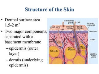
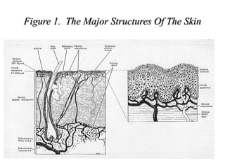
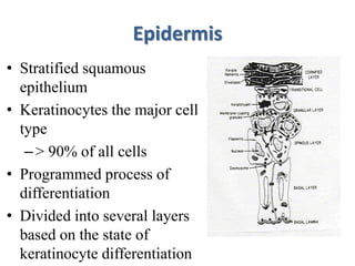
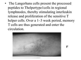
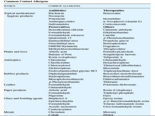
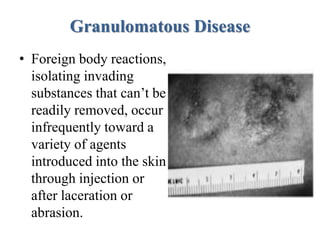
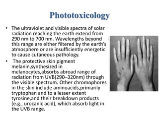
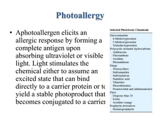
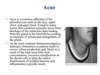
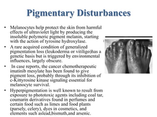
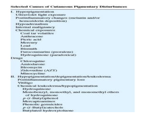
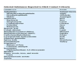
Skin Toxicology
Prepared by :- Krishna Champaneria
Institute Of Research And Development
Gujarat Forensic Sciences University
Prevalence Of Skin Disease
• Occupational skin diseases are the second
most common types of occupational disease
• 45,000 reported cases of occupational skin
disease in 2002
• 1983-1994: occupational skin diseases
increased by 26%, and 75% of workers with
occupational skin disease developed a chronic
skin disease
Introduction to:
• Structure and function of the skin
• Percutaneous absorption
• Metabolism
• Allergic contact dermatitis
Functions Of The Skin
• Environmental barrier
–Diffusion barrier
–Metabolic barrier
• Mechanical support
• Neurosensory reception
• Physiologically, skin participates directly in
thermal, metabolic, electrolyte, hormonal, and
immune regulation
1. Temperature regulation
• Regulation of blood flow,
• Hair and fur,
• Sweating
2. Metabolism
• Keratin,
• Collagen,
• Melanin,
• Lipid,
• Carbohydrate,
• Respiration,
• Biotransformation & Vitamin D
3. Apocrine/Eccrine/Sebaceous Glandular Secretion
4. Endocrine
5. Immunological Affector And Effector
Structure of the Skin
• Dermal surface area
1.5-2 m2
• Two major components,
separated with a
basement membrane
–epidermis (outer
layer)
–dermis (underlying
epidermis)
Figure 1. The Major Structures Of The Skin
Epidermis
• Stratified squamous
epithelium
• Keratinocytes the major cell
type
–> 90% of all cells
• Programmed process of
differentiation
• Divided into several layers
based on the state of
keratinocyte differentiation
Dermis
• Largest fraction of the skin
– approximately 90%
• Provides structural strength
– high content of collagen and elastin
• Nerve and vascular networks and appendages
required to support the epidermis
Contact Dermatitis
• In the occupational area,where records are
compiled on large workforces, contact
dermatitis is by far the largest category
(∼90%) of compensated skin disease.
• Using eczema of the hand as a sentinel
condition, since 80% of the total reported
dermatitis occurs at that location (10% on the
face), reveals a prevalence of 7–10% among
workers.
• Contact dermatitis falls into the two major
categories of irritant and allergic forms. Both
involve inflammatory processes and), in duration
(thickening and firmness), scaling (flaking), and
vesiculation(blistering)in areas of direct contact
with the chemical. Biopsies from affected sites
reveal a mixed-cell inflammatory infiltrate of
lymphocytes and eosinophils and spongiosis
(intercellular edema), but are insufficient to
distinguish the two conditions from each other or
from certain other common syndromes.
Irritant Dermatitis
• This condition arises
from the direct action of
chemicals on the skin. A
chemical in this category
is anticipated to give an
adverse reaction to
anyone if the
concentration is high
enough and exposure
time long enough.
• Certain chemicals at sufficient concentration
produce an acute irritation, sometimes called a
second-degree chemical burn, which can even
result in scarring in serious cases.
• These include strong acids and alkalies and
powerful oxidizing and reducing agents that
substantially disrupt the cornified layer, produce
cytotoxicity directly, and stimulate release of
proinflammatory cytokines.
More common is chronic cumulative irritation
from repeated exposures to mild irritants such as
soaps, detergents, solvents, and cutting oils.
Allergic Contact Dermatitis
• Allergic contact dermatitis is a delayed (T-cell
mediated) hypersensitive reaction. To induce
sensitization through the skin, chemical
haptens generally penetrate the lipid barrier
and, to be detected by the immune system,
become attached to carrier proteins.
• The complete antigens are then processed by
Langerhans cells(resident macrophages) and
displayed on their surfaces with major
histocompatibility complex II molecules.
• The Langerhans cells present the processed
peptides to Thelpertype1cells in regional
lymphnodes, thereby stimulating interleukin
release and proliferation of the sensitive T
helper cells. Over a 1–3 week period, memory
T cells are thus generated and enter the
circulation.
Granulomatous Disease
• Foreign body reactions,
isolating invading
substances that can’t be
readily removed, occur
infrequently toward a
variety of agents
introduced into the skin
through injection or
after laceration or
abrasion.
• Injection of paraffin or mineral oil in the skin or
contamination of wounds with starch powder cross
linked with epichlorohydrin for use in surgical
gloves may also result in granulomatous reactions.
Phototoxicology
• The ultraviolet and visible spectra of solar
radiation reaching the earth extend from
290 nm to 700 nm. Wavelengths beyond
this range are either filtered by the earth’s
atmosphere or are insufficiently energetic
to cause cutaneous pathology.
• The protective skin pigment
melanin,synthesized in
melanocytes,absorbs abroad range of
radiation from UVB(290–320nm) through
the visible spectrum. Other chromophores
in the skin include aminoacids,primarily
tryptophan and to a lesser extent
tyrosine,and their breakdown products
(e.g., urocanic acid), which absorb light in
the UVB range.
Photoallergy
• Aphotoallergen elicits an
allergic response by forming a
complete antigen upon
absorbing ultraviolet or visible
light. Light stimulates the
chemical either to assume an
excited state that can bind
directly to a carrier protein or to
yield a stable photoproduct that
becomes conjugated to a carrier.
Acne
• Acne is a common affliction of the
pilosebaceous units in the face, upper
chest, and upper back. Found in many
forms, this condition typically arises from
blockage of the sebaceous duct leading
from the gland to the hairfollicle,resulting
in retention of sebum,and enlargement of
the gland.
• In the most common form(acnevulgaris),
androgen stimulation at puberty leads to
excess sebum production and, likely as a
result of the high local fatty acid
concentration, excessive cornification of
the ductal cells to plug the orifice.
Proliferation of resident bacteria and
inflammation typically result.
Pigmentary Disturbances
• Melanocytes help protect the skin from harmful
effects of ultraviolet light by producing the
insoluble polymeric pigment melanin, starting
with the action of tyrosine hydroxylase.
• A rare acquired condition of generalized
pigmentation loss (leukoderma or vitiligo)has a
genetic basis but is triggered by environmental
influences, largely obscure.
• In case reports, the cancer chemotherapeutic
imatinib mesylate has been found to give
pigment loss, probably through its inhibition of
c-Kittyrosine kinase signaling essential for
melanocyte survival.
• Hyperpigmentation is well known to result from
exposure to phototoxic agents including coal tar,
coumarin derivatives found in perfumes and
certain food such as limes and food plants
(parsely, celery), dyes in cosmetics, and
elements such aslead,bismuth,and arsenic.
Urticaria
• For those allergens to which IgE antibodies have been
elicited by previous or ongoing exposure,subsequent contact
can lead to development of hives, typically in minutes,
through an immediate type I hypersensitivity reaction.
• Hives are raised wheals that usually itch or sting and may
appear reddish.
• Generally disappearing within hours and rarely lasting
longer than a day or two, the symptoms result from
degranulation of cutaneous mast cells by liganded IgE,
leading to release of histamine and other vasoactive
substances. Food allergies and pharmaceuticals are major
causes of acute urticaria.
Toxic Epidermal Necrolysis
• At the most severe end of a spectrum of adverse cutaneous hypersensitivity
reactions to drugs, this syndrome involves detachment of ≥30% of the
epidermal surface from the dermis, accompanied by severe erosions of the
mucous membranes and often pulmonary, intestinal, and ocular damage.
• Toxic epidermal necrolysis commonly resembles an upper respiratory tract
infection in the first several days (fever, cough, malaise), but prompt
diagnosis when the cutaneous lesions become evident several days later
improves survival chances.
• A characteristic feature of the syndrome is the large-scale apoptosis of
epidermal keratinocytes.
• Candidates for mediating apoptosis through cell surface death receptors
include tumor necrosis factor and FAS ligand,which appear
elevated;alternatively, drug-sensitized cytotoxic T lymphocytes could be
responsible for granzyme-induced cytolysis.
• Effective treatment may involve judicious immunosuppression, possibly
with cyclosporine or blockage of death receptors using intravenous
immunoglobulin therapy.
Skin Cancer
• Skin cancer is the most common neoplasm in humans, accounting for nearly one-
third of all cancers diagnosed each year. At present, the major cause of skin cancer
(half a million new cases per year in the United States) is sunlight, which damages
epidermal cell DNA. UVB (290–320 nm) induces pyrimidine dimers and 8-
oxoguanine modifications, thereby eliciting mutations in critical genes.
• The p53 tumor suppressor gene is a major target in which damage occurs early and
is detectable in most resulting squamous cell carcinomas.
• Because the p53 protein arrests cell cycling until DNA damage is repaired and may
induce apoptosis,its loss destabilizes the genome of initiated cells and gives them a
growth advantage.
• Individuals with xeroderma pigmentosum,who are deficient in repair of pyrimidine
dimers, must scrupulously avoid sun exposure to prevent the occurrence of
premalignant lesions that progress with continued exposure.
• Even when it does not cause cancer in normal individuals, sun exposure leads to
premature aging of the skin. For this reason, sunbathing is discouraged and the use
of sunblock lotions is encouraged, especially those that remove wavelengths up to
400 nm.
References
• Casarett and Doull’s Toxicology-The basic
science of poison
• http://www.cdc.gov/niosh/ocderm1.html

Skin toxicology

  • 1.
    Skin Toxicology Prepared by:- Krishna Champaneria Institute Of Research And Development Gujarat Forensic Sciences University
  • 2.
    Prevalence Of SkinDisease • Occupational skin diseases are the second most common types of occupational disease • 45,000 reported cases of occupational skin disease in 2002 • 1983-1994: occupational skin diseases increased by 26%, and 75% of workers with occupational skin disease developed a chronic skin disease
  • 3.
    Introduction to: • Structureand function of the skin • Percutaneous absorption • Metabolism • Allergic contact dermatitis
  • 4.
    Functions Of TheSkin • Environmental barrier –Diffusion barrier –Metabolic barrier • Mechanical support • Neurosensory reception • Physiologically, skin participates directly in thermal, metabolic, electrolyte, hormonal, and immune regulation
  • 5.
    1. Temperature regulation •Regulation of blood flow, • Hair and fur, • Sweating 2. Metabolism • Keratin, • Collagen, • Melanin, • Lipid, • Carbohydrate, • Respiration, • Biotransformation & Vitamin D
  • 6.
    3. Apocrine/Eccrine/Sebaceous GlandularSecretion 4. Endocrine 5. Immunological Affector And Effector
  • 7.
    Structure of theSkin • Dermal surface area 1.5-2 m2 • Two major components, separated with a basement membrane –epidermis (outer layer) –dermis (underlying epidermis)
  • 8.
    Figure 1. TheMajor Structures Of The Skin
  • 9.
    Epidermis • Stratified squamous epithelium •Keratinocytes the major cell type –> 90% of all cells • Programmed process of differentiation • Divided into several layers based on the state of keratinocyte differentiation
  • 10.
    Dermis • Largest fractionof the skin – approximately 90% • Provides structural strength – high content of collagen and elastin • Nerve and vascular networks and appendages required to support the epidermis
  • 12.
    Contact Dermatitis • Inthe occupational area,where records are compiled on large workforces, contact dermatitis is by far the largest category (∼90%) of compensated skin disease. • Using eczema of the hand as a sentinel condition, since 80% of the total reported dermatitis occurs at that location (10% on the face), reveals a prevalence of 7–10% among workers.
  • 13.
    • Contact dermatitisfalls into the two major categories of irritant and allergic forms. Both involve inflammatory processes and), in duration (thickening and firmness), scaling (flaking), and vesiculation(blistering)in areas of direct contact with the chemical. Biopsies from affected sites reveal a mixed-cell inflammatory infiltrate of lymphocytes and eosinophils and spongiosis (intercellular edema), but are insufficient to distinguish the two conditions from each other or from certain other common syndromes.
  • 14.
    Irritant Dermatitis • Thiscondition arises from the direct action of chemicals on the skin. A chemical in this category is anticipated to give an adverse reaction to anyone if the concentration is high enough and exposure time long enough.
  • 15.
    • Certain chemicalsat sufficient concentration produce an acute irritation, sometimes called a second-degree chemical burn, which can even result in scarring in serious cases. • These include strong acids and alkalies and powerful oxidizing and reducing agents that substantially disrupt the cornified layer, produce cytotoxicity directly, and stimulate release of proinflammatory cytokines. More common is chronic cumulative irritation from repeated exposures to mild irritants such as soaps, detergents, solvents, and cutting oils.
  • 17.
    Allergic Contact Dermatitis •Allergic contact dermatitis is a delayed (T-cell mediated) hypersensitive reaction. To induce sensitization through the skin, chemical haptens generally penetrate the lipid barrier and, to be detected by the immune system, become attached to carrier proteins. • The complete antigens are then processed by Langerhans cells(resident macrophages) and displayed on their surfaces with major histocompatibility complex II molecules.
  • 18.
    • The Langerhanscells present the processed peptides to Thelpertype1cells in regional lymphnodes, thereby stimulating interleukin release and proliferation of the sensitive T helper cells. Over a 1–3 week period, memory T cells are thus generated and enter the circulation.
  • 20.
    Granulomatous Disease • Foreignbody reactions, isolating invading substances that can’t be readily removed, occur infrequently toward a variety of agents introduced into the skin through injection or after laceration or abrasion.
  • 21.
    • Injection ofparaffin or mineral oil in the skin or contamination of wounds with starch powder cross linked with epichlorohydrin for use in surgical gloves may also result in granulomatous reactions.
  • 22.
    Phototoxicology • The ultravioletand visible spectra of solar radiation reaching the earth extend from 290 nm to 700 nm. Wavelengths beyond this range are either filtered by the earth’s atmosphere or are insufficiently energetic to cause cutaneous pathology. • The protective skin pigment melanin,synthesized in melanocytes,absorbs abroad range of radiation from UVB(290–320nm) through the visible spectrum. Other chromophores in the skin include aminoacids,primarily tryptophan and to a lesser extent tyrosine,and their breakdown products (e.g., urocanic acid), which absorb light in the UVB range.
  • 23.
    Photoallergy • Aphotoallergen elicitsan allergic response by forming a complete antigen upon absorbing ultraviolet or visible light. Light stimulates the chemical either to assume an excited state that can bind directly to a carrier protein or to yield a stable photoproduct that becomes conjugated to a carrier.
  • 24.
    Acne • Acne isa common affliction of the pilosebaceous units in the face, upper chest, and upper back. Found in many forms, this condition typically arises from blockage of the sebaceous duct leading from the gland to the hairfollicle,resulting in retention of sebum,and enlargement of the gland. • In the most common form(acnevulgaris), androgen stimulation at puberty leads to excess sebum production and, likely as a result of the high local fatty acid concentration, excessive cornification of the ductal cells to plug the orifice. Proliferation of resident bacteria and inflammation typically result.
  • 25.
    Pigmentary Disturbances • Melanocyteshelp protect the skin from harmful effects of ultraviolet light by producing the insoluble polymeric pigment melanin, starting with the action of tyrosine hydroxylase. • A rare acquired condition of generalized pigmentation loss (leukoderma or vitiligo)has a genetic basis but is triggered by environmental influences, largely obscure. • In case reports, the cancer chemotherapeutic imatinib mesylate has been found to give pigment loss, probably through its inhibition of c-Kittyrosine kinase signaling essential for melanocyte survival. • Hyperpigmentation is well known to result from exposure to phototoxic agents including coal tar, coumarin derivatives found in perfumes and certain food such as limes and food plants (parsely, celery), dyes in cosmetics, and elements such aslead,bismuth,and arsenic.
  • 27.
    Urticaria • For thoseallergens to which IgE antibodies have been elicited by previous or ongoing exposure,subsequent contact can lead to development of hives, typically in minutes, through an immediate type I hypersensitivity reaction. • Hives are raised wheals that usually itch or sting and may appear reddish. • Generally disappearing within hours and rarely lasting longer than a day or two, the symptoms result from degranulation of cutaneous mast cells by liganded IgE, leading to release of histamine and other vasoactive substances. Food allergies and pharmaceuticals are major causes of acute urticaria.
  • 29.
    Toxic Epidermal Necrolysis •At the most severe end of a spectrum of adverse cutaneous hypersensitivity reactions to drugs, this syndrome involves detachment of ≥30% of the epidermal surface from the dermis, accompanied by severe erosions of the mucous membranes and often pulmonary, intestinal, and ocular damage. • Toxic epidermal necrolysis commonly resembles an upper respiratory tract infection in the first several days (fever, cough, malaise), but prompt diagnosis when the cutaneous lesions become evident several days later improves survival chances. • A characteristic feature of the syndrome is the large-scale apoptosis of epidermal keratinocytes. • Candidates for mediating apoptosis through cell surface death receptors include tumor necrosis factor and FAS ligand,which appear elevated;alternatively, drug-sensitized cytotoxic T lymphocytes could be responsible for granzyme-induced cytolysis. • Effective treatment may involve judicious immunosuppression, possibly with cyclosporine or blockage of death receptors using intravenous immunoglobulin therapy.
  • 30.
    Skin Cancer • Skincancer is the most common neoplasm in humans, accounting for nearly one- third of all cancers diagnosed each year. At present, the major cause of skin cancer (half a million new cases per year in the United States) is sunlight, which damages epidermal cell DNA. UVB (290–320 nm) induces pyrimidine dimers and 8- oxoguanine modifications, thereby eliciting mutations in critical genes. • The p53 tumor suppressor gene is a major target in which damage occurs early and is detectable in most resulting squamous cell carcinomas. • Because the p53 protein arrests cell cycling until DNA damage is repaired and may induce apoptosis,its loss destabilizes the genome of initiated cells and gives them a growth advantage. • Individuals with xeroderma pigmentosum,who are deficient in repair of pyrimidine dimers, must scrupulously avoid sun exposure to prevent the occurrence of premalignant lesions that progress with continued exposure. • Even when it does not cause cancer in normal individuals, sun exposure leads to premature aging of the skin. For this reason, sunbathing is discouraged and the use of sunblock lotions is encouraged, especially those that remove wavelengths up to 400 nm.
  • 31.
    References • Casarett andDoull’s Toxicology-The basic science of poison • http://www.cdc.gov/niosh/ocderm1.html