Download to read offline
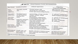


The document discusses dermatophytes, which are fungi causing superficial infections of skin, hair, and nails, collectively known as dermatomycosis. It outlines the main genera of dermatophytes (Trichophyton, Microsporum, and Epidermophyton), their transmission, clinical manifestations (like tinea), and laboratory diagnostic methods. The infections can be acquired from humans, animals, or soil, and are characterized by itchy, ring-like lesions.


