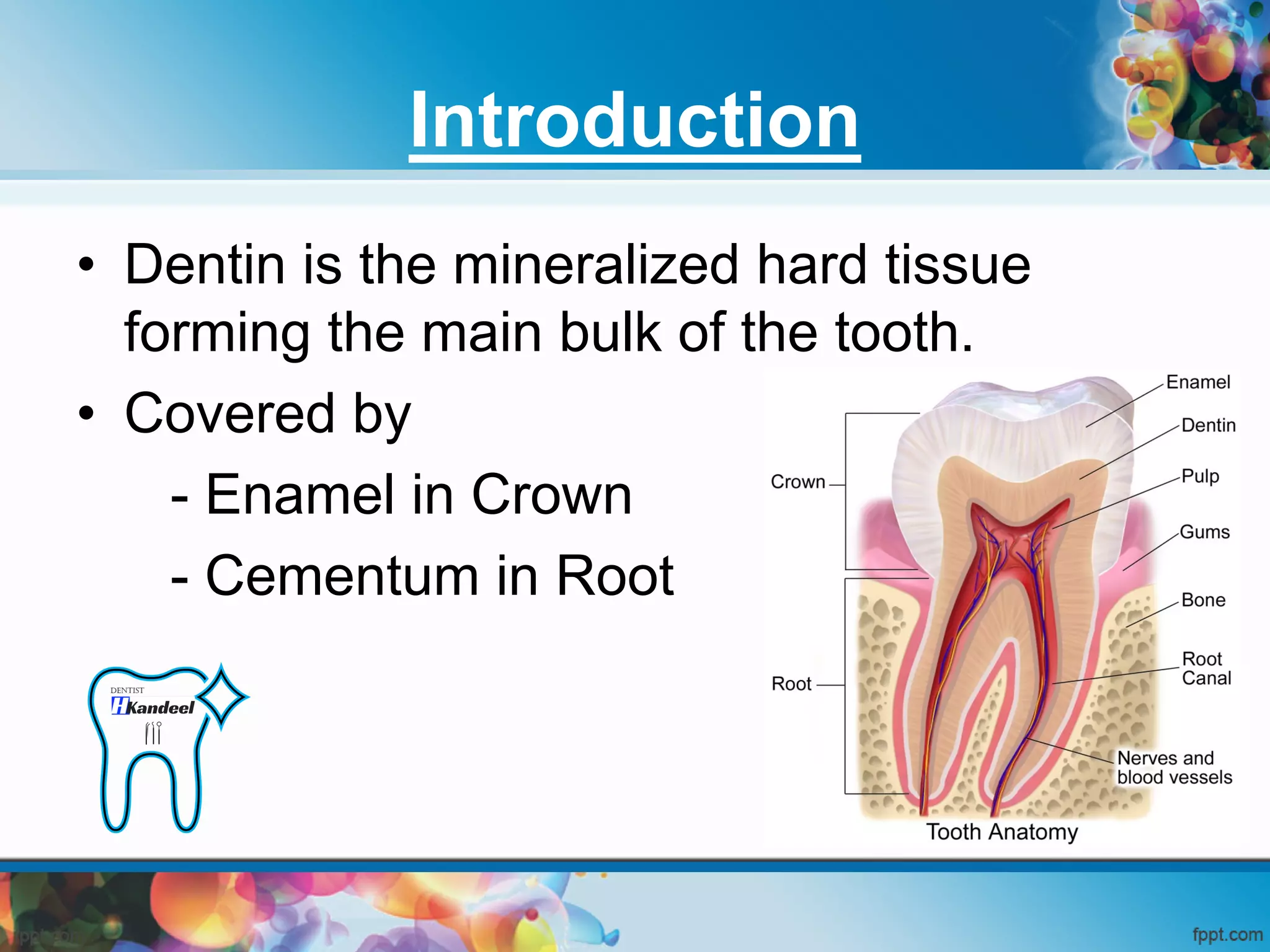
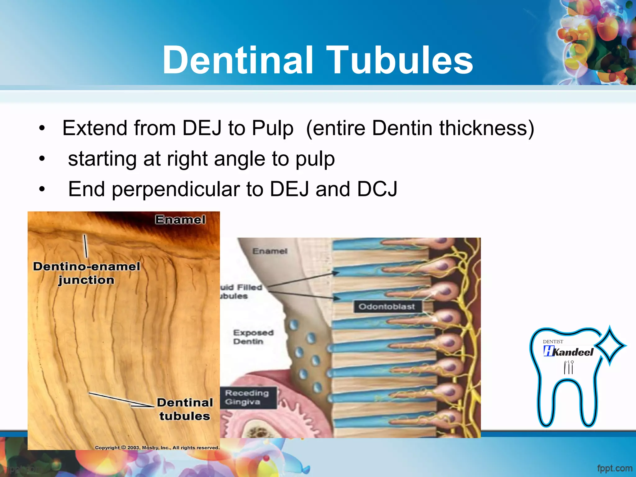
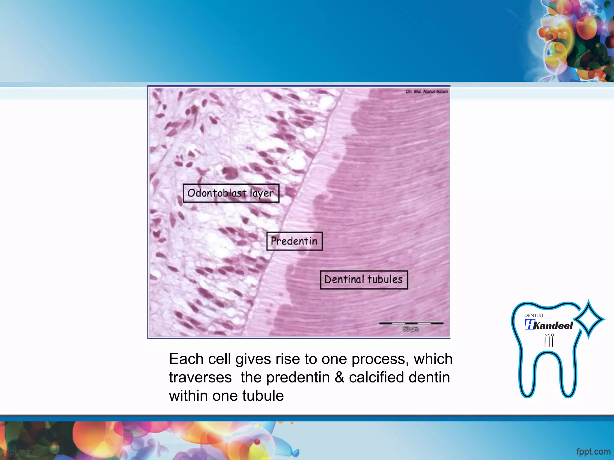
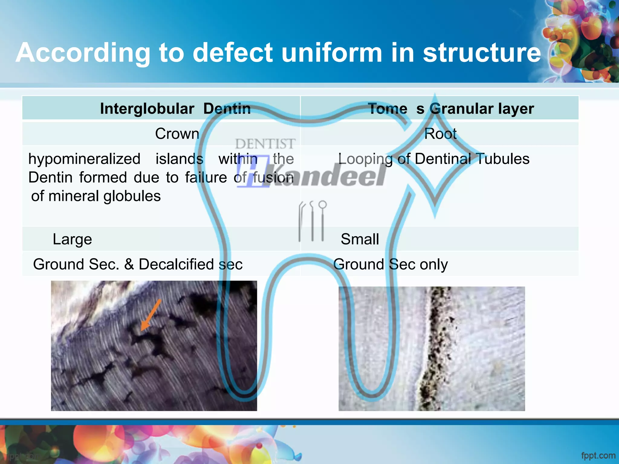
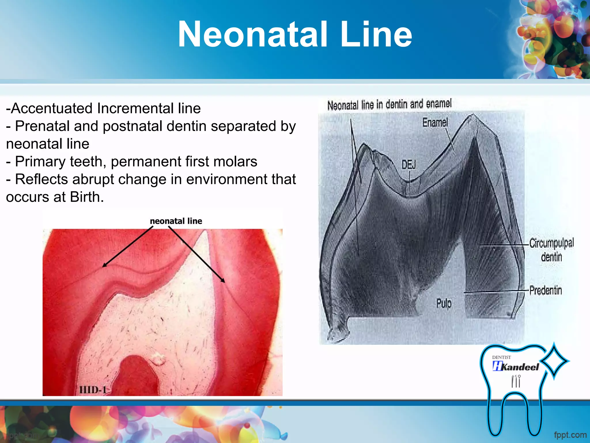
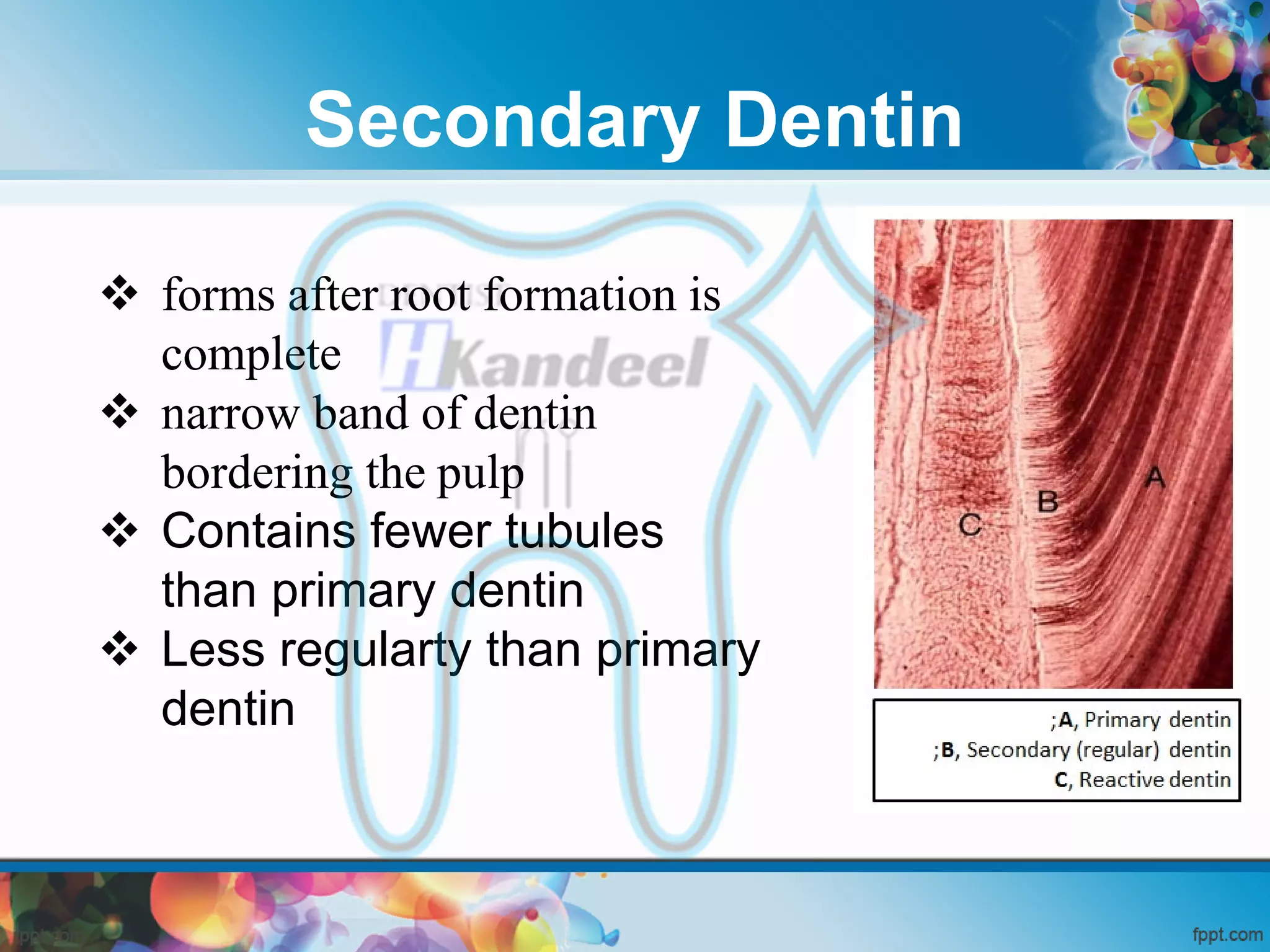
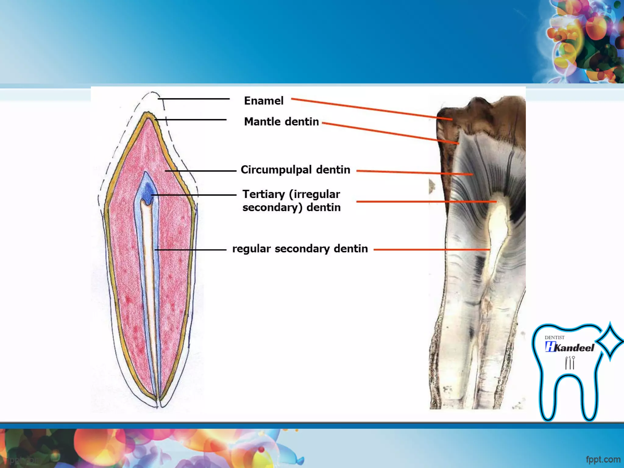
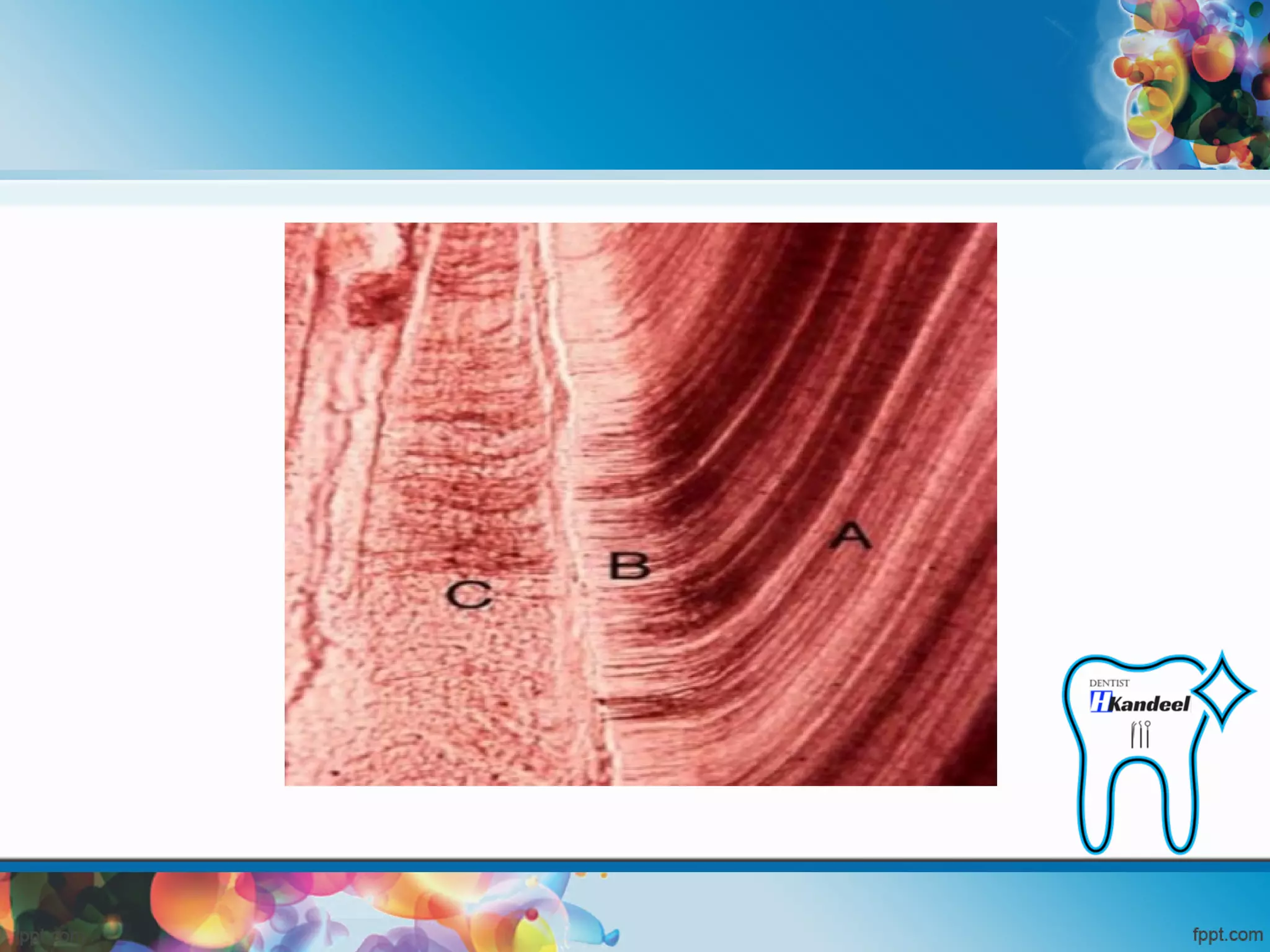
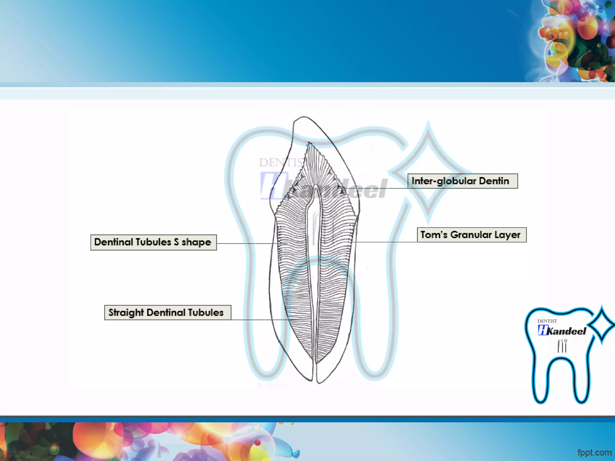
Introduction
• Dentin is the mineralized hard tissue
forming the main bulk of the tooth.
• Covered by
- Enamel in Crown
- Cementum in Root
Physical Properties
• It is light yellowish in color, becoming darker with age.
• Viscoelastic Material (Harder than bone but softer than
enamel)
• Lower content of mineral salts in dentin renders it more
radiolucent than enamel
Chemical Properties
70 %
30%
Organic Matter + Water
substance consists of
collagenous fibrils and a
ground substance of
mucopolysaccharides
(proteoglycans and glycos
aminoglycans
Inorganic Matter
consists of
hydroxyapetite as
in bone, cementum &
enamel
Dentinal Tubules
• Extend from DEJ to Pulp (entire Dentin thickness)
• starting at right angle to pulp
• End perpendicular to DEJ and DCJ
Curvatures Of Dentinal Tubules
1- Primary Curvature
2- Secondary Curvature
Dentinal Tubules
Primary Curvatures
• ‘S’ shaped curve.
(Sigmoid curvatures)
• More prominent in crown
• Straight at
- cusp tips
- incisal edge
Dentinal Tubules
Dentinal Tubules
Root Dentin
Coronal Dentin
Dentinal Tubules
Secondary Curvature
Spiral in Shape
Content Of Dentinal Tubules
Dental lymph
Content Of Dentinal Tubules
A, Dentinal tubule; B, DEJ;
C, Enamel; D, Enamel spindle
Each cell gives rise to one process, which
traverses the predentin & calcified dentin
within one tubule
Dentin Pulp
Types Of Dentin
• According to :
1- Defect uniform in structure
2- Ultra Structure
3- Age Change
According to defect uniform in structure
Tome s Granular layer
Interglobular Dentin
Root
Crown
Looping of Dentinal Tubules
hypomineralized islands within the
Dentin formed due to failure of fusion
of mineral globules
Small
Large
Ground Sec only
Ground Sec. & Decalcified sec
Interglobular Dentin
Tome s Granular layer
According to UltraStructure
Intertubular Dentin
Peritubular Dentin
Between D.T
Wall Of D.T
Less in Hardness
High in Hardness
Form main bulk of dentin
T.S. In Dentinal Tubules
INCREMENTAL LINES
- Von Ebner Line
- Contour Line of Owen
- Neonatal Line
Von Ebner Line
- Fine striations - perpendicular to tubules
- Shows the daily deposition of Dentin
Contour Line of Owen
- Due to the “Co-incidence of secondary curvatures”
- Accentuated Von ebnar incremental line
- Shows Hypo-mineralized areas
Neonatal Line
-Accentuated Incremental line
- Prenatal and postnatal dentin separated by
neonatal line
- Primary teeth, permanent first molars
- Reflects abrupt change in environment that
occurs at Birth.
Neonatal Line
According to Age Change
Primary dentin Secondary physiologic
dentin
Tertiary dentin or
reparative dentin or
reactionary dentin or
irregular secondary
Mantle Dentin Circumpulpal dentin
Primary Dentin
Circumpulpal
dentin
Mantle Dentin
After Root Completion
Before Root
Completion
Form Remaining Primary
Dentin
(Bulk Of Tooth )
First Formed Dentin layer
Most Peripheral part of
primary Dentin
Contain more mineral
Contain Less Mineral
Secondary Dentin
 forms after root formation is
complete
 narrow band of dentin
bordering the pulp
 Contains fewer tubules
than primary dentin
 Less regularty than primary
dentin
Tertiary dentin
 Reactive Dentin, Reparative Dentin, Irritation Dentin, Replacement Dentin, Defense
Dentin
 Localized formation of Dentin At pulp –Dentin Border in response to noxious
stimuli ( Caries, Trauma, Attrition , Cavity Preparation )
 No continuity with primary or secondary Dentin so there is decrease in Dentin
permeability
 Tubules may or may not be present
Sclerotic Dentin
 Dentinal tubules become
occluded with calcified
material
 Blocking of tubules- Defensive
reaction.
 Elderly people – Mostly in Roots
 Found specially in radicular
dentin (apical 3rd).
 Transparent or light in
transmitted Light
 dark in reflected light
 Cause : Mild Stimuli
Sclerotic Dentin
Dead tracts
 Complete Destruction of Odontoblastic process in the affected area
 Dark in transmitted Light
 Transparent or light in reflected light
 Cause : Sever stimuli
A, Dead tract;
B, teritary dentin
Test YourSelf ??
A
B
C
A
B
C
D
A
Thank You

Dentin

  • 2.
    Introduction • Dentin isthe mineralized hard tissue forming the main bulk of the tooth. • Covered by - Enamel in Crown - Cementum in Root
  • 3.
    Physical Properties • Itis light yellowish in color, becoming darker with age. • Viscoelastic Material (Harder than bone but softer than enamel) • Lower content of mineral salts in dentin renders it more radiolucent than enamel
  • 4.
    Chemical Properties 70 % 30% OrganicMatter + Water substance consists of collagenous fibrils and a ground substance of mucopolysaccharides (proteoglycans and glycos aminoglycans Inorganic Matter consists of hydroxyapetite as in bone, cementum & enamel
  • 6.
    Dentinal Tubules • Extendfrom DEJ to Pulp (entire Dentin thickness) • starting at right angle to pulp • End perpendicular to DEJ and DCJ
  • 7.
    Curvatures Of DentinalTubules 1- Primary Curvature 2- Secondary Curvature
  • 8.
    Dentinal Tubules Primary Curvatures •‘S’ shaped curve. (Sigmoid curvatures) • More prominent in crown • Straight at - cusp tips - incisal edge
  • 9.
  • 10.
  • 11.
  • 12.
    Content Of DentinalTubules Dental lymph
  • 13.
  • 14.
    A, Dentinal tubule;B, DEJ; C, Enamel; D, Enamel spindle
  • 15.
    Each cell givesrise to one process, which traverses the predentin & calcified dentin within one tubule
  • 16.
  • 18.
    Types Of Dentin •According to : 1- Defect uniform in structure 2- Ultra Structure 3- Age Change
  • 19.
    According to defectuniform in structure Tome s Granular layer Interglobular Dentin Root Crown Looping of Dentinal Tubules hypomineralized islands within the Dentin formed due to failure of fusion of mineral globules Small Large Ground Sec only Ground Sec. & Decalcified sec
  • 20.
  • 21.
  • 22.
    According to UltraStructure IntertubularDentin Peritubular Dentin Between D.T Wall Of D.T Less in Hardness High in Hardness Form main bulk of dentin
  • 25.
  • 26.
    INCREMENTAL LINES - VonEbner Line - Contour Line of Owen - Neonatal Line
  • 27.
    Von Ebner Line -Fine striations - perpendicular to tubules - Shows the daily deposition of Dentin
  • 28.
    Contour Line ofOwen - Due to the “Co-incidence of secondary curvatures” - Accentuated Von ebnar incremental line - Shows Hypo-mineralized areas
  • 31.
    Neonatal Line -Accentuated Incrementalline - Prenatal and postnatal dentin separated by neonatal line - Primary teeth, permanent first molars - Reflects abrupt change in environment that occurs at Birth.
  • 32.
  • 33.
    According to AgeChange Primary dentin Secondary physiologic dentin Tertiary dentin or reparative dentin or reactionary dentin or irregular secondary Mantle Dentin Circumpulpal dentin
  • 34.
    Primary Dentin Circumpulpal dentin Mantle Dentin AfterRoot Completion Before Root Completion Form Remaining Primary Dentin (Bulk Of Tooth ) First Formed Dentin layer Most Peripheral part of primary Dentin Contain more mineral Contain Less Mineral
  • 35.
    Secondary Dentin  formsafter root formation is complete  narrow band of dentin bordering the pulp  Contains fewer tubules than primary dentin  Less regularty than primary dentin
  • 38.
    Tertiary dentin  ReactiveDentin, Reparative Dentin, Irritation Dentin, Replacement Dentin, Defense Dentin  Localized formation of Dentin At pulp –Dentin Border in response to noxious stimuli ( Caries, Trauma, Attrition , Cavity Preparation )  No continuity with primary or secondary Dentin so there is decrease in Dentin permeability  Tubules may or may not be present
  • 41.
    Sclerotic Dentin  Dentinaltubules become occluded with calcified material  Blocking of tubules- Defensive reaction.  Elderly people – Mostly in Roots  Found specially in radicular dentin (apical 3rd).  Transparent or light in transmitted Light  dark in reflected light  Cause : Mild Stimuli
  • 42.
  • 43.
    Dead tracts  CompleteDestruction of Odontoblastic process in the affected area  Dark in transmitted Light  Transparent or light in reflected light  Cause : Sever stimuli A, Dead tract; B, teritary dentin
  • 44.
  • 45.
  • 46.
  • 50.
  • 63.