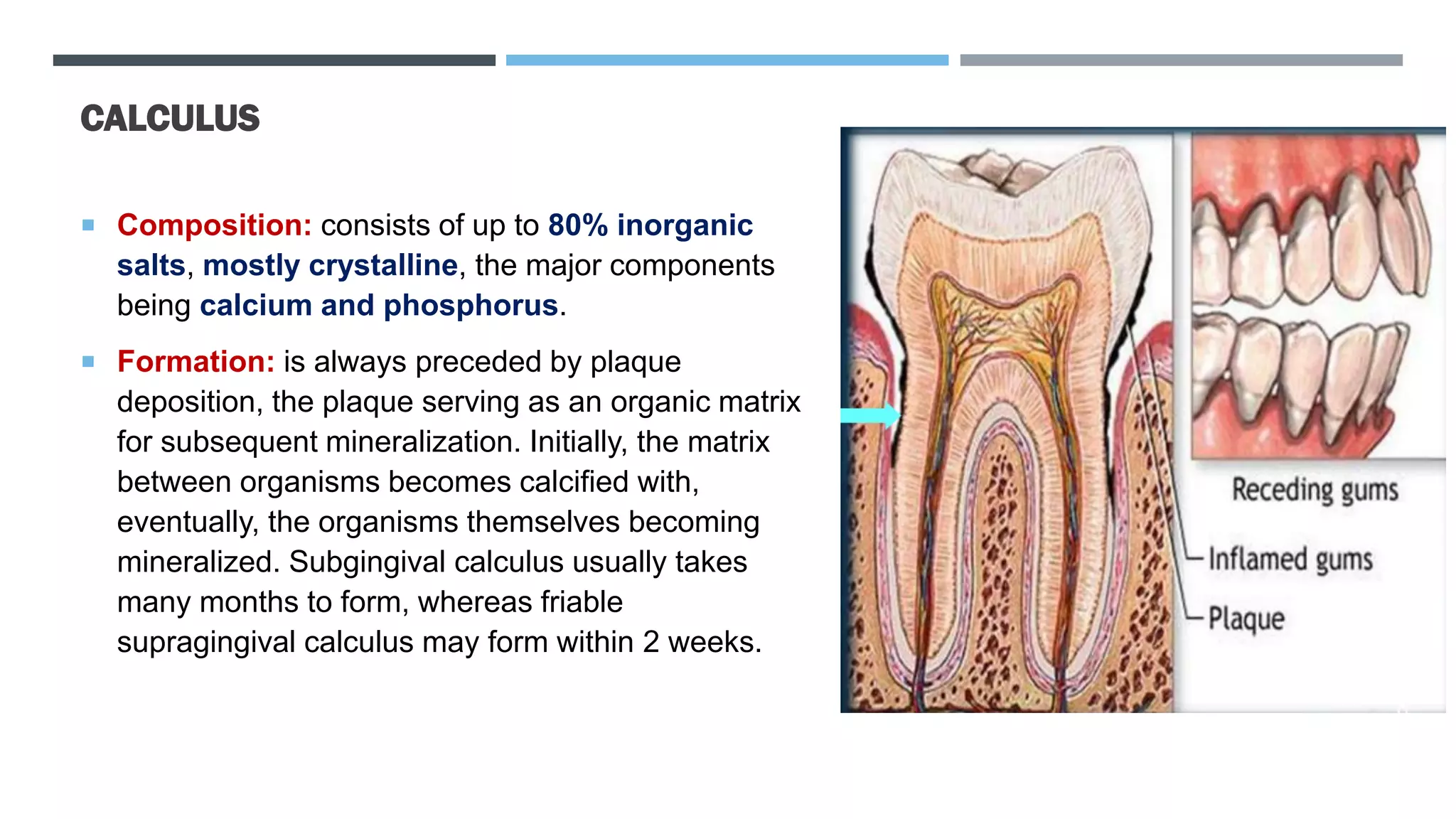
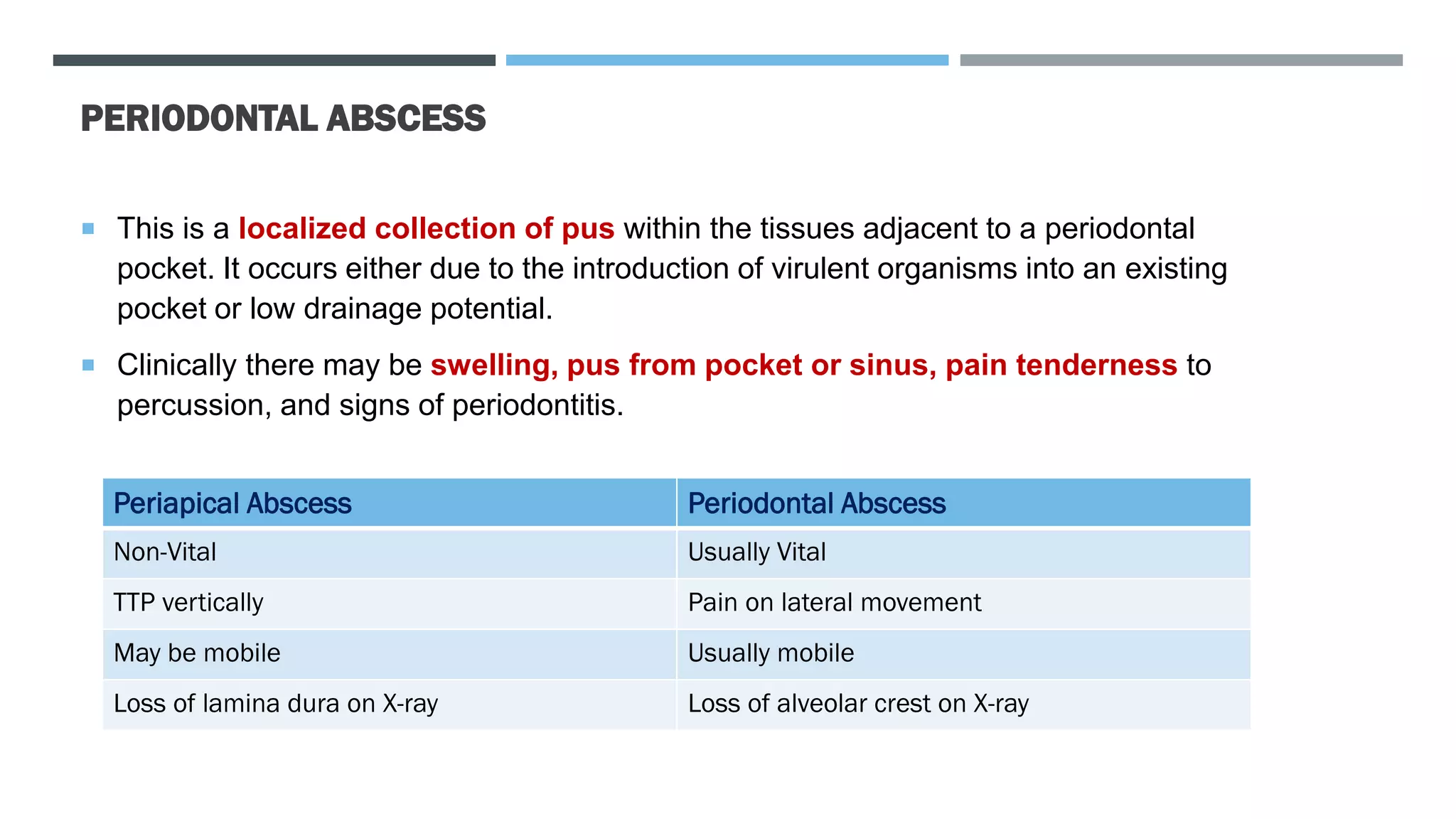
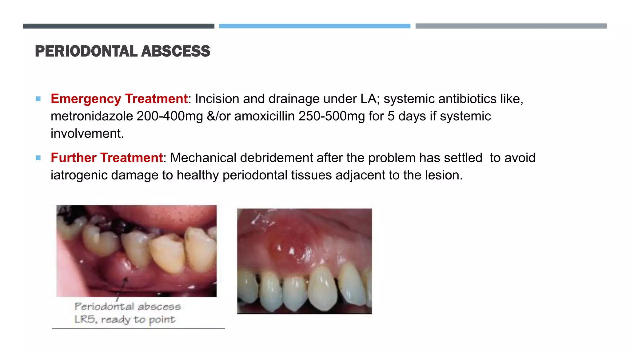
MEDENTA ACADEMY
PERIODONTOLOGY
CONTENT
1
Aetiology of
Periodontal
Diseases
2
Plaque
3
Calculus
4
Periodontal
Abscess
5
Treatment
AETIOLOGY OF PERIODONTAL DISEASES
 Primary etiology of all forms of periodontal diseases = Plaque
 Biofilm:
a thin layer of microorganisms adhering to the surface of a structure, which may be organic or inorganic, to
gether with the polymers that they secrete.
 Dental biofilm- a biofilm is a layer of bacteria attached to the supragingival or subgingival surface of a
tooth.
 Plaque - the visible accumulation of a supragingival or subgingival biofilm.
 Tartar is what accumulates on your teeth when plaque is not removed. If plaque is left on your teeth for too
long, it will harden into tartar and is much more difficult to remove. In fact, tartar can only be removed by a
dental professional–you can't get rid of it with regular brushing and flossing
AETIOLOGY OF PERIODONTAL DISEASES
 Plaque Biofilm cause gingivitis by inducing inflammatory host response.
 That inflammatory response – gingival pocket – ideal environment – for further bacterial
colonization.
 In Gingival Pocket – extremely low oxygen – develops of anaerobes and several other – periodontal
diseases.
 In 1996, three species (bacteria) – cause periodontitis = Aggregatibacter actinomycetemcomitans,
Porphyromonas gingivalis, and Tannerella forsythia.
 Virulence Factors: pathogens use a number of mechanisms to exert damage to host tissues:
adherence, proteases, bone resorption factors, cytotoxic metabolites, leukotoxins, and induction
of the inflammatory response via cytokines and chemotaxins.
PLAQUE
 Plaque Biofilm or dental biofilm – is an adherent mass of diverse micro-organisms in a muco-
polysaccharide matrix. It cannot be rinsed off but can be removed by brushing.
 Pellicle: one essential function of human saliva is the formation of the acquired dental pellicle, a thin
coating layer of high tenacity covering all solid surfaces in the oral cavity. It is composed of proteins and
glycoproteins (It consists of an aggregation of cells, proteins, and polymers). It lubricates the tooth
surface and serves as a barrier and buffer to demineralizing agents.
 The growth and development of biofilm are characterized by 4 stages: initial adherence, lag phase,
rapid growth, and steady state. Biofilm formation begins with the adherence of bacteria to a tooth
surface, followed by a lag phase in which changes in genetic expression (phenotypic shifts)occur.
 Four stages of dental plaque biofilm growth: (1) bacterial attachment to a surface, (2)
microcolony formation, (3) biofilm maturation and (4) detachment (also termed dispersal) of
bacteria which may then colonize new areas
PLAQUE
The process of biofilm formation in the oral cavity is divided into four stages: 1. acquired pellicle formation;
2. initial adhesion; 3. coaggregation; 4. maturation and diffusion.
PLAQUE
 Compositions of plaque: 20 to 30% Intercellular Matrix & 80% Microorganisms.
 Intercellular Matrix: Organic & Inorganic
 Microorganisms: one gram of plaque contains approximately 2*𝟏𝟎𝟏𝟏
bacteria
 More than 500 distinct microbial species found in dental plaque.
 Nonbacterial Organisms: MYCOPLASMA, YEAST, PROTOZOA, VIRUSES
FORMATION OF PLAQUE
Pellicle Formation on Tooth Surface
Initial Adhesion and Attachment
Colonization and Plaque Maturation
FORMATION OF PLAQUE
 `A fully established pellicle is found within 30 min. Within 24 hr, the pellicle is around 0.1-
0.8 µm in diameter.
 Derived from components of saliva and crevicular fluid as well as bacterial and host tissue
cell products and food debris.
 FUNCTIONS OF DENTAL PELLICLE:
1. Protective Barrier
2. Lubrication
3. Preventing Tissue Desiccation
4. Substrate to Which Bacteria Attaches
FORMATION OF PLAQUE
What Happen in First hour of Plaque Formation?
 Bacteria adhere to pellicle, and pellicle coats the enamel. (1Hr)
 Gram positive rods and cocci are laid down in the first hour. (1Hr)
24 to 48Hr
 Bacteria multiply and form mini-colonies in layers upon the pellicle. (24-48Hr)
 The bacteria adhere and increase in mass and thickness. (24-48Hr)
4 to 7 Days
 As the plaque thickens at the cervical area, the deeper layers incorporate more filaments and fusiforms,
eventually turning gram negative. (4-7days)
 The coronal plaque is a more simple early arrangement of rods and cocci. (4-7days)
 Bleeding on probing and erythema can be seen. (4-7days)
 The plaque becomes more gram negative and anaerobic in the deeper layers. The signs of inflammation
are more pronounced. (7 to 14days)
CALCULUS
 Calculus (Tartar) is a calcified deposit found on teeth or prosthesis and is formed by
mineralization of plaque deposits. The mineral content of supragingival calculus derives from
saliva, that for subgingival is from gingival crevicular fluid.
 It is classified as supragingival or subgingival, according to its relation to the gingival
margin.
 Supragingival Calculus: most often found opposite the openings of the salivary ducts, i.e.
76┴67 opposite the parotid (Stensen’s) duct or the lingual surfaces of the lower anterior teeth.
It is usually creamy-coloured, but can become stained a variety of colors.
 Subgingival Calculus: is found underneath the gingival margin and is firmly attached to tooth
roots. It tends to be brown or black, is extremely tenacious, and is most often found on
interproximal and lingual surfaces. It is associated with subsequent periodontitis. With gingival
recession it can become supragingival.
CALCULUS
 Supragingival
 Subgingival
CALCULUS
 Composition: consists of up to 80% inorganic
salts, mostly crystalline, the major components
being calcium and phosphorus.
 Formation: is always preceded by plaque
deposition, the plaque serving as an organic matrix
for subsequent mineralization. Initially, the matrix
between organisms becomes calcified with,
eventually, the organisms themselves becoming
mineralized. Subgingival calculus usually takes
many months to form, whereas friable
supragingival calculus may form within 2 weeks.
DEVELOPMENT OF PERIODONTAL DISEASES
PERIODONTAL ABSCESS
 This is a localized collection of pus within the tissues adjacent to a periodontal
pocket. It occurs either due to the introduction of virulent organisms into an existing
pocket or low drainage potential.
 Clinically there may be swelling, pus from pocket or sinus, pain tenderness to
percussion, and signs of periodontitis.
Periapical Abscess Periodontal Abscess
Non-Vital Usually Vital
TTP vertically Pain on lateral movement
May be mobile Usually mobile
Loss of lamina dura on X-ray Loss of alveolar crest on X-ray
PERIODONTAL ABSCESS
 Emergency Treatment: Incision and drainage under LA; systemic antibiotics like,
metronidazole 200-400mg &/or amoxicillin 250-500mg for 5 days if systemic
involvement.
 Further Treatment: Mechanical debridement after the problem has settled to avoid
iatrogenic damage to healthy periodontal tissues adjacent to the lesion.
THE END
Thanks/‫مننه‬

Plaque and Calculus

  • 1.
  • 2.
  • 3.
    AETIOLOGY OF PERIODONTALDISEASES  Primary etiology of all forms of periodontal diseases = Plaque  Biofilm: a thin layer of microorganisms adhering to the surface of a structure, which may be organic or inorganic, to gether with the polymers that they secrete.  Dental biofilm- a biofilm is a layer of bacteria attached to the supragingival or subgingival surface of a tooth.  Plaque - the visible accumulation of a supragingival or subgingival biofilm.  Tartar is what accumulates on your teeth when plaque is not removed. If plaque is left on your teeth for too long, it will harden into tartar and is much more difficult to remove. In fact, tartar can only be removed by a dental professional–you can't get rid of it with regular brushing and flossing
  • 4.
    AETIOLOGY OF PERIODONTALDISEASES  Plaque Biofilm cause gingivitis by inducing inflammatory host response.  That inflammatory response – gingival pocket – ideal environment – for further bacterial colonization.  In Gingival Pocket – extremely low oxygen – develops of anaerobes and several other – periodontal diseases.  In 1996, three species (bacteria) – cause periodontitis = Aggregatibacter actinomycetemcomitans, Porphyromonas gingivalis, and Tannerella forsythia.  Virulence Factors: pathogens use a number of mechanisms to exert damage to host tissues: adherence, proteases, bone resorption factors, cytotoxic metabolites, leukotoxins, and induction of the inflammatory response via cytokines and chemotaxins.
  • 5.
    PLAQUE  Plaque Biofilmor dental biofilm – is an adherent mass of diverse micro-organisms in a muco- polysaccharide matrix. It cannot be rinsed off but can be removed by brushing.  Pellicle: one essential function of human saliva is the formation of the acquired dental pellicle, a thin coating layer of high tenacity covering all solid surfaces in the oral cavity. It is composed of proteins and glycoproteins (It consists of an aggregation of cells, proteins, and polymers). It lubricates the tooth surface and serves as a barrier and buffer to demineralizing agents.  The growth and development of biofilm are characterized by 4 stages: initial adherence, lag phase, rapid growth, and steady state. Biofilm formation begins with the adherence of bacteria to a tooth surface, followed by a lag phase in which changes in genetic expression (phenotypic shifts)occur.  Four stages of dental plaque biofilm growth: (1) bacterial attachment to a surface, (2) microcolony formation, (3) biofilm maturation and (4) detachment (also termed dispersal) of bacteria which may then colonize new areas
  • 6.
    PLAQUE The process ofbiofilm formation in the oral cavity is divided into four stages: 1. acquired pellicle formation; 2. initial adhesion; 3. coaggregation; 4. maturation and diffusion.
  • 7.
    PLAQUE  Compositions ofplaque: 20 to 30% Intercellular Matrix & 80% Microorganisms.  Intercellular Matrix: Organic & Inorganic  Microorganisms: one gram of plaque contains approximately 2*𝟏𝟎𝟏𝟏 bacteria  More than 500 distinct microbial species found in dental plaque.  Nonbacterial Organisms: MYCOPLASMA, YEAST, PROTOZOA, VIRUSES
  • 8.
    FORMATION OF PLAQUE PellicleFormation on Tooth Surface Initial Adhesion and Attachment Colonization and Plaque Maturation
  • 9.
    FORMATION OF PLAQUE `A fully established pellicle is found within 30 min. Within 24 hr, the pellicle is around 0.1- 0.8 µm in diameter.  Derived from components of saliva and crevicular fluid as well as bacterial and host tissue cell products and food debris.  FUNCTIONS OF DENTAL PELLICLE: 1. Protective Barrier 2. Lubrication 3. Preventing Tissue Desiccation 4. Substrate to Which Bacteria Attaches
  • 10.
    FORMATION OF PLAQUE WhatHappen in First hour of Plaque Formation?  Bacteria adhere to pellicle, and pellicle coats the enamel. (1Hr)  Gram positive rods and cocci are laid down in the first hour. (1Hr) 24 to 48Hr  Bacteria multiply and form mini-colonies in layers upon the pellicle. (24-48Hr)  The bacteria adhere and increase in mass and thickness. (24-48Hr) 4 to 7 Days  As the plaque thickens at the cervical area, the deeper layers incorporate more filaments and fusiforms, eventually turning gram negative. (4-7days)  The coronal plaque is a more simple early arrangement of rods and cocci. (4-7days)  Bleeding on probing and erythema can be seen. (4-7days)  The plaque becomes more gram negative and anaerobic in the deeper layers. The signs of inflammation are more pronounced. (7 to 14days)
  • 11.
    CALCULUS  Calculus (Tartar)is a calcified deposit found on teeth or prosthesis and is formed by mineralization of plaque deposits. The mineral content of supragingival calculus derives from saliva, that for subgingival is from gingival crevicular fluid.  It is classified as supragingival or subgingival, according to its relation to the gingival margin.  Supragingival Calculus: most often found opposite the openings of the salivary ducts, i.e. 76┴67 opposite the parotid (Stensen’s) duct or the lingual surfaces of the lower anterior teeth. It is usually creamy-coloured, but can become stained a variety of colors.  Subgingival Calculus: is found underneath the gingival margin and is firmly attached to tooth roots. It tends to be brown or black, is extremely tenacious, and is most often found on interproximal and lingual surfaces. It is associated with subsequent periodontitis. With gingival recession it can become supragingival.
  • 12.
  • 13.
    CALCULUS  Composition: consistsof up to 80% inorganic salts, mostly crystalline, the major components being calcium and phosphorus.  Formation: is always preceded by plaque deposition, the plaque serving as an organic matrix for subsequent mineralization. Initially, the matrix between organisms becomes calcified with, eventually, the organisms themselves becoming mineralized. Subgingival calculus usually takes many months to form, whereas friable supragingival calculus may form within 2 weeks.
  • 14.
  • 15.
  • 16.
  • 17.
    PERIODONTAL ABSCESS  Thisis a localized collection of pus within the tissues adjacent to a periodontal pocket. It occurs either due to the introduction of virulent organisms into an existing pocket or low drainage potential.  Clinically there may be swelling, pus from pocket or sinus, pain tenderness to percussion, and signs of periodontitis. Periapical Abscess Periodontal Abscess Non-Vital Usually Vital TTP vertically Pain on lateral movement May be mobile Usually mobile Loss of lamina dura on X-ray Loss of alveolar crest on X-ray
  • 18.
    PERIODONTAL ABSCESS  EmergencyTreatment: Incision and drainage under LA; systemic antibiotics like, metronidazole 200-400mg &/or amoxicillin 250-500mg for 5 days if systemic involvement.  Further Treatment: Mechanical debridement after the problem has settled to avoid iatrogenic damage to healthy periodontal tissues adjacent to the lesion.
  • 19.