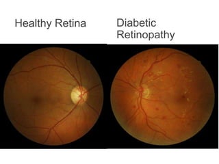
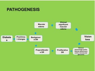
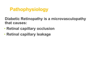
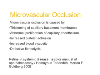
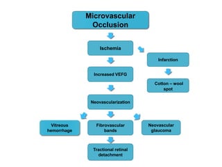
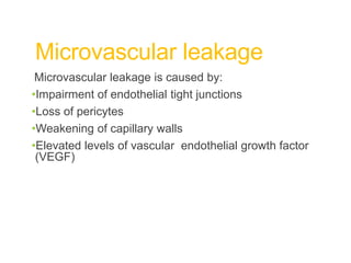
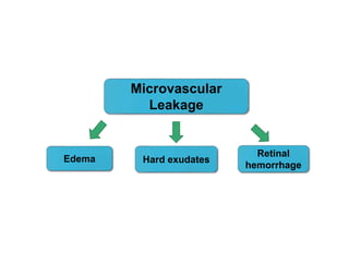
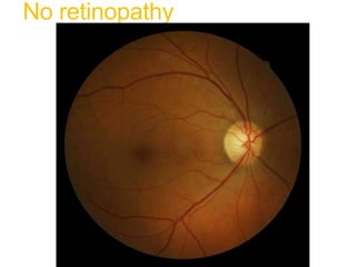
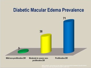
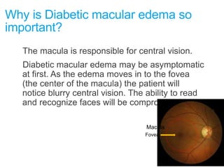
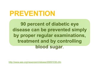
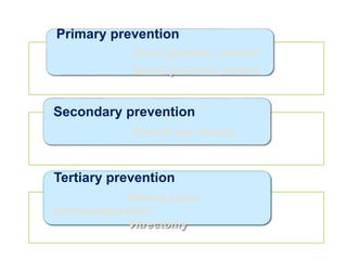
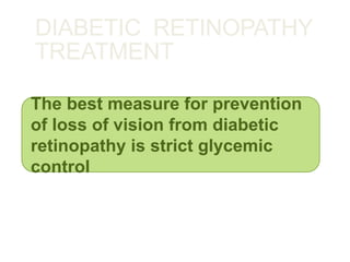
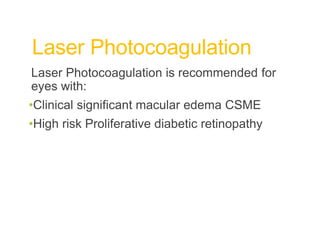
Department: Surgery
Казахский Национальный
Медицинский Университет
имени С.Д.Асфендиярова
С.Ж.Асфендияров
атындағы Қазақ Ұлттық
Медицина Университеті
Almaty 2017
DIABETIC RETINOPATHY
Checked by:
Done by: Nematullah Z.
Faculty: GM
Course: 4
Group: 77-2
Plan
•Recognize the importance of diabetic retinopathy as a
public
•Classification
•Pathogenesis
•Causes
•health problem
•Identify the risk factors for diabetic retinopathy
•Describe and distinguish between the stages of diabetic
retinopathy
•Understand the role of risk factor control and annual
dilated eye exams in the prevention of vision loss
•Treatment
Diabetes Mellitus
Diabetes Mellitus is a group of diseases characterized by
high blood glucose levels. Diabetes results from defects in
the body's ability to produce and/or use insulin.
•Type 1 diabetes is usually diagnosed in children and
young adults, and was previously known as juvenile
diabetes. In type 1 diabetes, the body does not produce
insulin. 5% of people with diabetes have this form of the
disease.
•In Type 2 diabetes, either the body does not produce
enough insulin or the cells ignore the insulin. This is the
most common form of diabetes.
Diabetic Retinopathy (DR)
Definition
•Progressive dysfunction of the retinal blood
vessels caused by chronic hyperglycemia.
•DR can be a complication of diabetes type 1 or
diabetes type 2.
•Initially, DR is asymptomatic, if not treated
though it can cause low vision and blindness.
Classification
Background Diabetic Retinopathy
Diabetic Maculopathy
Nonproliferative Diabetic Retinopathy
Proliferative Diabetic Retinopathy
Advanced Diabetic Eye Disease
What is THE Retina?
•The retina is a multilayered, light sensitive neural
tissue lining the inner eye ball. Light is focused
onto the retina and then transmitted to the brain
through the optic nerve.
•The macula is a highly sensitive area in the
center of the retina, responsible for central vision.
The macula is needed for reading, recognizing
faces and executing other activities that require
fine, sharp vision.
RETINA
Healthy Retina Diabetic
Retinopathy
Diabetic Retinopathy
Epidemiology
•The total number of people with diabetes is
projected to rise from 285 million in 2010 to 439
million in 2030.
•Diabetic retinopathy is responsible for 1.8 million
of the 37 million cases of blindness throughout
the world .
•Diabetic retinopathy (DR) is the leading cause of
blindness in people of working age in
industrialized countries.
Diabetic Retinopathy
Epidemiology
•The best predictor of diabetic retinopathy is the
duration of the disease
•After 20 years of diabetes, nearly 99% of patients with
type 1 diabetes and 60% with type 2 have some
degree on diabetic retinopathy
•33% of patients with diabetes have signs of diabetic
retinopathy
•People with diabetes are 25 times more likely to
become blind than the general population.
Diabetic retinopathy symptoms
Diabetic retinopathy is asymptomatic in early
stages of the disease
As the disease progresses symptoms may include
•Blurred vision
•Floaters
•Fluctuating vision
•Distorted vision
•Dark areas in the vision
•Poor night vision
•Impaired color vision
•Partial or total loss of vision
Risk factors
Duration of diabetes
Poor Blood Sugar control
HTN
Hyperlipidemia
Barriers to care
The Effect of Intensive Diabetes Treatment
On the Progression of Diabetic Retinopathy
In Insulin-Dependent Diabetes Mellitus
The Diabetes Control and Complications Trial
The Diabetes Control and Complications Trial Research Group
Intensive control reduced the risk of developing retinopathy by 76%
and slowed progression of retinopathy by 54%; intensive control
also reduced the risk of clinical neuropathy by 60% and albuminuria
by 54%.
Arch Ophthalmol. 1995; 113:36-51
Risk factors Diabetic Retinopathy
Duration of diabetes is a major risk
factor associated with the development
of diabetic retinopathy
The severity of hyperglycemia is the
key alterable risk factor associated with
the development of diabetic retinopathy
http://one.aao.org/CE/PracticeGuidelines/PPP_Content.aspx?cid=d0c853d3-219f-487b-a524-326ab3cecd9a
How diabetes cause vision
loss
PATHOGENESIS
Preclinica
l changes
Macular
edema
Proliferative
DR
Diabete
s
Backgroun
d DR
Clinical
significant
macular
edema
Vitreous
hemorrhage and/or
Retinal detachment
and/or neovascular
glaucoma
Preproliferativ
e DR
Vision
loss
Pathophysiology
Diabetic Retinopathy is a microvasculopathy
that causes:
• Retinal capillary occlusion
• Retinal capillary leakage
Microvascular Occlusion
Microvascular occlusion is caused by:
•Thickening of capillary basement membranes
•Abnormal proliferation of capillary endothelium
•Increased platelet adhesion
•Increased blood viscosity
•Defective fibrinolysis
Retina in systemic disease : a color manual of
ophthalmoscopy / Homayoun Tabandeh, Morton F.
Goldberg 2009
Cotton – wool
spot
Neovascularization
Ischemia
Neovascular
glaucoma
Microvascular
Occlusion
Fibrovascular
bands
Vitreous
hemorrhage
Increased VEFG
Tractional retinal
detachment
Infarction
Microvascular leakage
Microvascular leakage is caused by:
•Impairment of endothelial tight junctions
•Loss of pericytes
•Weakening of capillary walls
•Elevated levels of vascular endothelial growth factor
(VEGF)
Edema
Retinal
hemorrhage
Hard exudates
Microvascular
Leakage
No retinopathy
MILD NONPROLIFERATIVE
DIABETIC RETINOPATHY
Characteristics
•Microaneurysms only
MILD NONPROLIFERATIVE
DIABETIC RETINOPATHY
Microaneurysms
Moderate Nonproliferative
Diabetic Retinopathy (NPDR)
Characteristics
• More than just microaneurysms but less than severe
NPDR
•That increase the Eye pressure
•And this pressure decrease the blood flow
•And cause ischemia
Moderate Nonproliferative
Diabetic Retinopathy (NPDR)
Hard exudates
Flamed shaped
hemorrhage
Microaneurysm
Moderate Nonproliferative
Diabetic Retinopathy (NPDR)
Hard exudates
microaneurysm
Severe Nonproliferative Diabetic
Retinopathy (NPDR)
Any of the following:
•More than 20 intraretinal hemorrhages in each of
four quadrants
•Definite venous beading in two or more quadrants
•Prominent Intraretinal Microvascular Abnormalities
(IRMA) in one or more quadrants
•And no signs of proliferative retinopathy
Severe Nonproliferative Diabetic
Retinopathy (NPDR)
Venous beading
Proliferative Diabetic Retinopathy (PDR)
Characteristics
• Neovascularization
• Vitreous/preretinal
hemorrhage
PROLIFERATIVE
DIABETIC
RETINOPATHY
Neovascularization
Neovascularization
Hard exudate
Cotton-wool
spot
Blot hemorrhage
High-Risk Proliferative diabetic
retinopathy
At risk for serious vision loss
Any combination of three of the following four findings
•Presence of vitreous or preretinal hemorrhage.
•Presence of new vessels (neovascularization, NV)
•Location of NV on or near the optic disc.
•Moderate to severe extent of new vessels.
Basic and Clinical Science Course, Section 12: Retina and Vitreous
AAO
Diabetic macular edema
•Diabetic macular edema is the leading cause
of legal blindness in diabetics.
•Diabetic macular edema can be present at any
stage of the disease, but is more common in
patients with proliferative diabetic retinopathy.
Meta analysis and review on the effect on bevacizumab id diabetic macular edema
Graefes Arch Clin Exp Ophthalmol(2011) 249:15-27
Why is Diabetic macular edema so
important?
The macula is responsible for central vision.
Diabetic macular edema may be asymptomatic
at first. As the edema moves in to the fovea
(the center of the macula) the patient will
notice blurry central vision. The ability to read
and recognize faces will be compromised.
Macula
Fovea
NORMAL MACULAR EDEMA
Clinically significant
macular edema (CSME)
•Thickening of the retina at or within 500 µm of
the center of the macula.
•Hard exudates at or within 500 µm of the
center of the macula, if associated with
thickening of the adjacent retina.
•Area of retinal thickening 1 disc area or larger,
within 1 disc diameter of the center of the
macula.
ETDRS
Imaging of macular edema with optical
coherence tomography
PREVENTION
http://www.aao.org/newsroom/release/20091030.cfm
90 percent of diabetic eye
disease can be prevented simply
by proper regular examinations,
treatment and by controlling
blood sugar.
Primary prevention
Strict glycemic control
Blood pressure control
Secondary prevention
Annual eye exams
Tertiary prevention
Retinal Laser
photocoagulation
Vitrectomy
DIABETIC RETINOPATHY
TREATMENT
The best measure for prevention
of loss of vision from diabetic
retinopathy is strict glycemic
control
Laser Photocoagulation
Laser Photocoagulation is recommended for
eyes with:
•Clinical significant macular edema CSME
•High risk Proliferative diabetic retinopathy
DIABETIC RETINOPATHY TREATMENT
Once DR threatens vision treatments can include:
Laser therapy to seal leaking blood
vessels (focal laser)
Laser therapy to reduce retinal
oxygen demand (scatter laser)
Surgical removal of blood from the
eye (vitrectomy)
DIABETIC RETINOPATHY TREATMENT
NEWER DEVELOPMENTS:
The use of anti-vascular endothelial
growth factor antibodies has been
shown to be useful in the treatment
of DR
Anti-VEGF antibody treatment
appears to be useful for both
macular edema and proliferative
retinopathy
Studies to determine the exact role
of anti-VEGF treatment in relation
to laser treatment in specific
situations are underway.
http://drcrnet.jaeb.org
CONCLUSIONS
Diabetic Retinopathy is
preventable through strict
glycemic control and annual
dilated eye exams by an
ophthalmologist.
References
•Retina in systemic disease : a color manual of ophthalmoscopy /
Homayoun Tabandeh, Morton F. Goldberg 2009
•Goyal S, Laavalley M, Subramanian ML, Meta analysis and
review on the effect on bevacizumab in diabetic macular edema,
Graefes Arch Clin Exp Ophthalmol(2011) 249:15-27
•C. P. Wilkinson, MD,1 Frederick L. Ferris, III, MD,2 Ronald E.
Klein, MD, MPH,3 Paul P. Lee, MD, JD,4 Carl David Agardh, MD,5
Matthew Davis, MD,3 Diana Dills, MD,6 Anselm Kampik, MD,7 R.
Pararajasegaram, MD,8 Juan T. Verdaguer, MD,9 representing the
Global Diabetic Retinopathy Project Group, Proposed
International Clinical Diabetic, Retinopathy and Diabetic Macular
Edema Disease Severity Scales Ophthalmology Volume 110,
Number 9, September 2003 Proposed international clinical
diabetic retinopathy and diabetic macular edema disease
severity scales
References
•Preferred Practice Patterns, Diabetic retinopathy, America
Academy of Ophthalmology 2008.
http://one.aao.org/CE/PracticeGuidelines/PPP_Content.aspx?cid=
d0c853d3-219f-487b-a524-326ab3cecd9a
•Brett J. Rosenblatt and William E. Benson Diabetic Retinopathy Yanoff &
Duker: Ophthalmology, 3rd ed.
http://www.mdconsult.com/das/book/pdf/282715756-3/978-0-323-04332-8/4-
u1.0-B978-0-323- 04332- 8..00092-5..DOCPDF.pdf?isbn=978-0-323-04332-
8&eid=4-u1.0-B978-0-323-04332-8..00092- 5..DOCPDF
•Resnikoff S, Pascolini D, Etya'ale D, Kocur I, Pararajasegaram R, Pokharel
GP, Mariotti SP. Global data on visual impairment in the year 2002. Bull
World Health Organ. 2004 Nov;82(11):844-51. Epub 2004 Dec 14.
•Basic and Clinical Science Course, Section 12: Retina and Vitreous AAO,
2011-2012.
•The Effect of Intensive Diabetes Treatment On the Progression of Diabetic
Retinopathy In Insulin-Dependent Diabetes Mellitus, The Diabetes Control
and Complications Trial Research Group, Arch Ophthalmol. 1995; 113:36-51
References
• http://www.ncbi.nlm.nih.gov/pubmed/19896746
• http://www.aao.org/eyecare/news/upload/Eye-Health-Fact-
Sheet.pdf
• http://www.who.int/bulletin/volumes/82/11/en/844.pdf
• http://jama.ama-assn.org/content/304/6/649.short?rss=1
• http://www.aao.org/newsroom/release/20091030.cfm
• http://www.diabetes.org/diabetes-basics/?loc=GlobalNavDB
• http://www.ophed.com/group/2205
DIABETIC RETINOPATHY

DIABETIC RETINOPATHY

  • 1.
    Department: Surgery Казахский Национальный МедицинскийУниверситет имени С.Д.Асфендиярова С.Ж.Асфендияров атындағы Қазақ Ұлттық Медицина Университеті Almaty 2017 DIABETIC RETINOPATHY Checked by: Done by: Nematullah Z. Faculty: GM Course: 4 Group: 77-2
  • 2.
    Plan •Recognize the importanceof diabetic retinopathy as a public •Classification •Pathogenesis •Causes •health problem •Identify the risk factors for diabetic retinopathy •Describe and distinguish between the stages of diabetic retinopathy •Understand the role of risk factor control and annual dilated eye exams in the prevention of vision loss •Treatment
  • 3.
    Diabetes Mellitus Diabetes Mellitusis a group of diseases characterized by high blood glucose levels. Diabetes results from defects in the body's ability to produce and/or use insulin. •Type 1 diabetes is usually diagnosed in children and young adults, and was previously known as juvenile diabetes. In type 1 diabetes, the body does not produce insulin. 5% of people with diabetes have this form of the disease. •In Type 2 diabetes, either the body does not produce enough insulin or the cells ignore the insulin. This is the most common form of diabetes.
  • 4.
    Diabetic Retinopathy (DR) Definition •Progressivedysfunction of the retinal blood vessels caused by chronic hyperglycemia. •DR can be a complication of diabetes type 1 or diabetes type 2. •Initially, DR is asymptomatic, if not treated though it can cause low vision and blindness.
  • 5.
    Classification Background Diabetic Retinopathy DiabeticMaculopathy Nonproliferative Diabetic Retinopathy Proliferative Diabetic Retinopathy Advanced Diabetic Eye Disease
  • 6.
    What is THERetina? •The retina is a multilayered, light sensitive neural tissue lining the inner eye ball. Light is focused onto the retina and then transmitted to the brain through the optic nerve. •The macula is a highly sensitive area in the center of the retina, responsible for central vision. The macula is needed for reading, recognizing faces and executing other activities that require fine, sharp vision.
  • 7.
  • 8.
  • 9.
    Diabetic Retinopathy Epidemiology •The totalnumber of people with diabetes is projected to rise from 285 million in 2010 to 439 million in 2030. •Diabetic retinopathy is responsible for 1.8 million of the 37 million cases of blindness throughout the world . •Diabetic retinopathy (DR) is the leading cause of blindness in people of working age in industrialized countries.
  • 10.
    Diabetic Retinopathy Epidemiology •The bestpredictor of diabetic retinopathy is the duration of the disease •After 20 years of diabetes, nearly 99% of patients with type 1 diabetes and 60% with type 2 have some degree on diabetic retinopathy •33% of patients with diabetes have signs of diabetic retinopathy •People with diabetes are 25 times more likely to become blind than the general population.
  • 11.
    Diabetic retinopathy symptoms Diabeticretinopathy is asymptomatic in early stages of the disease As the disease progresses symptoms may include •Blurred vision •Floaters •Fluctuating vision •Distorted vision •Dark areas in the vision •Poor night vision •Impaired color vision •Partial or total loss of vision
  • 12.
    Risk factors Duration ofdiabetes Poor Blood Sugar control HTN Hyperlipidemia Barriers to care
  • 13.
    The Effect ofIntensive Diabetes Treatment On the Progression of Diabetic Retinopathy In Insulin-Dependent Diabetes Mellitus The Diabetes Control and Complications Trial The Diabetes Control and Complications Trial Research Group Intensive control reduced the risk of developing retinopathy by 76% and slowed progression of retinopathy by 54%; intensive control also reduced the risk of clinical neuropathy by 60% and albuminuria by 54%. Arch Ophthalmol. 1995; 113:36-51
  • 14.
    Risk factors DiabeticRetinopathy Duration of diabetes is a major risk factor associated with the development of diabetic retinopathy The severity of hyperglycemia is the key alterable risk factor associated with the development of diabetic retinopathy http://one.aao.org/CE/PracticeGuidelines/PPP_Content.aspx?cid=d0c853d3-219f-487b-a524-326ab3cecd9a
  • 15.
    How diabetes causevision loss PATHOGENESIS Preclinica l changes Macular edema Proliferative DR Diabete s Backgroun d DR Clinical significant macular edema Vitreous hemorrhage and/or Retinal detachment and/or neovascular glaucoma Preproliferativ e DR Vision loss
  • 16.
    Pathophysiology Diabetic Retinopathy isa microvasculopathy that causes: • Retinal capillary occlusion • Retinal capillary leakage
  • 17.
    Microvascular Occlusion Microvascular occlusionis caused by: •Thickening of capillary basement membranes •Abnormal proliferation of capillary endothelium •Increased platelet adhesion •Increased blood viscosity •Defective fibrinolysis Retina in systemic disease : a color manual of ophthalmoscopy / Homayoun Tabandeh, Morton F. Goldberg 2009
  • 18.
  • 19.
    Microvascular leakage Microvascular leakageis caused by: •Impairment of endothelial tight junctions •Loss of pericytes •Weakening of capillary walls •Elevated levels of vascular endothelial growth factor (VEGF)
  • 20.
  • 21.
  • 22.
  • 23.
  • 24.
    Moderate Nonproliferative Diabetic Retinopathy(NPDR) Characteristics • More than just microaneurysms but less than severe NPDR •That increase the Eye pressure •And this pressure decrease the blood flow •And cause ischemia
  • 25.
    Moderate Nonproliferative Diabetic Retinopathy(NPDR) Hard exudates Flamed shaped hemorrhage Microaneurysm
  • 26.
    Moderate Nonproliferative Diabetic Retinopathy(NPDR) Hard exudates microaneurysm
  • 27.
    Severe Nonproliferative Diabetic Retinopathy(NPDR) Any of the following: •More than 20 intraretinal hemorrhages in each of four quadrants •Definite venous beading in two or more quadrants •Prominent Intraretinal Microvascular Abnormalities (IRMA) in one or more quadrants •And no signs of proliferative retinopathy
  • 28.
  • 29.
    Proliferative Diabetic Retinopathy(PDR) Characteristics • Neovascularization • Vitreous/preretinal hemorrhage
  • 30.
  • 31.
    High-Risk Proliferative diabetic retinopathy Atrisk for serious vision loss Any combination of three of the following four findings •Presence of vitreous or preretinal hemorrhage. •Presence of new vessels (neovascularization, NV) •Location of NV on or near the optic disc. •Moderate to severe extent of new vessels. Basic and Clinical Science Course, Section 12: Retina and Vitreous AAO
  • 32.
    Diabetic macular edema •Diabeticmacular edema is the leading cause of legal blindness in diabetics. •Diabetic macular edema can be present at any stage of the disease, but is more common in patients with proliferative diabetic retinopathy.
  • 33.
    Meta analysis andreview on the effect on bevacizumab id diabetic macular edema Graefes Arch Clin Exp Ophthalmol(2011) 249:15-27
  • 34.
    Why is Diabeticmacular edema so important? The macula is responsible for central vision. Diabetic macular edema may be asymptomatic at first. As the edema moves in to the fovea (the center of the macula) the patient will notice blurry central vision. The ability to read and recognize faces will be compromised. Macula Fovea
  • 35.
  • 36.
    Clinically significant macular edema(CSME) •Thickening of the retina at or within 500 µm of the center of the macula. •Hard exudates at or within 500 µm of the center of the macula, if associated with thickening of the adjacent retina. •Area of retinal thickening 1 disc area or larger, within 1 disc diameter of the center of the macula. ETDRS
  • 37.
    Imaging of macularedema with optical coherence tomography
  • 38.
    PREVENTION http://www.aao.org/newsroom/release/20091030.cfm 90 percent ofdiabetic eye disease can be prevented simply by proper regular examinations, treatment and by controlling blood sugar.
  • 39.
    Primary prevention Strict glycemiccontrol Blood pressure control Secondary prevention Annual eye exams Tertiary prevention Retinal Laser photocoagulation Vitrectomy
  • 40.
    DIABETIC RETINOPATHY TREATMENT The bestmeasure for prevention of loss of vision from diabetic retinopathy is strict glycemic control
  • 41.
    Laser Photocoagulation Laser Photocoagulationis recommended for eyes with: •Clinical significant macular edema CSME •High risk Proliferative diabetic retinopathy
  • 42.
    DIABETIC RETINOPATHY TREATMENT OnceDR threatens vision treatments can include: Laser therapy to seal leaking blood vessels (focal laser) Laser therapy to reduce retinal oxygen demand (scatter laser) Surgical removal of blood from the eye (vitrectomy)
  • 43.
    DIABETIC RETINOPATHY TREATMENT NEWERDEVELOPMENTS: The use of anti-vascular endothelial growth factor antibodies has been shown to be useful in the treatment of DR Anti-VEGF antibody treatment appears to be useful for both macular edema and proliferative retinopathy Studies to determine the exact role of anti-VEGF treatment in relation to laser treatment in specific situations are underway. http://drcrnet.jaeb.org
  • 44.
    CONCLUSIONS Diabetic Retinopathy is preventablethrough strict glycemic control and annual dilated eye exams by an ophthalmologist.
  • 45.
    References •Retina in systemicdisease : a color manual of ophthalmoscopy / Homayoun Tabandeh, Morton F. Goldberg 2009 •Goyal S, Laavalley M, Subramanian ML, Meta analysis and review on the effect on bevacizumab in diabetic macular edema, Graefes Arch Clin Exp Ophthalmol(2011) 249:15-27 •C. P. Wilkinson, MD,1 Frederick L. Ferris, III, MD,2 Ronald E. Klein, MD, MPH,3 Paul P. Lee, MD, JD,4 Carl David Agardh, MD,5 Matthew Davis, MD,3 Diana Dills, MD,6 Anselm Kampik, MD,7 R. Pararajasegaram, MD,8 Juan T. Verdaguer, MD,9 representing the Global Diabetic Retinopathy Project Group, Proposed International Clinical Diabetic, Retinopathy and Diabetic Macular Edema Disease Severity Scales Ophthalmology Volume 110, Number 9, September 2003 Proposed international clinical diabetic retinopathy and diabetic macular edema disease severity scales
  • 46.
    References •Preferred Practice Patterns,Diabetic retinopathy, America Academy of Ophthalmology 2008. http://one.aao.org/CE/PracticeGuidelines/PPP_Content.aspx?cid= d0c853d3-219f-487b-a524-326ab3cecd9a •Brett J. Rosenblatt and William E. Benson Diabetic Retinopathy Yanoff & Duker: Ophthalmology, 3rd ed. http://www.mdconsult.com/das/book/pdf/282715756-3/978-0-323-04332-8/4- u1.0-B978-0-323- 04332- 8..00092-5..DOCPDF.pdf?isbn=978-0-323-04332- 8&eid=4-u1.0-B978-0-323-04332-8..00092- 5..DOCPDF •Resnikoff S, Pascolini D, Etya'ale D, Kocur I, Pararajasegaram R, Pokharel GP, Mariotti SP. Global data on visual impairment in the year 2002. Bull World Health Organ. 2004 Nov;82(11):844-51. Epub 2004 Dec 14. •Basic and Clinical Science Course, Section 12: Retina and Vitreous AAO, 2011-2012. •The Effect of Intensive Diabetes Treatment On the Progression of Diabetic Retinopathy In Insulin-Dependent Diabetes Mellitus, The Diabetes Control and Complications Trial Research Group, Arch Ophthalmol. 1995; 113:36-51
  • 47.
    References • http://www.ncbi.nlm.nih.gov/pubmed/19896746 • http://www.aao.org/eyecare/news/upload/Eye-Health-Fact- Sheet.pdf •http://www.who.int/bulletin/volumes/82/11/en/844.pdf • http://jama.ama-assn.org/content/304/6/649.short?rss=1 • http://www.aao.org/newsroom/release/20091030.cfm • http://www.diabetes.org/diabetes-basics/?loc=GlobalNavDB • http://www.ophed.com/group/2205

Editor's Notes

  • #4  http://www.diabetes.org/diabetes-basics/?loc=GlobalNavDB
  • #5 http://www.mdconsult.com/das/book/pdf/282715756-3/978-0-323-04332-8/4-u1.0-B978-0-323-04332-8..00092-5..DOCPDF.pdf?isbn=978-0-323-04332-8&eid=4-u1.0-B978-0-323-04332-8..00092-5..DOCPDF
  • #10  http://www.who.int/bulletin/volumes/82/11/en/844.pdf http://www.ncbi.nlm.nih.gov/pubmed/19896746
  • #11 Ophthalmology Myron Yanoff MD and Jay S. Duker Basic and Clinical Science Course, Section 12: Retina and Vitreous AAO http://www.aao.org/eyecare/news/upload/Eye-Health-Fact-Sheet.pdf -
  • #14 Knowing that worse sugar control is associated with worse retinopathy is not quite the same as knowing that good sugar control can alter the natural history of retinal disease. And so the Diabetes Control and Complications Trial Research Group set out to answer this question prospectively. They looked at over 700 diabetic pts with no retinopathy and over 700 pts with mild to moderate retinopathy and treated some with conventional therapy bid shots and some with intensive control qid shots or a pump and found that intensive control reduced risk of developing retinoapthy by 75% and reduced the risk of progression by 54% and that neuropathy and nephropathy were reduced as well. Risk factor control is key.
  • #15  http://one.aao.org/CE/PracticeGuidelines/PPP_Content.aspx?cid=d0c853d3-219f-487b-a524-326ab3cecd9a
  • #39 http://www.aao.org/newsroom/release/20091030.cfm
  • #44 http://drcrnet.jaeb.org
  • #46  Proposed international clinical diabetic retinopathy and diabetic macular edema disease severity scales
  • #47 Preferred Practice Patterns, Diabetic retinopathy, America Academy of Ophthalmology 2008. http://one.aao.org/CE/PracticeGuidelines/PPP_Content.aspx?cid=d0c853d3-219f-487b-a524-326ab3cecd9a Brett J. Rosenblatt and William E. Benson Diabetic Retinopathy Yanoff & Duker: Ophthalmology, 3rd ed. http://www.mdconsult.com/das/book/pdf/282715756-3/978-0-323-04332-8/4-u1.0-B978-0-323- 04332- 8..00092-5..DOCPDF.pdf?isbn=978-0-323-04332-8&eid=4-u1.0-B978-0-323-04332-8..00092- 5..DOCPDF
  • #48 http://www.ncbi.nlm.nih.gov/pubmed/19896746 http://www.aao.org/eyecare/news/upload/Eye-Health-Fact-Sheet.pdf http://www.who.int/bulletin/volumes/82/11/en/844.pdf http://jama.ama-assn.org/content/304/6/649.short?rss=1 http://www.aao.org/newsroom/release/20091030.cfm http://www.diabetes.org/diabetes-basics/?loc=GlobalNavDB http://www.ophed.com/group/2205