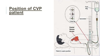
VIVEKANANDA COLLEGE OF NURSING
MICRO-TEACHING PRACTICE
PRESENTED BY- APURVA DWIVEDI [M.Sc. NSG. 1ST YR.]
Central venous pressure
Overview
Introduction
Measurement of CVP
Factors affecting CVP
Low central venous pressure
Elevated central venous
pressure
Why do we measure CVP?
Methods to measure CVP
CVP-transducer set-up
Set up of equipment
Position of CVP patient
Access of CVP
Nurses role
Introduction
Central venous pressure, which is a measure of pressure in the vena cava, can be
used as an estimation of preload and right atrial pressure.
Central venous pressure is often used as an assessment of hemodynamic status,
particularly in the intensive care unit.
The central venous pressure can be measured using a central venous catheter
advanced via the internal jugular vein and placed in the superior vena cava near the
right atrium.
A normal central venous pressure reading is between 8 to 12 mmHg. This value is
altered by volume status and/or venous compliance.
Measurement
Normal CVP in patients can be measured from two points of reference:
•Sternum: 0–14 cm H2O
•Midaxillary line: 8–15 cm H2O
CVP can be measured by connecting the patient's central venous catheter to
a special infusion set which is connected to a small diameter water column.
If the water column is calibrated properly the height of the column indicates
the CVP.
In most intensive care units, facilities are available to measure CVP
continuously.
Normal values vary between 4 and 12 cmH2O.
Site
Normal
pressure range
(in mmHg)[5]
Central venous pressure 3–8
Right ventricular pressure
systolic 15–30
diastolic 3–8
Pulmonary artery pressure
systolic 15–30
diastolic 4–12
Pulmonary vein/
Pulmonary capillary wedge pressure
2–15
Left ventricular pressure
systolic 100–140
diastolic 3–12
Factors affecting CVP
Factors that increase CVP include:
•Cardiac tamponade
•Decreased cardiac output
•Forced exhalation
•Heart failure
•Hypervolemia
•Mechanical ventilation and the application of positive end-expiratory pressure (PEEP)
•Pleural effusion
•Pulmonary embolism
•Pulmonary hypertension
•Tension pneumothorax
Conti..
Factors that decrease CVP include:
•Deep inhalation
•Distributive shock
•Hypovolemia
Low central venous pressure
Some factors that can decrease central venous pressure are hypovolemia or
vasodilation.
Either of these would decrease venous return and thus decrease the central venous
pressure.
A decrease in central venous pressure is noted when there is more than 10% of blood
loss or shift of blood volume.
A decrease in intrathoracic pressure caused by forced inspiration causes the vena cave
to collapse which decreases the venous return and, in turn, decreases the central
venous pressure.
Elevated central venous pressure
Elevated Central Venous Pressure can occur in heart failure due to decreased
contractility, valve abnormalities, and dysrhythmias.
Any patients on ventilator assistance that have excessive positive end-expiratory
pressure would have an increase in pulmonary arterial resistance which causes an
increase in central venous pressure.
However, an increased central venous pressure caused by increased pulmonary
arterial resistance can also be affected by a decrease in the fraction of inspired
oxygen, an increase in ventilation/perfusion abnormalities in the lung, an increase
in pericardial pressure, or an increase in intra-abdominal pressure which would
increase thoracic pressure.
Increased juxta-cardiac pressure - tension pneumothorax, pericardial tamponade,
right ventricular infarct, right ventricular outflow obstruction - can also
decrease venous return.
Why do we measure CVP?
To serve as a guide of fluid balance in
critically ill patients
To estimate the circulating blood volume
To determine the function of the right side of
the heart
To assist in monitoring circulatory failure
None of these variables are measured
directly; they must be interpreted.
Methods to measure CVP
CVP Measurement
Method
Direct
Using manometer
Using transducer
Indirect
Inspection of Juglar
venous pulsations
in the neck
CVP-transducer set-up
✓500ml NS
✓ Pressure bag
✓ Transducer KIT
✓ Transducer holder
✓ Transducer pressure cable
Set up of equipment
Maintain aseptic technique
Open the transducer set and tighten all the connections
Hang the 500 ml saline in the pressure bag, and spike it.
Inflate the pressure bag to 300 mm hg and turn stopcock to upwards position.
Clamp iv tubing
Place the transducer holder on IV pole
Place transducer in transducer holder attach the IV tubing and the PM line in the
transducer kit
Unclamp the tubing and remove air from the tubing by activating the flush device.
Point stopcock of the transducer horizontal and do the tubing's
Position of CVP
patient
Access of CVP
Nurses role
Monitor for the signs of complications.
Assess for patency of the CVP line.
Sterile dressing should be done to prevent infection( CVP care
per the hospital protocol)
The length of the indwelling catheter should be recorded and
regularly monitored.
Summary
We have learned about Introduction, Measurement of CVP,
Factors affecting CVP, Low central venous pressure, Elevated
central venous pressure, Why do we measure CVP?, Methods to
measure CVP, CVP-transducer set-up, Set up of equipment,
Position of CVP patient, Access of CVP and Nurses role.
Conclusion
Central venous pressure estimates the right atrial pressure
pulmonary capillary wedge pressure estimates the left atrial
pressure. The values reflect end-diastolic volume, or pre load, of
the right and left ventricles, respectively. Central venous pressure
and pulmonary capillary wedge pressure both would have been
decreased because of the loss of blood volume from the venous
side.
Bibliography
Book reference
‘‘SUDDARTH’S AND BRUNNER’’, Text book of medical surgical
nursing,volume-1,edition-13,wolters Kluwer publisher, Page no
‘‘CHINTAMANI, MRINALINI, HARINDARJEET GOYAL;’’ Lewis’s medical
surgical nursing ,volume2,1st edition-2011, Elsevier publisher, page no.
Net reference
https://www.ncbi.nlm.nih.gov/books/NBK519493/#:~:text=Central%20venous
%20pressure%2C%20which%20is,in%20the%20intensive%20care%20unit.

CVP micro teaching.pptx

  • 1.
    VIVEKANANDA COLLEGE OFNURSING MICRO-TEACHING PRACTICE PRESENTED BY- APURVA DWIVEDI [M.Sc. NSG. 1ST YR.]
  • 2.
  • 3.
    Overview Introduction Measurement of CVP Factorsaffecting CVP Low central venous pressure Elevated central venous pressure Why do we measure CVP? Methods to measure CVP CVP-transducer set-up Set up of equipment Position of CVP patient Access of CVP Nurses role
  • 4.
    Introduction Central venous pressure,which is a measure of pressure in the vena cava, can be used as an estimation of preload and right atrial pressure. Central venous pressure is often used as an assessment of hemodynamic status, particularly in the intensive care unit. The central venous pressure can be measured using a central venous catheter advanced via the internal jugular vein and placed in the superior vena cava near the right atrium. A normal central venous pressure reading is between 8 to 12 mmHg. This value is altered by volume status and/or venous compliance.
  • 5.
    Measurement Normal CVP inpatients can be measured from two points of reference: •Sternum: 0–14 cm H2O •Midaxillary line: 8–15 cm H2O CVP can be measured by connecting the patient's central venous catheter to a special infusion set which is connected to a small diameter water column. If the water column is calibrated properly the height of the column indicates the CVP. In most intensive care units, facilities are available to measure CVP continuously. Normal values vary between 4 and 12 cmH2O.
  • 6.
    Site Normal pressure range (in mmHg)[5] Centralvenous pressure 3–8 Right ventricular pressure systolic 15–30 diastolic 3–8 Pulmonary artery pressure systolic 15–30 diastolic 4–12 Pulmonary vein/ Pulmonary capillary wedge pressure 2–15 Left ventricular pressure systolic 100–140 diastolic 3–12
  • 7.
    Factors affecting CVP Factorsthat increase CVP include: •Cardiac tamponade •Decreased cardiac output •Forced exhalation •Heart failure •Hypervolemia •Mechanical ventilation and the application of positive end-expiratory pressure (PEEP) •Pleural effusion •Pulmonary embolism •Pulmonary hypertension •Tension pneumothorax
  • 8.
    Conti.. Factors that decreaseCVP include: •Deep inhalation •Distributive shock •Hypovolemia
  • 9.
    Low central venouspressure Some factors that can decrease central venous pressure are hypovolemia or vasodilation. Either of these would decrease venous return and thus decrease the central venous pressure. A decrease in central venous pressure is noted when there is more than 10% of blood loss or shift of blood volume. A decrease in intrathoracic pressure caused by forced inspiration causes the vena cave to collapse which decreases the venous return and, in turn, decreases the central venous pressure.
  • 10.
    Elevated central venouspressure Elevated Central Venous Pressure can occur in heart failure due to decreased contractility, valve abnormalities, and dysrhythmias. Any patients on ventilator assistance that have excessive positive end-expiratory pressure would have an increase in pulmonary arterial resistance which causes an increase in central venous pressure. However, an increased central venous pressure caused by increased pulmonary arterial resistance can also be affected by a decrease in the fraction of inspired oxygen, an increase in ventilation/perfusion abnormalities in the lung, an increase in pericardial pressure, or an increase in intra-abdominal pressure which would increase thoracic pressure. Increased juxta-cardiac pressure - tension pneumothorax, pericardial tamponade, right ventricular infarct, right ventricular outflow obstruction - can also decrease venous return.
  • 11.
    Why do wemeasure CVP? To serve as a guide of fluid balance in critically ill patients To estimate the circulating blood volume To determine the function of the right side of the heart To assist in monitoring circulatory failure None of these variables are measured directly; they must be interpreted.
  • 12.
    Methods to measureCVP CVP Measurement Method Direct Using manometer Using transducer Indirect Inspection of Juglar venous pulsations in the neck
  • 13.
    CVP-transducer set-up ✓500ml NS ✓Pressure bag ✓ Transducer KIT ✓ Transducer holder ✓ Transducer pressure cable
  • 14.
    Set up ofequipment Maintain aseptic technique Open the transducer set and tighten all the connections Hang the 500 ml saline in the pressure bag, and spike it. Inflate the pressure bag to 300 mm hg and turn stopcock to upwards position. Clamp iv tubing Place the transducer holder on IV pole Place transducer in transducer holder attach the IV tubing and the PM line in the transducer kit Unclamp the tubing and remove air from the tubing by activating the flush device. Point stopcock of the transducer horizontal and do the tubing's
  • 15.
  • 16.
  • 17.
    Nurses role Monitor forthe signs of complications. Assess for patency of the CVP line. Sterile dressing should be done to prevent infection( CVP care per the hospital protocol) The length of the indwelling catheter should be recorded and regularly monitored.
  • 18.
    Summary We have learnedabout Introduction, Measurement of CVP, Factors affecting CVP, Low central venous pressure, Elevated central venous pressure, Why do we measure CVP?, Methods to measure CVP, CVP-transducer set-up, Set up of equipment, Position of CVP patient, Access of CVP and Nurses role.
  • 19.
    Conclusion Central venous pressureestimates the right atrial pressure pulmonary capillary wedge pressure estimates the left atrial pressure. The values reflect end-diastolic volume, or pre load, of the right and left ventricles, respectively. Central venous pressure and pulmonary capillary wedge pressure both would have been decreased because of the loss of blood volume from the venous side.
  • 20.
    Bibliography Book reference ‘‘SUDDARTH’S ANDBRUNNER’’, Text book of medical surgical nursing,volume-1,edition-13,wolters Kluwer publisher, Page no ‘‘CHINTAMANI, MRINALINI, HARINDARJEET GOYAL;’’ Lewis’s medical surgical nursing ,volume2,1st edition-2011, Elsevier publisher, page no. Net reference https://www.ncbi.nlm.nih.gov/books/NBK519493/#:~:text=Central%20venous %20pressure%2C%20which%20is,in%20the%20intensive%20care%20unit.