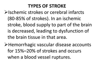
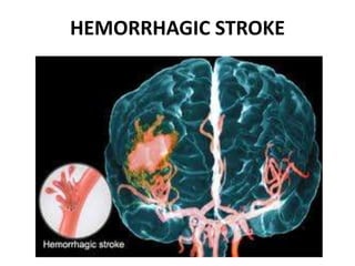
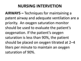
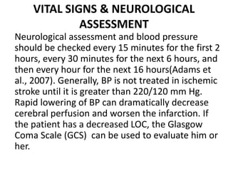
CEREBROVASCULAR ACCIDENT
(STROKE): CHALLENGES IN NURSING
CARE
PRESENTINGDEPARTMENT: NURSINGSERVICES
PRESENTER: MR TAHAV KERSHIO
19th FEBRUARY, 2015
DEFINITION
A cerebrovascular accident (CVA), an
ischemic stroke or “brain attack,” is a
sudden loss of brain function
resulting from a disruption of the
blood supply to a part of the brain.
TYPES OF STROKE
Ischemic strokes or cerebral infarcts
(80-85% of strokes). In an ischemic
stroke, blood supply to part of the brain
is decreased, leading to dysfunction of
the brain tissue in that area.
Hemorrhagic vascular disease accounts
for 15%–20% of strokes and occurs
when a blood vessel ruptures.
HEMORRHAGIC STROKE
ISCHEMIC STROKE
OTHER SUB TYPES
Watershed stroke: when blood supply to
these areas is compromised(hypoperfussion).
Silent stroke: is a stroke that does not have
any outward symptoms, and the patients are
typically unaware they have suffered a stroke.
Transient Ischemic Attack: temporary
disruption in the blood supply to part of the
brain.
STATISTICS
 There are 15 million people worldwide
who suffer a stroke each year.
 According to the WHO, stroke is the
second leading cause of death for
people above the age of 60 years, and
the fifth leading cause in people aged
15 to 59 years old.
 Each year, nearly six million people
worldwide die from stroke.
 One in six people worldwide will have a
stroke in their lifetime.
 Every six seconds, stroke kills
someone!!!
 Hemorrhagic strokes are more severe,
and mortality rates are higher than
ischemic strokes.
In fact, stroke continues to be
responsible for more deaths annually
than those attributed to AIDS,
tuberculosis and malaria combined.
The burden of stroke now
disproportionately affects individuals
living in resource-poor countries.
RISK FACTORS
MODIFIABLE FACTORS
NON-MODIFIABLE FACTORS
MODIFIABLE FACTORS
Hypertension
Atrial fibrillation
Hyperlipidemia
Obesity
Smoking
Diabetes
Periodontal disease
NON-MODIFIABLE FACTORS
• Age (older than 55 years)
• Gender (Male)
• Race (African American)
PATHOPHHYSIOLOGY
Cerebrovascular accident (CVA,
“stroke” or “brain attack”) is injury or
death to parts of the brain caused by
an interruption in the blood supply
to that area causing disability, such
as paralysis or speech impairment.
CLINICAL MANIFESTATIONS
General signs and symptoms include
numbness or weakness of face, arm,
or leg (especially on one side of
body); confusion or change in mental
status; trouble speaking or
understanding speech; visual
disturbances; loss of balance,
dizziness, difficulty walking; or
sudden severe headache.
INVESTIGATIONS
• CT scan: Demonstrates structural
abnormalities, edema, hematomas, ischemia,
and infarctions.
• MRI: Shows areas of infarction, hemorrhage,
AV malformations; and areas of ischemia.
• Cerebral angiography: Helps determine
specific cause of stroke, e.g., hemorrhage or
obstructed artery, pinpoints site of occlusion
or rupture.
PET scan: (positron emission tomography)
Provides data on cerebral metabolism and
blood flow changes.
Lumbar puncture: Pressure is usually normal
and CSF is clear in cerebral thrombosis,
embolism, and TIA. Pressure elevation and
grossly bloody fluid suggest subarachnoid and
intracerebral hemorrhage.
Trans-cranial Doppler ultrasonography:
Evaluates the velocity of blood flow through
major intracranial vessels.
EEG: Identifies problems based on reduced
electrical activity in specific areas of
infarction; and can differentiate seizure
activity from CVA damage.
X-rays (skull): May show shift of pineal gland
to the opposite side from an expanding mass;
calcifications of the internal carotid may be
visible in cerebral thrombosis; partial
calcification of walls of an aneurysm may be
noted in subarachnoid hemorrhage.
• Laboratory studies to rule out systemic
causes: CBC, platelet and clotting studies,
VDRL/RPR, erythrocyte sedimentation rate
(ESR), chemistries (glucose, sodium).
• ECG, chest x-ray, and echocardiography: To
rule out cardiac origin as source of embolus
(20% of strokes are the result of blood or
vegetative emboli associated with valvular
disease, dysrhythmias, or endocarditis).
• Physical Examination.
MANAGEMENT OF CVA (STROKE):
MEDICAL MANAGEMENT
SURGICAL MANAGEMENT
NURSING MANAGEMENT
STROKE UNIT
Ideally, people who have had a stroke are
admitted to a "stroke unit", a ward or dedicated
area in hospital staffed by nurses and therapists
with experience in stroke treatment. It has been
shown that people admitted to a stroke unit
have a higher chance of surviving than those
admitted elsewhere in hospital, even if they are
being cared for by doctors without experience in
stroke.
TREATMENT
Approaches in stroke vary widely because of the huge
variety of presentations. They will depend on:
 The site of occlusion or aneurismal rapture
 The degree and extent of the ischemia or
haemorrhage
 The effectiveness of medical and nursing intervention
 The patient’s response
 The aims are to prevent further brain damage, reduce
the risk factors, provide supportive care and regain
functional independence.
MEDICAL MANAGEMENT
Recombinant tissue plasminogen activator,
unless contraindicated; monitor for bleeding
Anticoagulation therapy
Antiplatelet agents
Management of increased intracranial pressure
(ICP): osmotic diuretics, elevate the head of bed
to promote venous drainage and to lower
increased ICP
Possible hemicraniectomy for increased ICP
from brain edema in a very large stroke
Intubation with an endotracheal tube to
establish a patent airway, if necessary
Continuous hemodynamic monitoring
Neurologic assessment to determine if the
stroke is evolving and if other acute
complications are developing
CONSERVATIVE RX
• ANTICOAGULANTS -E.g. warfarin, although doubt
has been cast on the usefulness of anticoagulant
as it poses a risk of further heamorrhage into the
infracted brain.
• ANTIFIBRONOLYTIC - Their use is thought to
prevent re-bleeding by delaying dissolution of the
clot around the aneurysm, but their effort on the
overall outcome is questionable.
ANTIPLATELET AGENTS – The use of
aspirin as an anti platelet agent has
received attention in recent years.
Other factors – pre-existing
contributory disorders may be treated
with drugs therapy, e.g.
antihypertensive agents and diuretics
may be used in the patient with raised
blood pressure
SURGICAL MANAGEMENT: USES TWO
TECHNIQUES
 Carotid endarterectomy, which involves the
removal of stenosing or ulcerating atheromatous
lesions at the bifurcation of the common carotid
arteries.
 A superficial temporal to middle cerebral artery
anastomosis , which provides an artificial
collateral blood supply to the affected part of the
brain.
NURSING PRIORITIES AND
MANAGEMENT
Prevention: One of the most important aspect of
stroke management is prevention, by identifying
at-risk individuals and dealing with early
predisposing factors such as hypertension.
NURSING CARE FOLLOWING A STROKE
A successful outcome is more likely
when the optimum techniques and
resources are utilized, encompassing
every member of the multidisciplinary
team. The outcome can also be
influenced by other factors such as
recognizing the need to start the
rehabilitation process as soon as
possible.
INITIAL TREATMENT CONCERN
The initial plan for the management of a
patient with acute stroke is to control vital
signs, prevent deterioration of the patient,
and prevent medical complications of the
stroke that worsen the patient’s outcome.
Medical complications include respiratory
failure, hypertension, hyperglycemia, cerebral
edema, and fever. The nurse caring for the
patient must coordinate the activities of an
interdisciplinary team to provide high-quality
RISK ASSESSMENT
Nurses are generally responsible for
ensuring that risk assessments are carried
out soon after patient admission. These can
include assessing the risks of moving and
handling, nutrition, pressure ulcers, falls and
DVT.
NURSING DIAGNOSES
• Impaired physical mobility related to
hemiparesis, loss of balance and coordination.
• Deficient self-care (bathing, hygiene, toileting,
dressing, grooming, and feeding).
• Impaired urinary elimination related to flaccid
bladder, detrusor instability, confusion, or
difficulty in communicating.
• Disturbed thought processes related to brain
damage.
• Impaired verbal communication related to brain
damage.
Risk for impaired skin integrity related to
hemiparesis or hemiplegia, decreased mobility
• Interrupted family processes related to
catastrophic illness and caregiving burdens.
• Sexual dysfunction related to neurologic deficits
or fear of failure.
NURSING INTERVETION
AIRWAYS – Techniques for maintaining a
patent airway and adequate ventilation are a
priority. An oxygen saturation monitor
should be used to evaluate the patient’s
oxygenation. If the patient’s oxygen
saturation is less than 90%, the patient
should be placed on oxygen titrated at 2–4
liters per minute to maintain an oxygen
saturation of 90%.
VITAL SIGNS & NEUROLOGICAL
ASSESSMENT
Neurological assessment and blood pressure
should be checked every 15 minutes for the first 2
hours, every 30 minutes for the next 6 hours, and
then every hour for the next 16 hours(Adams et
al., 2007). Generally, BP is not treated in ischemic
stroke until it is greater than 220/120 mm Hg.
Rapid lowering of BP can dramatically decrease
cerebral perfusion and worsen the infarction. If
the patient has a decreased LOC, the Glasgow
Coma Scale (GCS) can be used to evaluate him or
her.
IMPROVING MOBILITY &
PREVENTING DEFORMITIES
Position to prevent contractures; use measures to
relieve pressure, assist in maintaining good body
alignment.
Elevate affected arm to prevent edema and
fibrosis.
Change position every 2 hours; place patient in a
prone position for 15 to 30 minutes several times
a day.
EATING AND DRINKING
Initially the patient’s fluid intake is likely to be via
IV infusion(this maintains arterial BP and, in turn,
prevents cerebral ischemia and infarction)
Maintain accurate fluid balance
Subsequently, oral diet – through NG tube or as
the case may be, will be introduced
Regular oral inspection and oral hygiene should
be carried out.
IMPROVING COMMUNICATION
Speech impairment or loss can be a
frightening experience for the patient and his
family. Early referral to a speech therapist is
important in order for an expert assessment
can be performed and a strategy identified. It
is crucial to ascertain the type and nature of
the speech deficit, e.g. whether the patient’s
difficulties are related to expression or
comprehension.
IMPROVING COMMUNICATION CONT.
• Maintain patient’s attention when talking
with patient, speak slowly, and give one
instruction at a time; allow patient time to
process.
• Make the atmosphere conducive to
communication, remaining sensitive to
patient’s reactions and needs
MAINTAINGN SKIN INTEGRITY
• Frequently assess skin for signs of breakdown,
with emphasis on bony areas and dependent
body parts.
• Employ pressure relieving devices; continue
regular turning and positioning (every 2 hours
minimally); minimize shear and friction when
positioning.
• Keep skin clean and dry, gently massage healthy
dry skin, and maintain adequate nutrition.
ESTABLISHING AN EXERCISE
PROGRAMME
• Provide full range of motion 4 or 5 times a day
to maintain joint mobility, regain motor control,
prevent contractures in the paralyzed extremity.
• Observe for signs of pulmonary embolus or
excessive cardiac workload during exercise
period.
• Supervise and support patient during exercises;
plan frequent short periods of exercise.
PREPARING FOR AMBULATION
• Start an active rehabilitation program when
consciousness returns.
• Teach patient to maintain balance in a sitting
position, then to balance while standing (use a
tilt table if needed).
• Begin walking as soon as standing balance is
achieved.
• Keep training periods for ambulation short
and frequent.
IMPROVING FAMILY COPING
• Provide counseling and support to family.
• Involve others in patient’s care; teach stress
management techniques and maintenance of
personal health for family coping.
• Give family information about the expected
outcome of the stroke, and counsel them to avoid
doing things for patient that he or she can do.
• Encourage everyone to approach patient with a
supportive and optimistic attitude,
ELIMINATION
Interruption of the patient’s elimination
pattern is due to loss of consciousness and
enforced immobility. Urinary incontinence is
best dealt with by retraining the patient to
use bedpans or urinals at specified intervals,
rather than resorting to catheterization.
Condom-type urinary appliances may be
suitable for male patients, but no successful
female equivalent is yet available.
ELIMINATION CONT.
• Analyze voiding pattern and offer urinal or
bedpan on patient’s voiding schedule.
• Assist the male patient to an upright posture
for voiding.
• Provide highfiber diet and adequate fluid
intake (2 to 3 L/day), unless contraindicated.
HELPING TO COPE WITH SEXUAL
DYSFUNCTION
• Perform in-depth assessment to determine
sexual history before and after the stroke.
• Counseling regarding coping skills, suggestions
for alternative sexual positions, and a means of
sexual expression and satisfaction.
REHABILITATION
This is the process by which those with disabling
strokes undergo treatment to help return to
normal as much as possible regaining and learning
the skills of everyday living. It also aims to help
the survivor understand and adapt to difficulties,
prevents secondary complication and educates
family to play a supporting role. It’s usually
multidisciplinary as it involves staff with different
skills working together to help the person.
These include nursing staff,
physiotherapists, occupational therapists,
speech and language therapists, orthotists
and usually a physician trained in
rehabilitation medicine. Some teams may
include psychologists, social workers, and
pharmacists. Validated instrument such as
the Barthel Scale may be used to assess the
likelihood of a stroke patient being managed
at home with or without support
subsequent to discharge from hospital.
PROGNOSIS
• Disability affects 75% of stroke survivors
enough to decrease their employability. Stroke
can affect people physically, mentally,
emotionally, or a combination of the three.
The result of stroke vary widely depending on
size or location. Dysfunction corresponds to
areas in the brain that have been affected.
PROGNOSIS CONT.
• Some of the physical disabilities that can result
from stroke include muscle weakness, numbness,
pressure sores, pneumonia, incontinence, apraxia
(inability to perform learned movement),
difficulties carrying out daily activities, appetite
loss, speech loss, vision loss and pain. If the
stroke is severe enough, or in a certain location
such as parts of the brain-stem, coma or death
can result.
SUGGESTIONS/RECOMMENDATIONS
• In view of the enormous and numerous
challenges involved in the caring of CVA
patients, I wish to make the following
suggestions/recommendations:
• Need for adequate man-power/staff,
especially in the field of Nursing.
• Provision of necessary equipment in the
stroke unit.
• Creation of a stroke unit/ward with a
tag-team comprising; orthotist,
speech/language therapists,
occupational therapists,
psychologists, social workers,
physicians trained in rehabilitation
medicine and nurses who are
specially trained on how to care for
CVA patients.
CONCLUSION
• In view of the above, it can be seen that the
challenges associated with the care of patients
with CVA are quite numerous, enormous and
require special training.
• CVA (Stroke) is a life-threatening condition with
high morbidity & mortality rate, and knows no
limit to race.
• Requires multi-disciplinary & team approach.
In spite of these huge challenges, if we do our
part very well and leave the rest to GOD, we
will put some smiles on the faces of our
patients.
Remember, anyone can be a victim!!!
THANK YOU FOR LISTENING

Presentationon cva

  • 1.
    CEREBROVASCULAR ACCIDENT (STROKE): CHALLENGESIN NURSING CARE PRESENTINGDEPARTMENT: NURSINGSERVICES PRESENTER: MR TAHAV KERSHIO 19th FEBRUARY, 2015
  • 2.
    DEFINITION A cerebrovascular accident(CVA), an ischemic stroke or “brain attack,” is a sudden loss of brain function resulting from a disruption of the blood supply to a part of the brain.
  • 3.
    TYPES OF STROKE Ischemicstrokes or cerebral infarcts (80-85% of strokes). In an ischemic stroke, blood supply to part of the brain is decreased, leading to dysfunction of the brain tissue in that area. Hemorrhagic vascular disease accounts for 15%–20% of strokes and occurs when a blood vessel ruptures.
  • 4.
  • 5.
  • 6.
    OTHER SUB TYPES Watershedstroke: when blood supply to these areas is compromised(hypoperfussion). Silent stroke: is a stroke that does not have any outward symptoms, and the patients are typically unaware they have suffered a stroke. Transient Ischemic Attack: temporary disruption in the blood supply to part of the brain.
  • 7.
    STATISTICS  There are15 million people worldwide who suffer a stroke each year.  According to the WHO, stroke is the second leading cause of death for people above the age of 60 years, and the fifth leading cause in people aged 15 to 59 years old.
  • 8.
     Each year,nearly six million people worldwide die from stroke.  One in six people worldwide will have a stroke in their lifetime.  Every six seconds, stroke kills someone!!!  Hemorrhagic strokes are more severe, and mortality rates are higher than ischemic strokes.
  • 9.
    In fact, strokecontinues to be responsible for more deaths annually than those attributed to AIDS, tuberculosis and malaria combined. The burden of stroke now disproportionately affects individuals living in resource-poor countries.
  • 10.
  • 11.
  • 12.
    NON-MODIFIABLE FACTORS • Age(older than 55 years) • Gender (Male) • Race (African American)
  • 13.
    PATHOPHHYSIOLOGY Cerebrovascular accident (CVA, “stroke”or “brain attack”) is injury or death to parts of the brain caused by an interruption in the blood supply to that area causing disability, such as paralysis or speech impairment.
  • 14.
    CLINICAL MANIFESTATIONS General signsand symptoms include numbness or weakness of face, arm, or leg (especially on one side of body); confusion or change in mental status; trouble speaking or understanding speech; visual disturbances; loss of balance, dizziness, difficulty walking; or sudden severe headache.
  • 15.
    INVESTIGATIONS • CT scan:Demonstrates structural abnormalities, edema, hematomas, ischemia, and infarctions. • MRI: Shows areas of infarction, hemorrhage, AV malformations; and areas of ischemia. • Cerebral angiography: Helps determine specific cause of stroke, e.g., hemorrhage or obstructed artery, pinpoints site of occlusion or rupture.
  • 16.
    PET scan: (positronemission tomography) Provides data on cerebral metabolism and blood flow changes. Lumbar puncture: Pressure is usually normal and CSF is clear in cerebral thrombosis, embolism, and TIA. Pressure elevation and grossly bloody fluid suggest subarachnoid and intracerebral hemorrhage. Trans-cranial Doppler ultrasonography: Evaluates the velocity of blood flow through major intracranial vessels.
  • 17.
    EEG: Identifies problemsbased on reduced electrical activity in specific areas of infarction; and can differentiate seizure activity from CVA damage. X-rays (skull): May show shift of pineal gland to the opposite side from an expanding mass; calcifications of the internal carotid may be visible in cerebral thrombosis; partial calcification of walls of an aneurysm may be noted in subarachnoid hemorrhage.
  • 18.
    • Laboratory studiesto rule out systemic causes: CBC, platelet and clotting studies, VDRL/RPR, erythrocyte sedimentation rate (ESR), chemistries (glucose, sodium). • ECG, chest x-ray, and echocardiography: To rule out cardiac origin as source of embolus (20% of strokes are the result of blood or vegetative emboli associated with valvular disease, dysrhythmias, or endocarditis). • Physical Examination.
  • 19.
    MANAGEMENT OF CVA(STROKE): MEDICAL MANAGEMENT SURGICAL MANAGEMENT NURSING MANAGEMENT
  • 20.
    STROKE UNIT Ideally, peoplewho have had a stroke are admitted to a "stroke unit", a ward or dedicated area in hospital staffed by nurses and therapists with experience in stroke treatment. It has been shown that people admitted to a stroke unit have a higher chance of surviving than those admitted elsewhere in hospital, even if they are being cared for by doctors without experience in stroke.
  • 21.
    TREATMENT Approaches in strokevary widely because of the huge variety of presentations. They will depend on:  The site of occlusion or aneurismal rapture  The degree and extent of the ischemia or haemorrhage  The effectiveness of medical and nursing intervention  The patient’s response  The aims are to prevent further brain damage, reduce the risk factors, provide supportive care and regain functional independence.
  • 22.
    MEDICAL MANAGEMENT Recombinant tissueplasminogen activator, unless contraindicated; monitor for bleeding Anticoagulation therapy Antiplatelet agents Management of increased intracranial pressure (ICP): osmotic diuretics, elevate the head of bed to promote venous drainage and to lower increased ICP
  • 23.
    Possible hemicraniectomy forincreased ICP from brain edema in a very large stroke Intubation with an endotracheal tube to establish a patent airway, if necessary Continuous hemodynamic monitoring Neurologic assessment to determine if the stroke is evolving and if other acute complications are developing
  • 24.
    CONSERVATIVE RX • ANTICOAGULANTS-E.g. warfarin, although doubt has been cast on the usefulness of anticoagulant as it poses a risk of further heamorrhage into the infracted brain. • ANTIFIBRONOLYTIC - Their use is thought to prevent re-bleeding by delaying dissolution of the clot around the aneurysm, but their effort on the overall outcome is questionable.
  • 25.
    ANTIPLATELET AGENTS –The use of aspirin as an anti platelet agent has received attention in recent years. Other factors – pre-existing contributory disorders may be treated with drugs therapy, e.g. antihypertensive agents and diuretics may be used in the patient with raised blood pressure
  • 26.
    SURGICAL MANAGEMENT: USESTWO TECHNIQUES  Carotid endarterectomy, which involves the removal of stenosing or ulcerating atheromatous lesions at the bifurcation of the common carotid arteries.  A superficial temporal to middle cerebral artery anastomosis , which provides an artificial collateral blood supply to the affected part of the brain.
  • 27.
    NURSING PRIORITIES AND MANAGEMENT Prevention:One of the most important aspect of stroke management is prevention, by identifying at-risk individuals and dealing with early predisposing factors such as hypertension.
  • 28.
    NURSING CARE FOLLOWINGA STROKE A successful outcome is more likely when the optimum techniques and resources are utilized, encompassing every member of the multidisciplinary team. The outcome can also be influenced by other factors such as recognizing the need to start the rehabilitation process as soon as possible.
  • 29.
    INITIAL TREATMENT CONCERN Theinitial plan for the management of a patient with acute stroke is to control vital signs, prevent deterioration of the patient, and prevent medical complications of the stroke that worsen the patient’s outcome. Medical complications include respiratory failure, hypertension, hyperglycemia, cerebral edema, and fever. The nurse caring for the patient must coordinate the activities of an interdisciplinary team to provide high-quality
  • 30.
    RISK ASSESSMENT Nurses aregenerally responsible for ensuring that risk assessments are carried out soon after patient admission. These can include assessing the risks of moving and handling, nutrition, pressure ulcers, falls and DVT.
  • 31.
    NURSING DIAGNOSES • Impairedphysical mobility related to hemiparesis, loss of balance and coordination. • Deficient self-care (bathing, hygiene, toileting, dressing, grooming, and feeding). • Impaired urinary elimination related to flaccid bladder, detrusor instability, confusion, or difficulty in communicating.
  • 32.
    • Disturbed thoughtprocesses related to brain damage. • Impaired verbal communication related to brain damage. Risk for impaired skin integrity related to hemiparesis or hemiplegia, decreased mobility • Interrupted family processes related to catastrophic illness and caregiving burdens. • Sexual dysfunction related to neurologic deficits or fear of failure.
  • 33.
    NURSING INTERVETION AIRWAYS –Techniques for maintaining a patent airway and adequate ventilation are a priority. An oxygen saturation monitor should be used to evaluate the patient’s oxygenation. If the patient’s oxygen saturation is less than 90%, the patient should be placed on oxygen titrated at 2–4 liters per minute to maintain an oxygen saturation of 90%.
  • 34.
    VITAL SIGNS &NEUROLOGICAL ASSESSMENT Neurological assessment and blood pressure should be checked every 15 minutes for the first 2 hours, every 30 minutes for the next 6 hours, and then every hour for the next 16 hours(Adams et al., 2007). Generally, BP is not treated in ischemic stroke until it is greater than 220/120 mm Hg. Rapid lowering of BP can dramatically decrease cerebral perfusion and worsen the infarction. If the patient has a decreased LOC, the Glasgow Coma Scale (GCS) can be used to evaluate him or her.
  • 35.
    IMPROVING MOBILITY & PREVENTINGDEFORMITIES Position to prevent contractures; use measures to relieve pressure, assist in maintaining good body alignment. Elevate affected arm to prevent edema and fibrosis. Change position every 2 hours; place patient in a prone position for 15 to 30 minutes several times a day.
  • 36.
    EATING AND DRINKING Initiallythe patient’s fluid intake is likely to be via IV infusion(this maintains arterial BP and, in turn, prevents cerebral ischemia and infarction) Maintain accurate fluid balance Subsequently, oral diet – through NG tube or as the case may be, will be introduced Regular oral inspection and oral hygiene should be carried out.
  • 37.
    IMPROVING COMMUNICATION Speech impairmentor loss can be a frightening experience for the patient and his family. Early referral to a speech therapist is important in order for an expert assessment can be performed and a strategy identified. It is crucial to ascertain the type and nature of the speech deficit, e.g. whether the patient’s difficulties are related to expression or comprehension.
  • 38.
    IMPROVING COMMUNICATION CONT. •Maintain patient’s attention when talking with patient, speak slowly, and give one instruction at a time; allow patient time to process. • Make the atmosphere conducive to communication, remaining sensitive to patient’s reactions and needs
  • 39.
    MAINTAINGN SKIN INTEGRITY •Frequently assess skin for signs of breakdown, with emphasis on bony areas and dependent body parts. • Employ pressure relieving devices; continue regular turning and positioning (every 2 hours minimally); minimize shear and friction when positioning. • Keep skin clean and dry, gently massage healthy dry skin, and maintain adequate nutrition.
  • 40.
    ESTABLISHING AN EXERCISE PROGRAMME •Provide full range of motion 4 or 5 times a day to maintain joint mobility, regain motor control, prevent contractures in the paralyzed extremity. • Observe for signs of pulmonary embolus or excessive cardiac workload during exercise period. • Supervise and support patient during exercises; plan frequent short periods of exercise.
  • 41.
    PREPARING FOR AMBULATION •Start an active rehabilitation program when consciousness returns. • Teach patient to maintain balance in a sitting position, then to balance while standing (use a tilt table if needed). • Begin walking as soon as standing balance is achieved. • Keep training periods for ambulation short and frequent.
  • 42.
    IMPROVING FAMILY COPING •Provide counseling and support to family. • Involve others in patient’s care; teach stress management techniques and maintenance of personal health for family coping. • Give family information about the expected outcome of the stroke, and counsel them to avoid doing things for patient that he or she can do. • Encourage everyone to approach patient with a supportive and optimistic attitude,
  • 43.
    ELIMINATION Interruption of thepatient’s elimination pattern is due to loss of consciousness and enforced immobility. Urinary incontinence is best dealt with by retraining the patient to use bedpans or urinals at specified intervals, rather than resorting to catheterization. Condom-type urinary appliances may be suitable for male patients, but no successful female equivalent is yet available.
  • 44.
    ELIMINATION CONT. • Analyzevoiding pattern and offer urinal or bedpan on patient’s voiding schedule. • Assist the male patient to an upright posture for voiding. • Provide highfiber diet and adequate fluid intake (2 to 3 L/day), unless contraindicated.
  • 45.
    HELPING TO COPEWITH SEXUAL DYSFUNCTION • Perform in-depth assessment to determine sexual history before and after the stroke. • Counseling regarding coping skills, suggestions for alternative sexual positions, and a means of sexual expression and satisfaction.
  • 46.
    REHABILITATION This is theprocess by which those with disabling strokes undergo treatment to help return to normal as much as possible regaining and learning the skills of everyday living. It also aims to help the survivor understand and adapt to difficulties, prevents secondary complication and educates family to play a supporting role. It’s usually multidisciplinary as it involves staff with different skills working together to help the person.
  • 47.
    These include nursingstaff, physiotherapists, occupational therapists, speech and language therapists, orthotists and usually a physician trained in rehabilitation medicine. Some teams may include psychologists, social workers, and pharmacists. Validated instrument such as the Barthel Scale may be used to assess the likelihood of a stroke patient being managed at home with or without support subsequent to discharge from hospital.
  • 48.
    PROGNOSIS • Disability affects75% of stroke survivors enough to decrease their employability. Stroke can affect people physically, mentally, emotionally, or a combination of the three. The result of stroke vary widely depending on size or location. Dysfunction corresponds to areas in the brain that have been affected.
  • 49.
    PROGNOSIS CONT. • Someof the physical disabilities that can result from stroke include muscle weakness, numbness, pressure sores, pneumonia, incontinence, apraxia (inability to perform learned movement), difficulties carrying out daily activities, appetite loss, speech loss, vision loss and pain. If the stroke is severe enough, or in a certain location such as parts of the brain-stem, coma or death can result.
  • 50.
    SUGGESTIONS/RECOMMENDATIONS • In viewof the enormous and numerous challenges involved in the caring of CVA patients, I wish to make the following suggestions/recommendations: • Need for adequate man-power/staff, especially in the field of Nursing. • Provision of necessary equipment in the stroke unit.
  • 51.
    • Creation ofa stroke unit/ward with a tag-team comprising; orthotist, speech/language therapists, occupational therapists, psychologists, social workers, physicians trained in rehabilitation medicine and nurses who are specially trained on how to care for CVA patients.
  • 52.
    CONCLUSION • In viewof the above, it can be seen that the challenges associated with the care of patients with CVA are quite numerous, enormous and require special training. • CVA (Stroke) is a life-threatening condition with high morbidity & mortality rate, and knows no limit to race. • Requires multi-disciplinary & team approach.
  • 53.
    In spite ofthese huge challenges, if we do our part very well and leave the rest to GOD, we will put some smiles on the faces of our patients. Remember, anyone can be a victim!!!
  • 54.
    THANK YOU FORLISTENING