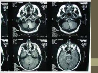
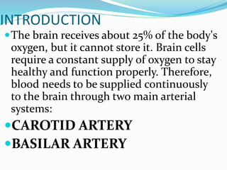
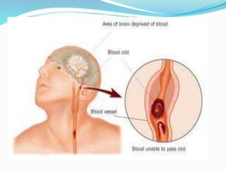
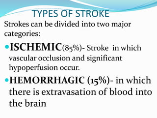
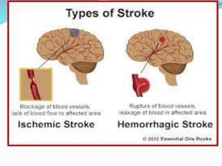
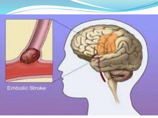
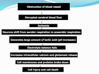
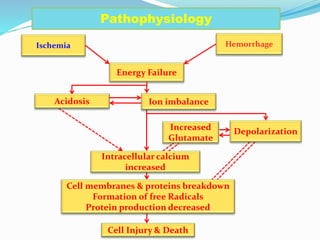
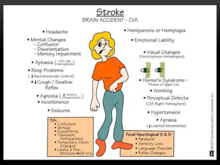
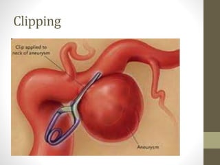
STROKE
OR
CVA
(CEREBROVASCULAR
ACCIDENT)
INTRODUCTION
The brain receives about 25% of the body's
oxygen, but it cannot store it. Brain cells
require a constant supply of oxygen to stay
healthy and function properly. Therefore,
blood needs to be supplied continuously
to the brain through two main arterial
systems:
CAROTID ARTERY
BASILAR ARTERY
CONT..
The carotid arteries come up
through either side of the front of
the neck.
The basilar artery forms at the base
of the skull from the vertebral
arteries, which run up along the
spine, join, and come up through
the rear of the neck.
Definition:
Stroke occurs when there is
ischemia (inadequate blood
flow) to a part of the brain or
hemorrhage into the brain
that results in death of brain
cells.
RISK FACTORS
RISK FACTORS ARE DIVIDED INTO :
 NON MODIFIABLE
 MODIFIABLE
NONMODIFIABLE RISK FACTORS INCLUDE:
AGE- Stroke increase with age
GENDER- Equal in men and women
RACE-
HEREDITY- a family history of stroke
Modifiable
Asymptomatic carotid
stenosis
D.M
Heart diseases
Atrial fibrillation
Excessive Alcohol
consumption
Hypercoagulability
Hyperlipidemia
Cont…
HTN
Obesity
Oral contraceptive use
Physical inactivity
Sickle cell
disease(drepanocytosis)
Smoking
TYPES OF STROKE
Strokes can be divided into two major
categories:
ISCHEMIC(85%)- Stroke in which
vascular occlusion and significant
hypoperfusion occur.
HEMORRHAGIC (15%)- in which
there is extravasation of blood into
the brain
Ischemic stroke
Ischemic stroke results from
inadequate blood flow to the brain
from partial or complete occlusion of an
artery. It is further divided into:
Thrombotic stroke.
Embolic stroke.
THROMBOTIC
Thrombotic strokes are due to atherosclerotic
plaques in the large blood vessels of the brain.
Thrombus formation and occlusion at the site
of the atherosclerosis result in ischemia and
infarction. Thrombotic strokes have been
preceded by TIA (transient ischemic attack ).
TIA is a temporary loss of neurologic function
caused by ischemia of one of the vascular
territories of the brain due to micro emboli
that temporarily block the blood flow.
EMBOLIC STROKE
An embolus is a blood clot or
other debris circulating in the
blood. When it reaches an
artery in the brain that is too
narrow to pass through, it
lodges there and blocks the flow
of blood.
Hemorrhagic Stroke
A burst of blood vessel may allow
blood to seep into and damage the
brain tissues until clotting shuts off
the leak. It is further divided into two.
 Intra-cerebral hemorrhage(intra-
parenchymal)
Subarachnoid hemorrhage (intra-
ventricular )
Intra-cerebral hemorrhage
It is the bleeding within the brain caused
by a rupture of a vessel. Hypertension is
the most important cause of intra-
cerebral hemorrhage. There is most often
a sudden onset of symptoms, with
progression over minutes to hours
because of ongoing bleeding. Symptoms
include neurologic deficits, headache,
nausea, vomiting, decreased level of
consciousness and hypertension.
Subarachnoid hemorrhage
It occurs when there is intracranial
bleeding into cerebrospinal fluid filled
space between the arachnoid and pia
mater membranes on the surface of the
brain. It commonly caused by rupture of a
cerebral aneurysm. Common symptoms
includes severe headache, sudden
comatose, focal neurologic deficits,
nausea, vomiting, seizures and stiff neck
PATHOPHYSIOLOGY
Obstruction of blood vessel
Disrupted cerebral blood flow
Ischemia
Neurons shift from aerobic respiration to anaerobic respiration
Generates large amount of lactic acid (pH increases)
Electrolyte balance fails
Increases intracellular calcium and glutamate release
Cell membranes and proteins brake down
Cell injury and cell death
Ischemia
Energy Failure
Ion imbalance
Cell Injury & Death
Cell membranes & proteins breakdown
Formation of free Radicals
Protein production decreased
Pathophysiology
Intracellular calcium
increased
Depolarization
Increased
Glutamate
Hemorrhage
Acidosis
CLINICAL MANIFESTATIONS
• A stroke can cause a wide variety of
neurologic deficits ,depending on the
location of the lesion (which vessels
are obstructed),the size of the area of
inadequate perfusion, and the amount
of secondary or accessory blood
flow. Ischemia or haemorrhage to one
side of the brain can often result in
signs and symptoms on the opposite
side of the body.
THE PATIENT MAY PRESENT WITH ANY OF THE
FOLLOWING SIGNS OR SYMPTOMS:
• Numbness or weakness of the face, arm, or
leg, especially on one side of the body
• Confusion or change in mental status
• Trouble speaking or understanding speech
• Visual disturbances
• Difficulty walking, dizziness, or loss of
balance or coordination
• Sudden severe headache
VISUAL FIELD DEFICITS
• Hemianopsia -Loss of half of the visual
field
• Loss of peripheral vision - Difficulty seeing
at night. Unaware of objects or the borders
of objects
• Diplopia - Double vision
HEMIANOPSIA
DIPLOPIA
MOTOR DEFICITS
• Hemiparesis –Weakness of the face, arm, and leg
on the same side (due to a lesion in the opposite
hemisphere)
• Hemiplegia – Paralysis of the face, arm, and leg on
the same side (due to a lesion in the opposite
hemisphere).
• Ataxia - Staggering, unsteady gait .Unable to keep
feet together; needs a broad base to stand
• Dysarthria - Difficulty in forming words
• Dysphagia- Difficulty in swallowing.
SENSORY DEFICITS
• Paresthesia - Numbness and
tingling of extremity.
VERBAL DEFICITS ( APHASIA)
• Expressive aphasia - Unable to form words
that are understandable; may be able to
speak in single-word responses
• Receptive aphasia -Unable to comprehend
the spoken word; can speak but may not
make sense
• Global (mixed) aphasia -Combination of
both receptive and expressive aphasia
COGNITIVE DEFICITS
• Short- and long-term memory loss
• Decreased attention span
• Impaired ability to concentrate
• Poor abstract reasoning
• Altered judgment
• Apraxia
EMOTIONAL DEFICITS
• Loss of self-control
• Emotional liability
• Decreased tolerance to stressful situations
• Depression
• Withdrawal
• Fear, hostility, and anger
• Feelings of isolation
• Paralysis or weakness on left
side of body
• Left visual field deficit
• Spatial-perceptual deficits
• Increased distractibility
• Impulsive behavior and poor
judgment
• Lack of awareness of deficits
• Paralysis or weakness on
right side of body
• Right visual field deficit
• Aphasia (expressive,
receptive, or global)
• Altered intellectual ability
• Slow, cautious behavior
COMPARISON OF LEFT AND RIGHT
HEMISPHERIC STROKES
LEFT HEMISPHERIC STROKE RIGHT HEMISPHERIC STROKE
DIAGNOSTIC STUDIES
DIAGNOSIS STUDIES
Diagnostic studies are done to:
•Diagnosis of stroke ,including
extent of involvement
•To measure cerebral blood flow
• To assess cardiac involvement
Diagnostic studies
• Computed tomography angiography.
• Magnetic resonance angiography
• Single photon emission computed
tomography (SPECT)
• Positron emission tomography (PET)
• Magnetic resonance spectroscopy (MRS)
• Xenon CT
• Electroencephalogram
• Cerebral angiography
• Cerebrospinal fluid analysis
•Digital subtraction angiography
•Doppler ultrasonography
•Trans-cranial Doppler
•Carotid duplex
•Carotid angiography
Cardiac assessment
•Electrocardiogram
•Chest x-ray
•Cardiac enzymes
• Echocardiaography
•Holter monitoring
Additional studies
•Cbc/ FBC
•Platelets, PT, Activated partial
thromboplastin time.
•Electrolytes, Blood glucose
•Renal and hepatic studies
•Lipid profile
•ABG
MANAGEMENT
PHARMACOLOGICAL MANAGEMENT
• ANTI PLATELET DRUG
e.g.: ASPIRIN,CLOPIDOGREL (dose:81-325
mg/day).
• ANTICOAGULANT
E.g.: warfarin
• THROMBOLYTIC THERAPY
Recombinant t-PA thrombolytic substance made
naturally by the body works by binding to fibrin
and converting plasminogen to plasmin, which
stimulates fibrinolysis of the atherosclerotic
lesion.
Cont..
• CALCIUM CHANNEL BLOCKER
Nimodipine is given to patients with
subarachnoid hemorrhage to decrease the
effects of vasospasm and minimize cerebral
damage.
• ANTISEIZURE DRUGS
Phenytoin is given if seizure occurs.
• ANALGESICS
• codeine, acetaminophen may be prescribed
for head and neck pain.
Cont…
• DIURETIC DRUGS
it is used to reduce cerebral edema and it also
helps in lowering ICP
E.g.: MANNITOL , FUROSEMIDE.
NON-PHARMACOLOGICAL MANAGEMENT
•Maintain ABC.
•Reduce salt and sodium intake
•Maintain a normal body weight
•Maintain a normal blood pressure
•Increase level of physical exercise
•Avoid cigarette smoking or tobacco
products
Cont…
•Limit consumption of alcohol to
moderate levels
•Use a diet that is low in saturated fat
and dietary cholesterol and high in
fruits and vegetables.
•The patient is fitted with elastic
compression stockings to prevent
deep vein thrombosis, a threat to any
patient on bed rest.
SURGICAL MANAGEMENT
• Carotid endarterectomy
The artheromatous lesion is removed from the
carotid artery and to improve blood flow.
• Transluminal angioplasty -
It is the insertion of a balloon to open a
stenosed artery and improve blood flow.
• EC-IC bypass
It involves anastomosing a branch of an
extracranial artery to an intracranial artery beyond
the area of obstruction with the goal of increasing
cerebral perfusion.
Merci retriever-it removes the blood clots by going
into the artery that is blocked and pulls the clot out.
Carotid enderectomy
Merci retriever
For aneurysm and hemorrhage
• CLIPPING AND WRAPPING.
It’s a traditional method to prevent rebleeding.
• COILING
It’s a endovascular procedure done by inserting a
metal coil (GDC’S) into the lumen and thus
provides immediate protection against bleeding
and reduce the pulsation within the aneurysm.
Clipping
coiling
NURSING MANAGEMENT OF CVA
a. Nursing Responsibilities
The role of a nurse in taking care of a
patient with CVA are:
1. Improve physical mobility and prevent
joint deformities by providing correct
positioning and assist the patient in
maintain good body alignment.
2. The arm of the patient is slightly flexed
with a pillow under the arm where elbow
is positioned higher than shoulder and the
wrist higher than elbow to prevent edema.
Cont…
3.Change the position of the
patient every 2 hours to
prevent pressure sores.
4.Provide passive exercises to
affected extremities four or five
times a day to maintain
mobility, motor control which
helps in preventing venous
stasis.
CVA positioning in bed..
Cont..
5. Ambulate the patient as soon as
possible when the patient can be
assisted out of bed.
6. The nurse should never lift or
pull the patient by the affected
shoulder . The proper positioning
and range of exercise reduce
shoulder pain.
Cont..
7.The nurse should encourage the
patient to do self care activities.
8.The role of a nurse is to support
the client in improving thought
process in collaboration with
primary care physician
neuropsychologist and other
professionals.
Cont..
9. The patient skin must be kept
clean and dry .Maintain
normal skin and tissue
integrity by providing
adequate nutrition.
10.Educate and counsel the
family members who play an
important role in the patients
recovery.
Priority based
Nursing diagnosis
CVA diagnosis are:
1. Ineffective cerebral tissue
perfusion related to decreased
cerebral blood flow as evidenced
by decreased GCS.
2. Impaired physical mobility
related to hemiparesis, loss of
balance and co-ordination and
brain injury as evidenced by
limited ability to perform
activities.
Cont..
3. Impaired verbal
communication related to
brain damage as evidenced
by inability to speak or find
words.
4. Self care deficit related to
stroke squeals as evidenced
by inability in performing
daily course.
assignment: NURSING
CARE PLAN ON CVA

Stroke

  • 1.
  • 3.
    INTRODUCTION The brain receivesabout 25% of the body's oxygen, but it cannot store it. Brain cells require a constant supply of oxygen to stay healthy and function properly. Therefore, blood needs to be supplied continuously to the brain through two main arterial systems: CAROTID ARTERY BASILAR ARTERY
  • 4.
    CONT.. The carotid arteriescome up through either side of the front of the neck. The basilar artery forms at the base of the skull from the vertebral arteries, which run up along the spine, join, and come up through the rear of the neck.
  • 5.
    Definition: Stroke occurs whenthere is ischemia (inadequate blood flow) to a part of the brain or hemorrhage into the brain that results in death of brain cells.
  • 7.
    RISK FACTORS RISK FACTORSARE DIVIDED INTO :  NON MODIFIABLE  MODIFIABLE NONMODIFIABLE RISK FACTORS INCLUDE: AGE- Stroke increase with age GENDER- Equal in men and women RACE- HEREDITY- a family history of stroke
  • 8.
    Modifiable Asymptomatic carotid stenosis D.M Heart diseases Atrialfibrillation Excessive Alcohol consumption Hypercoagulability Hyperlipidemia
  • 9.
    Cont… HTN Obesity Oral contraceptive use Physicalinactivity Sickle cell disease(drepanocytosis) Smoking
  • 10.
    TYPES OF STROKE Strokescan be divided into two major categories: ISCHEMIC(85%)- Stroke in which vascular occlusion and significant hypoperfusion occur. HEMORRHAGIC (15%)- in which there is extravasation of blood into the brain
  • 12.
    Ischemic stroke Ischemic strokeresults from inadequate blood flow to the brain from partial or complete occlusion of an artery. It is further divided into: Thrombotic stroke. Embolic stroke.
  • 13.
    THROMBOTIC Thrombotic strokes aredue to atherosclerotic plaques in the large blood vessels of the brain. Thrombus formation and occlusion at the site of the atherosclerosis result in ischemia and infarction. Thrombotic strokes have been preceded by TIA (transient ischemic attack ). TIA is a temporary loss of neurologic function caused by ischemia of one of the vascular territories of the brain due to micro emboli that temporarily block the blood flow.
  • 15.
    EMBOLIC STROKE An embolusis a blood clot or other debris circulating in the blood. When it reaches an artery in the brain that is too narrow to pass through, it lodges there and blocks the flow of blood.
  • 17.
    Hemorrhagic Stroke A burstof blood vessel may allow blood to seep into and damage the brain tissues until clotting shuts off the leak. It is further divided into two.  Intra-cerebral hemorrhage(intra- parenchymal) Subarachnoid hemorrhage (intra- ventricular )
  • 18.
    Intra-cerebral hemorrhage It isthe bleeding within the brain caused by a rupture of a vessel. Hypertension is the most important cause of intra- cerebral hemorrhage. There is most often a sudden onset of symptoms, with progression over minutes to hours because of ongoing bleeding. Symptoms include neurologic deficits, headache, nausea, vomiting, decreased level of consciousness and hypertension.
  • 19.
    Subarachnoid hemorrhage It occurswhen there is intracranial bleeding into cerebrospinal fluid filled space between the arachnoid and pia mater membranes on the surface of the brain. It commonly caused by rupture of a cerebral aneurysm. Common symptoms includes severe headache, sudden comatose, focal neurologic deficits, nausea, vomiting, seizures and stiff neck
  • 20.
  • 21.
    Obstruction of bloodvessel Disrupted cerebral blood flow Ischemia Neurons shift from aerobic respiration to anaerobic respiration Generates large amount of lactic acid (pH increases) Electrolyte balance fails Increases intracellular calcium and glutamate release Cell membranes and proteins brake down Cell injury and cell death
  • 22.
    Ischemia Energy Failure Ion imbalance CellInjury & Death Cell membranes & proteins breakdown Formation of free Radicals Protein production decreased Pathophysiology Intracellular calcium increased Depolarization Increased Glutamate Hemorrhage Acidosis
  • 23.
    CLINICAL MANIFESTATIONS • Astroke can cause a wide variety of neurologic deficits ,depending on the location of the lesion (which vessels are obstructed),the size of the area of inadequate perfusion, and the amount of secondary or accessory blood flow. Ischemia or haemorrhage to one side of the brain can often result in signs and symptoms on the opposite side of the body.
  • 24.
    THE PATIENT MAYPRESENT WITH ANY OF THE FOLLOWING SIGNS OR SYMPTOMS: • Numbness or weakness of the face, arm, or leg, especially on one side of the body • Confusion or change in mental status • Trouble speaking or understanding speech • Visual disturbances • Difficulty walking, dizziness, or loss of balance or coordination • Sudden severe headache
  • 25.
    VISUAL FIELD DEFICITS •Hemianopsia -Loss of half of the visual field • Loss of peripheral vision - Difficulty seeing at night. Unaware of objects or the borders of objects • Diplopia - Double vision
  • 26.
  • 27.
  • 28.
    MOTOR DEFICITS • Hemiparesis–Weakness of the face, arm, and leg on the same side (due to a lesion in the opposite hemisphere) • Hemiplegia – Paralysis of the face, arm, and leg on the same side (due to a lesion in the opposite hemisphere). • Ataxia - Staggering, unsteady gait .Unable to keep feet together; needs a broad base to stand • Dysarthria - Difficulty in forming words • Dysphagia- Difficulty in swallowing.
  • 29.
    SENSORY DEFICITS • Paresthesia- Numbness and tingling of extremity.
  • 30.
    VERBAL DEFICITS (APHASIA) • Expressive aphasia - Unable to form words that are understandable; may be able to speak in single-word responses • Receptive aphasia -Unable to comprehend the spoken word; can speak but may not make sense • Global (mixed) aphasia -Combination of both receptive and expressive aphasia
  • 31.
    COGNITIVE DEFICITS • Short-and long-term memory loss • Decreased attention span • Impaired ability to concentrate • Poor abstract reasoning • Altered judgment • Apraxia
  • 32.
    EMOTIONAL DEFICITS • Lossof self-control • Emotional liability • Decreased tolerance to stressful situations • Depression • Withdrawal • Fear, hostility, and anger • Feelings of isolation
  • 33.
    • Paralysis orweakness on left side of body • Left visual field deficit • Spatial-perceptual deficits • Increased distractibility • Impulsive behavior and poor judgment • Lack of awareness of deficits • Paralysis or weakness on right side of body • Right visual field deficit • Aphasia (expressive, receptive, or global) • Altered intellectual ability • Slow, cautious behavior COMPARISON OF LEFT AND RIGHT HEMISPHERIC STROKES LEFT HEMISPHERIC STROKE RIGHT HEMISPHERIC STROKE
  • 35.
  • 36.
    DIAGNOSIS STUDIES Diagnostic studiesare done to: •Diagnosis of stroke ,including extent of involvement •To measure cerebral blood flow • To assess cardiac involvement
  • 37.
    Diagnostic studies • Computedtomography angiography. • Magnetic resonance angiography • Single photon emission computed tomography (SPECT) • Positron emission tomography (PET) • Magnetic resonance spectroscopy (MRS) • Xenon CT • Electroencephalogram • Cerebral angiography • Cerebrospinal fluid analysis
  • 38.
    •Digital subtraction angiography •Dopplerultrasonography •Trans-cranial Doppler •Carotid duplex •Carotid angiography
  • 39.
    Cardiac assessment •Electrocardiogram •Chest x-ray •Cardiacenzymes • Echocardiaography •Holter monitoring
  • 40.
    Additional studies •Cbc/ FBC •Platelets,PT, Activated partial thromboplastin time. •Electrolytes, Blood glucose •Renal and hepatic studies •Lipid profile •ABG
  • 41.
  • 42.
    PHARMACOLOGICAL MANAGEMENT • ANTIPLATELET DRUG e.g.: ASPIRIN,CLOPIDOGREL (dose:81-325 mg/day). • ANTICOAGULANT E.g.: warfarin • THROMBOLYTIC THERAPY Recombinant t-PA thrombolytic substance made naturally by the body works by binding to fibrin and converting plasminogen to plasmin, which stimulates fibrinolysis of the atherosclerotic lesion.
  • 43.
    Cont.. • CALCIUM CHANNELBLOCKER Nimodipine is given to patients with subarachnoid hemorrhage to decrease the effects of vasospasm and minimize cerebral damage. • ANTISEIZURE DRUGS Phenytoin is given if seizure occurs. • ANALGESICS • codeine, acetaminophen may be prescribed for head and neck pain.
  • 44.
    Cont… • DIURETIC DRUGS itis used to reduce cerebral edema and it also helps in lowering ICP E.g.: MANNITOL , FUROSEMIDE.
  • 45.
    NON-PHARMACOLOGICAL MANAGEMENT •Maintain ABC. •Reducesalt and sodium intake •Maintain a normal body weight •Maintain a normal blood pressure •Increase level of physical exercise •Avoid cigarette smoking or tobacco products
  • 46.
    Cont… •Limit consumption ofalcohol to moderate levels •Use a diet that is low in saturated fat and dietary cholesterol and high in fruits and vegetables. •The patient is fitted with elastic compression stockings to prevent deep vein thrombosis, a threat to any patient on bed rest.
  • 47.
    SURGICAL MANAGEMENT • Carotidendarterectomy The artheromatous lesion is removed from the carotid artery and to improve blood flow. • Transluminal angioplasty - It is the insertion of a balloon to open a stenosed artery and improve blood flow. • EC-IC bypass It involves anastomosing a branch of an extracranial artery to an intracranial artery beyond the area of obstruction with the goal of increasing cerebral perfusion. Merci retriever-it removes the blood clots by going into the artery that is blocked and pulls the clot out.
  • 48.
  • 49.
  • 50.
    For aneurysm andhemorrhage • CLIPPING AND WRAPPING. It’s a traditional method to prevent rebleeding. • COILING It’s a endovascular procedure done by inserting a metal coil (GDC’S) into the lumen and thus provides immediate protection against bleeding and reduce the pulsation within the aneurysm.
  • 51.
  • 52.
  • 53.
  • 54.
    a. Nursing Responsibilities Therole of a nurse in taking care of a patient with CVA are: 1. Improve physical mobility and prevent joint deformities by providing correct positioning and assist the patient in maintain good body alignment. 2. The arm of the patient is slightly flexed with a pillow under the arm where elbow is positioned higher than shoulder and the wrist higher than elbow to prevent edema.
  • 55.
    Cont… 3.Change the positionof the patient every 2 hours to prevent pressure sores. 4.Provide passive exercises to affected extremities four or five times a day to maintain mobility, motor control which helps in preventing venous stasis.
  • 56.
  • 57.
    Cont.. 5. Ambulate thepatient as soon as possible when the patient can be assisted out of bed. 6. The nurse should never lift or pull the patient by the affected shoulder . The proper positioning and range of exercise reduce shoulder pain.
  • 58.
    Cont.. 7.The nurse shouldencourage the patient to do self care activities. 8.The role of a nurse is to support the client in improving thought process in collaboration with primary care physician neuropsychologist and other professionals.
  • 59.
    Cont.. 9. The patientskin must be kept clean and dry .Maintain normal skin and tissue integrity by providing adequate nutrition. 10.Educate and counsel the family members who play an important role in the patients recovery.
  • 60.
  • 61.
    CVA diagnosis are: 1.Ineffective cerebral tissue perfusion related to decreased cerebral blood flow as evidenced by decreased GCS. 2. Impaired physical mobility related to hemiparesis, loss of balance and co-ordination and brain injury as evidenced by limited ability to perform activities.
  • 62.
    Cont.. 3. Impaired verbal communicationrelated to brain damage as evidenced by inability to speak or find words. 4. Self care deficit related to stroke squeals as evidenced by inability in performing daily course.
  • 63.