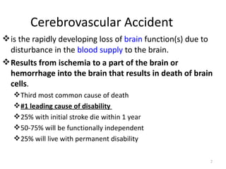
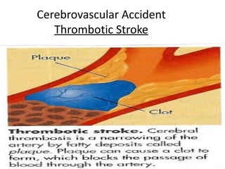
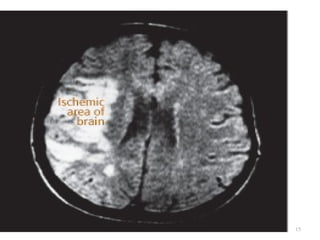
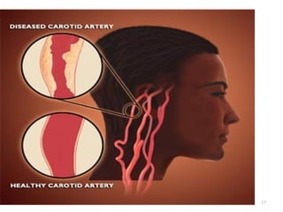
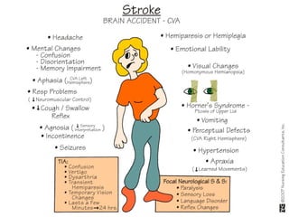
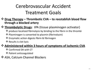
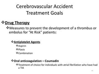
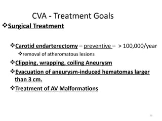
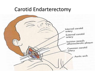
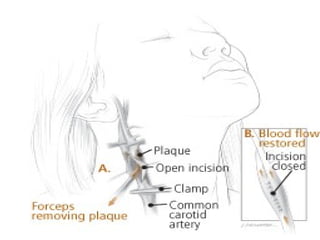
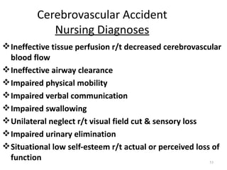
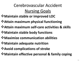
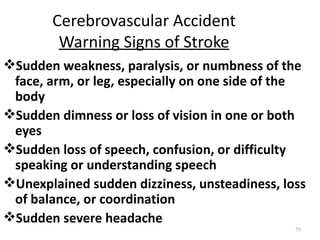
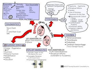
A cerebrovascular accident (CVA), also known as a stroke, is caused by the loss of blood flow to an area of the brain resulting in cell death. It is a leading cause of death and disability. The majority of strokes are ischemic and caused by blockage of an artery by a clot or debris. Hemorrhagic strokes occur when a blood vessel ruptures, causing bleeding into or around the brain. Treatment focuses on prevention through risk factor control as well as acute interventions like clot-busting drugs. Rehabilitation aims to maximize recovery of function and independence.
Dr. Pradeep Mandal presented on the evidence-based management of haemorrhagic stroke. Haemorrhagic strokes account for 10-20% of all strokes and have a higher mortality than ischemic strokes. For the 74-year-old female patient presented with right-sided weakness and altered sensorium, her ICH scale score of 3 indicates a 30-day mortality risk of 26-72%. Medical management is recommended over surgery based on the STICH trials. Her blood pressure needs to be controlled below 180/130 mmHg to prevent hematoma growth. Seizure prophylaxis, deep vein thrombosis prevention, and glycemic control are also important aspects of conservative management. The presentation concluded that
A stroke occurs when blood supply to the brain is interrupted, depriving brain cells of oxygen. Strokes can be either ischemic (88%), caused by clot or embolism blocking an artery, or hemorrhagic (12%), caused by bleeding within the brain. Diagnosis involves physical exam, blood tests, CT/MRI scans of the brain, and tests of the heart and arteries. Ischemic strokes result from plaque buildup in arteries leading to clot formation, while hemorrhagic strokes damage brain tissue through blood toxicity. Prompt treatment is needed to minimize brain injury from loss of oxygen and blood toxicity.
1) The document discusses strokes (cerebrovascular accidents or CVAs), their causes, types, symptoms, diagnosis and treatment.
2) Strokes are caused by disturbances in the vascular system and can be ischemic (caused by clot or blockage) or hemorrhagic (caused by bleeding).
3) Common symptoms of strokes include weakness or numbness on one side of the body, speech problems, and visual issues. The specific symptoms depend on the area of the brain affected.
This document discusses cerebrovascular disease and stroke. It provides classifications and descriptions of different types of strokes including transient ischemic attacks, hemorrhagic strokes, and thrombotic vs. embolic ischemic strokes. It also summarizes the clinical assessment and presentation of strokes, differential diagnoses, pathophysiology of cerebral infarction, and general risk factors.
Cerebral edema and intracranial hypertension after traumatic brain injury can be managed through various interventions to control increased intracranial pressure. These include cerebral resuscitation, intracranial pressure monitoring, hyperosmolar therapy with mannitol or hypertonic saline, mild hyperventilation, CSF drainage, temperature control, surgical decompression, and in refractory cases high-dose barbiturates or calcium channel blockers. Nutritional support and anti-seizure prophylaxis may also be considered as part of the management approach.
This document provides information about stroke, including definitions, statistics, risk factors, signs and symptoms, treatments, and the stroke program at PGI, Chd. Some key points:
- Stroke is defined as a sudden loss of brain function caused by an interruption of blood flow to the brain. It is the second most common cause of death worldwide.
- India has a high burden of stroke, with over 5000 new cases reported daily. Risk factors include hypertension, diabetes, smoking, heart disease, prior stroke or TIA, and high cholesterol.
- Signs of stroke include sudden numbness, confusion, vision problems, trouble walking or talking. The acronym FAST is used to help remember common
Head injury can range from minor scalp lacerations to serious brain injury. It is a leading cause of death from trauma. The most common causes are motor vehicle accidents, falls, assaults, and sports-related injuries. Injuries can be blunt or penetrating. Types of brain injuries include concussions, diffuse axonal injury, contusions, lacerations, and hemorrhages such as epidural, subdural, subarachnoid, intracerebral, and intraventricular hemorrhages. Clinical presentation depends on the type and severity of injury.
This document summarizes the pathophysiology of ischemic stroke and reperfusion injury. It describes how a reduction in blood flow during stroke can lead to tissue damage through various mechanisms like depletion of ATP, ionic imbalances, and free radical accumulation. It also discusses cerebral autoregulation and how its impairment during stroke contributes to low oxygen delivery. The consequences of reduced blood flow like focal ischemia, cell injury pathways like excitotoxicity and inflammation are summarized. Risk factors for reperfusion syndrome following revascularization like hypertension, dysautoregulation and ischemia-reperfusion are highlighted. Methods to assess risk and prevent reperfusion injury through blood pressure control are also summarized.
There are two main types of stroke: ischemic and hemorrhagic. Ischemic strokes are caused by blockage of an artery while hemorrhagic strokes result from bleeding. The goal of acute stroke treatment is to revive brain tissue at risk of infarction by rapidly restoring blood flow. Alteplase is a thrombolytic drug approved for treatment of acute ischemic stroke within 4.5 hours of symptom onset to dissolve clots. It works by activating plasminogen into plasmin, which breaks down fibrin clots.
Stroke occurs when blood supply to the brain is interrupted or reduced, causing brain cells to die. The most common type is ischemic stroke, which accounts for 80% of cases and occurs when a blood vessel is blocked. A stroke is a medical emergency and prompt treatment is important to minimize damage. Guidelines recommend administering thrombolysis within 3.5 hours, carefully controlling blood pressure, ordering diagnostic tests, and monitoring for potential complications. Lifestyle factors like controlling hypertension, cholesterol levels, diabetes, and use of anticoagulants can affect risk and outcomes of stroke.
Is characterized by the sudden loss of blood circulation to an area of the brain, resulting in a corresponding loss of neurologic function. Acute ischemic stroke is caused by thrombotic or embolic occlusion of a cerebral artery and is more common than hemorrhagic stroke.
It can occur
in the carotid
artery of the
neck as well as
other arteries.
When an artery is acutely occluded by thrombus or embolus, the area of the CNS supplied by it will undergo infarction if there is no adequate collateral blood supply.
Surrounding a central necrotic zone, an ‘ischemic penumbra’ remains viable for a time, i.e. it may recover function if blood flow is restored.
CNS ischemia may be accompanied by swelling for two reasons:
● cytotoxic oedema – accumulation of water in damaged glial cells and neurones,
● vasogenic oedema – extracellular fluid accumulation as a result of breakdown of the blood–brain barrier.
In the brain, this swelling may be sufficient to produce clinical deterioration in the days following a major stroke, as a result of a rise in intracranial pressure and compression of adjacent structures.
This document summarizes the management and complications of stroke. It discusses the medical management of acute ischemic stroke, which falls into six categories: medical support, IV thrombolysis, endovascular techniques, antithrombotic treatment, neuroprotection, and stroke centers and rehabilitation. It also outlines common complications of stroke seen in studies such as falls, urinary tract infections, chest infections, pressure sores, and depression. Serious complications discussed include various types of pneumonia, heart failure, gastrointestinal bleeding, cardiac arrest, and deep venous thrombosis.
Emergency treatment of stroke involves several steps:
1. Rapid diagnosis through imaging such as CT or MRI to determine if the stroke is ischemic or hemorrhagic.
2. For ischemic strokes within 3 hours, treatment with rTPA (recombinant tissue plasminogen activator) can dissolve clots and reduce long-term disability if eligibility criteria are met.
3. Intensive monitoring is required after rTPA to control blood pressure and watch for bleeding complications.
4. Surgery may be considered for large hemorrhagic strokes or subarachnoid hemorrhage from aneurysms to relieve pressure on the brain.
This document discusses increased intracranial pressure (ICP) by outlining the physiology of normal ICP and factors that increase ICP. It describes how the skull volume is occupied by brain, blood, and cerebrospinal fluid. Any increase in these contents can elevate ICP based on the Monroe-Kellie doctrine. Compensatory mechanisms aim to reduce ICP by decreasing CSF or blood volume, but herniation may occur if pressure rises beyond compensation. Clinical signs of increased ICP range from headache to herniation and death. Management focuses on reducing pressure through various medical or surgical interventions and monitoring ICP.
A head injury can range from mild to severe and is caused by blunt force trauma or penetrating injuries to the skull and brain. Symptoms of a serious head injury include loss of consciousness, persistent headaches, vomiting, or abnormal behavior. Treatment depends on severity but may include monitoring for deterioration, supporting circulation and lowering intracranial pressure by evacuating hematomas or reducing brain swelling.
Ischaemic stroke is caused by blockage of arteries in the brain and accounts for 87% of all strokes. The main types are thrombosis, embolism, and hypoperfusion. Risk factors include atherosclerosis, small vessel disease, and cardiogenic embolism from conditions like atrial fibrillation. Symptoms depend on the affected brain region and may include weakness, sensory loss, speech problems, and visual issues. Treatment involves stabilizing vital functions, managing blood pressure and glucose, and administering thrombolysis within 4.5 hours or revascularization procedures for selected patients to restore blood flow. Secondary prevention focuses on controlling risk factors and long-term anticoagulation or antiplatelet therapy.
This document provides information on head injuries. It begins by defining different types of head injuries from minor scalp lacerations to major trauma involving brain contusions and lacerations. It then discusses causes, presentations, investigations, management strategies and complications for various head injury types including skull fractures, epidural and subdural hematomas, subarachnoid hemorrhage, and intracerebral hemorrhage. Nursing management focuses on airway protection, maintaining cerebral perfusion, preventing secondary injuries, and supporting recovery.
Cerebrovascular accident refers to a stroke, which occurs when blood flow to the brain is interrupted. The document discusses the causes, types, symptoms, risk factors, and diagnostic evaluation of strokes. It also summarizes hypertension, myocardial infarction, and coronary artery disease - all of which can increase the risk of strokes if not properly managed.
This document discusses head injuries, including their epidemiology, pathophysiology, types, and management. Head injuries are a major public health problem worldwide and are mostly caused by road traffic accidents and assaults. The main types of head injuries discussed are cerebral contusions, diffuse axonal injury, cerebral edema, traumatic intracranial hematomas such as extradural, subdural, subarachnoid and intracerebral hemorrhages, and concussions. Initial management focuses on preventing secondary brain damage through measures such as neurological observation, immobilization, intubation if needed, and transport to a dedicated neurological facility for patients with more severe injuries.
The document discusses the epidemiology, pathophysiology, risk factors, clinical presentation, and treatment options for hemorrhagic stroke. It recognizes the importance of controlling risk factors like hypertension and anticoagulant use in treatment, and stabilizing vital signs and correcting coagulopathies. The document also evaluates benefits and risks of different pharmacological and surgical interventions for acute hemorrhagic stroke.
The document provides information about stroke, including definitions, classifications, symptoms, investigations, and management. It defines stroke as a focal neurological deficit lasting more than 24 hours caused by interrupted blood flow to the brain. Strokes are classified as ischemic (caused by blockage) or hemorrhagic (caused by bleeding). Common signs include weakness on one side of the body and speech problems. Investigations include CT, MRI, and angiography. Treatment focuses on rapidly restoring blood flow through thrombolysis or other recanalization strategies.
This document discusses normal and increased intracranial pressure. It begins by stating normal ICP ranges in adults and babies. ICP is influenced by several factors and shows pressure fluctuations with cardiac systole and respiration. Increased ICP can be caused by obstruction of venous outflow, increasing blood and CSF volume in the cranium. Clinical signs of increased ICP include Cushing's response of hypertension and bradycardia. The document then discusses methods of monitoring ICP, both invasive and non-invasive.
The document provides information about stroke including:
1. Stroke is caused by disrupted blood flow to the brain and can be ischemic (87%) due to blockage or hemorrhagic (13%) due to bleeding. Symptoms depend on the affected region and may include weakness, speech problems, or visual issues.
2. Risk factors include modifiable factors like hypertension and non-modifiable like age. Diagnosis involves assessing symptoms like facial droop or arm weakness using BEFAST. Treatment aims to restore blood flow and prevent further issues.
3. The pathophysiology of ischemic stroke involves reduced blood flow leading to cell death. There is an ischemic core and penumbra that may be salvage
Dr. Syed Muhammad Ali Shah provides an overview of ischemic stroke. Key points include:
- Stroke is defined as rapid onset of neurological deficit lasting over 24 hours caused by a vascular issue.
- Risk factors include atrial fibrillation, hypertension, smoking, obesity, and high cholesterol.
- Diagnosis involves investigations like CT scans and MRI. Treatment depends on the cause but may include thrombolysis within 4.5 hours, aspirin, rehabilitation, and preventing future strokes through controlling risk factors.
- Future advances include endovascular therapies to remove clots and research on neuroprotection strategies. Prevention through lifestyle changes and medications can reduce stroke risk.
This document provides an overview of ischaemic stroke, including its definition, risk factors, pathophysiology, clinical presentation, diagnosis and management. Key points include:
- Ischaemic stroke accounts for 80% of strokes and results from focal brain infarction due to obstruction of cerebral blood flow.
- Major risk factors include hypertension, atrial fibrillation, diabetes, hyperlipidemia and previous stroke or TIA.
- Clinical syndromes depend on the location of brain infarction and can include motor/sensory deficits, aphasia and visual field cuts.
- Diagnosis involves neuroimaging such as CT, MRI and vascular imaging to identify the cause.
- Acute
Autoregulation of cerebral blood flow part 1/2Sameep Koshti
This document summarizes several topics related to cerebral blood flow regulation, including:
1. Autoregulation allows cerebral blood flow to remain constant over a range of blood pressures through changes in cerebral vascular resistance.
2. Carbon dioxide is a potent vasodilator and changes in CO2 levels are the primary driver of physiological chemoregulation of cerebral blood flow.
3. Oxygen, neurotransmitters, astrocytes, and other vasoactive substances also play roles in regulating cerebral blood flow and coupling flow to metabolic demand.
1) The document defines terms related to strokes including stroke, transient ischemic attack, progressive stroke, and completed stroke.
2) It discusses the epidemiology of strokes, noting they are a leading cause of death and disability worldwide. Prevalence and risk factors like age, gender, and medical conditions are outlined.
3) Strokes are classified as either ischemic (caused by clot or blockage) or hemorrhagic (caused by bleeding). The major types and causes of ischemic and hemorrhagic strokes are summarized.
This document provides an overview of strokes, including:
1) Strokes are caused by an interruption of blood flow to the brain and are a leading cause of death and disability in the US.
2) The two main types of strokes are ischemic, caused by blockage of arteries, and hemorrhagic, caused by bleeding in the brain.
3) Warning signs of a stroke include sudden weakness, confusion, trouble speaking, and vision changes. Acting FAST (Face, Arms, Speech, Time) and calling 911 immediately can help reduce stroke damage.
This document provides information on cerebrovascular accidents (strokes). It defines a stroke as occurring when blood supply to the brain is interrupted, usually due to a blood clot or burst blood vessel. Strokes can be ischemic, caused by a clot cutting off blood flow, or hemorrhagic, caused by a ruptured blood vessel. Warning signs include sudden weakness, numbness, trouble speaking, and loss of vision. Acting FAST (Facial drooping, Arm weakness, Speech difficulties, Time to call for help) can help identify a stroke and get immediate medical attention, as rapid treatment improves outcomes. Risk factors include age, race, family history, high blood pressure, diabetes, smoking and more.
The document discusses cerebrovascular accidents (strokes) including definitions, blood supply to the brain, conditions caused by occlusion of different arteries, classification of strokes, diagnostic studies, and management of strokes. It covers topics such as transient ischemic attacks, the circle of Willis, effects of reduced cerebral blood flow, intravenous thrombolysis with tPA, complications of strokes, and risk factors. Rehabilitation goals and emerging therapies are also mentioned.
This document discusses cerebrovascular accidents (CVAs), also known as strokes. It defines a stroke as the rapidly developing loss of brain function due to a disturbance in blood supply to the brain caused by a blockage or hemorrhage. Risk factors include hypertension, smoking, diabetes, heart disease, and heredity. Strokes are classified as ischemic, resulting from a blockage, or hemorrhagic, resulting from a bleeding event in the brain. Signs and symptoms depend on the area of brain affected but may include weakness, numbness, vision issues, and altered mental status. Treatment involves restoring blood flow through thrombolysis or angioplasty for ischemic strokes and neurosurgery for hemorrhag
A stroke occurs when blood flow to the brain is disrupted, depriving brain tissue of oxygen and nutrients. It is a leading cause of death and disability. Risk factors include hypertension, diabetes, heart disease, smoking, and older age. Symptoms depend on the affected brain region but may include weakness, numbness, vision/speech problems, and impaired coordination. Treatment focuses on restoring blood flow and minimizing brain damage through medications, surgery, rehabilitation, and lifestyle changes to reduce long-term effects and risk of recurrence.
The document discusses cerebrovascular accidents (strokes), including types of strokes, risk factors, signs and symptoms, nursing management in the acute and hyperacute phases, and collaborative care approaches for prevention, diagnosis, and treatment. It provides an overview of strokes, their causes, impact, and the critical role of nurses in monitoring patients, administering treatments, and coordinating multidisciplinary care.
Cerebrovascular Accident CVA (Stroke), Angin Ahmar (malay)edge ryan
A cerebrovascular accident (CVA), also known as a stroke, occurs when blood flow to the brain is interrupted by a blockage or rupture of a blood vessel, depriving brain cells of oxygen and nutrients. It is the leading cause of disability. Risk factors include age, heredity, high blood pressure, diabetes, smoking, obesity, and physical inactivity. Treatment focuses on prevention through lifestyle changes and medication to prevent clots, as well as rehabilitation after a stroke to regain functions that may be lost.
This document provides information about cerebrovascular accidents (strokes), including:
1. Strokes occur when blood supply to the brain is disrupted, causing brain cells to die from lack of oxygen and nutrients.
2. There are two main types of strokes - ischemic (lack of blood flow) and hemorrhagic (bleeding in the brain).
3. Signs and symptoms of strokes vary depending on the area of the brain affected but may include weakness, confusion, vision problems, and headaches.
A stroke occurs when a blood vessel in the brain is blocked or ruptures, interrupting blood flow and oxygen to the brain. This causes brain cells to die. The main types of stroke are ischemic, caused by a clot blocking an artery, and hemorrhagic, caused by a ruptured blood vessel. Risk factors include hypertension, diabetes, heart disease, smoking, obesity, and head injuries. Symptoms include sudden numbness, weakness, confusion, trouble seeing or walking. Diagnosis involves CT scans, MRI, or lumbar puncture. Treatment includes medications, surgery, and rehabilitation to regain functions and prevent complications.
This document provides an overview of cerebrovascular disease and stroke. It defines stroke as an abrupt neurological deficit caused by a focal vascular issue. Strokes are classified as either ischemic, due to reduced blood flow, or hemorrhagic, caused by bleeding in the brain. The major risk factors for stroke include age, high blood pressure, heart disease, diabetes, and smoking. Diagnosis involves assessing clinical neurological signs and symptoms, and imaging tests like CT/MRI to determine if the stroke is ischemic or hemorrhagic and identify any underlying vascular abnormalities.
There are two main types of strokes: ischemic and hemorrhagic. Ischemic strokes occur when a blood clot blocks blood flow to the brain, while hemorrhagic strokes happen when a blood vessel in the brain ruptures. Symptoms depend on which area of the brain is affected but may include weakness on one side of the body, speech problems, or slurred speech. Treatment aims to restore blood flow if given within hours of symptom onset but cannot be used for hemorrhagic strokes due to the risk of bleeding.
cerebrovascular accident, commonly known as stroke is one of the most common health problems of the world. in the developing world, its increasing incidence is a matter of concern among the health workers across the globe. thus adequate knowledge about this medical condition is a must to deal with it effectively.
The document provides information about protocols for treating patients exhibiting signs of an acute stroke or CVA. It outlines assessments and tests to determine if a patient is having a stroke, including the Cincinnati Pre-hospital Stroke Scale (CPSS) and Rapid Arterial Occlusion Evaluation (RACE). It describes treating potential underlying causes of stroke symptoms like hypoglycemia. It also notes the differences between primary and comprehensive stroke centers in terms of capabilities for treating patients.
Vestibular assessment from the physiotherapy perspective SCGH ED CME
This document discusses vestibular assessment from a physiotherapy perspective. It begins with anatomy of the extraocular eye muscles and semicircular canals. Vestibular dysfunction can cause vertigo and imbalance, and the cause may be central or peripheral. A subjective history focuses on symptoms, tempo, and circumstances. Objective assessment includes eye movement testing, cerebellar tests, Rhomberg testing, and gait observation. Specific tests like Dix-Hallpike and roll tests evaluate the semicircular canals. Differential diagnoses and treatments like canalith repositioning maneuvers are also reviewed.
1. The document discusses emergency treatment options for stroke, including thrombolytic therapy and endovascular procedures administered within narrow time windows from symptom onset.
2. Thrombolytic drugs like rtPA, streptokinase, and urokinase were administered intravenously or intra-arterially to selected patients within 3 to 6 hours of stroke onset to reperfuse occluded arteries.
3. Outcomes were better for patients who received intra-arterial thrombolysis, had minor or posterior circulation strokes, and had normal CT scans prior to treatment. Hemorrhagic complications were related to drug dose.
Cerebrovascular accident (CVA), also known as stroke, is caused by disruption of blood flow to the brain, usually due to cerebrovascular disease. There are two main types of strokes - ischemic, caused by blood clots blocking arteries, and hemorrhagic, caused by bleeding in the brain. Symptoms vary depending on the area of brain affected but can include weakness, confusion, trouble speaking, and visual/sensory disturbances. Diagnosis involves neurological exams, imaging tests, and bloodwork. Treatment focuses on restoring blood flow, preventing further complications, and rehabilitation. Nursing care is aimed at improving mobility, communication, skin integrity and family support.
CVA (cerebrovascular accident), also known as stroke, and TIA (transient ischemic attack) are disruptions in blood flow to the brain. A CVA is caused by ischemia or hemorrhage in the brain and results in cell death, while a TIA's disruption is temporary without cell death. Risk factors include atherosclerosis, hypertension, cardiac issues, and diabetes. Symptoms depend on the location and size of the affected area but may include paralysis, impaired speech/vision, and sensory changes. Treatment focuses on prevention by controlling risk factors and potentially using blood thinners. Nursing care revolves around monitoring for complications and maximizing recovery of functions.
Congestive heart failure is a condition where the heart cannot pump enough blood to meet the body's needs. It can be caused by issues with the heart muscle itself or from other health problems. Common symptoms include shortness of breath, fatigue, and swelling in the legs. Treatment involves medications to help the heart function better like diuretics, vasodilators, and digitalis. Nurses play an important role in managing symptoms, monitoring medication effects, providing education to patients, and assisting with diet and activity levels. Prognosis depends on the underlying cause and can range from full recovery if the cause is treated to long-term management if heart damage is more severe.
The document provides information about strokes and cerebrovascular accidents. It defines different types of strokes including transient ischemic attack (TIA), reversible ischemic neurological deficit (RIND), and completed stroke. It discusses how to diagnose strokes by determining the neurological deficit, lesion location, type of lesion, and cause. The document also covers stroke risk factors, common locations for strokes including the middle cerebral artery (MCA), anatomy of the brain and blood supply, and types of strokes such as infarction and hemorrhage.
The document discusses cerebrovascular accidents (strokes), including the anatomy of cerebral circulation, risk factors, types (ischemic and hemorrhagic), clinical manifestations, diagnosis, and treatment including drug therapy, surgical interventions, and nursing management goals such as maintaining circulation and reducing intracranial pressure. Strokes occur when blood flow to the brain is disrupted, causing neurological deficits, and quick treatment is important to minimize damage.
Stroke results from a disruption in blood flow to the brain. It is a leading cause of death and disability. Risk factors include hypertension, smoking, heart disease, diabetes, and older age. There are two main types - ischemic caused by blockage and hemorrhagic caused by bleeding. Treatment depends on the type but may include blood thinners, clot busters, or surgery. Physical therapy focuses on regaining mobility and function through exercises, gait training, and positioning. Prevention emphasizes controlling risk factors like blood pressure, cholesterol, diabetes, and lifestyle changes like quitting smoking.
This document defines stroke as an acute CNS injury resulting from reduced blood flow to the brain. Strokes can be ischemic, caused by blockage of a blood vessel, or hemorrhagic, caused by rupture of a blood vessel. The main types of ischemic stroke are thrombotic, caused by clots within blood vessels, and embolic, caused by clots or debris traveling from elsewhere. Cardioembolic strokes are a type of embolic stroke where clots originate from the heart. Risk factors, signs and symptoms, treatment, and the effects of left versus right brain strokes are described. Transient ischemic attacks are also summarized.
1. Stroke is defined as a nontraumatic brain injury caused by occlusion or rupture of cerebral blood vessels that results in sudden neurological deficits.
2. The most common types of stroke are ischemic (85%) and hemorrhagic (15%). Ischemic strokes are further classified as thrombotic, embolic, or lacunar.
3. Major risk factors for stroke include hypertension, heart disease, diabetes, smoking, and older age. Location of brain injury determines the specific neurological symptoms, such as deficits on one side of the body for middle cerebral artery strokes.
Stroke is the third leading cause of death in the US. The main types are ischemic (85%) and hemorrhagic (15%). Ischemic strokes are caused by thrombosis, embolism, or small vessel disease. Common risk factors include hypertension, diabetes, smoking, heart disease, and prior TIA. Strokes cause neurological deficits depending on the affected brain region's blood supply. Accurate diagnosis relies on clinical exam and imaging studies. Prompt treatment aims to restore blood flow and prevent complications.
This document summarizes pathology related to cerebrovascular disease. It discusses different types of strokes including ischemic, hemorrhagic, and transient ischemic attacks. Specific factors that can contribute to tissue damage in ischemic strokes are explained. Locations and appearances of infarcts over time are described. Causes and presentations of hemorrhagic strokes including those from aneurysms and arteriovenous malformations are covered.
750,000 strokes occur in the US annually, making it the third leading cause of death and the leading cause of disability. A stroke is caused by ischemia or hemorrhage in the brain resulting in death of brain cells. Risk factors include age, gender, race, family history, hypertension, diabetes, smoking, heart disease, and atrial fibrillation. Treatment involves stabilizing the patient, administering fibrinolytic drugs if applicable, treating underlying causes, and long-term rehabilitation.
This document discusses several types of neurological pathophysiology including edema in the central nervous system, increased intracranial pressure, brain trauma, and cerebrovascular disease. It provides details on vasogenic and cytotoxic edema, the stages of increased intracranial pressure, types of brain injuries including contusions and hematomas, and the different types of strokes including thrombotic, embolic, and hemorrhagic. Treatment options are also mentioned for managing edema, increased intracranial pressure, brain injuries, and the various types of strokes.
This document provides an overview of cerebrovascular accidents (CVAs), also known as strokes. It defines CVAs, describes the blood supply to the brain, and classifies the major types of strokes as ischemic (caused by reduced blood flow) or hemorrhagic (caused by bleeding). It discusses the causes, risk factors, clinical presentation, progression, and management of different stroke subtypes, including transient ischemic attacks, infarction, and intracerebral hemorrhage.
Cerebrovascular accident (CVA), also known as stroke, results from ischemia or hemorrhage in the brain and is a leading cause of death and disability. Approximately 750,000 strokes occur in the US each year. Risk factors include age, gender, race, family history, hypertension, diabetes, smoking, heart disease, and atrial fibrillation. Treatment involves stabilizing the patient, performing diagnostic imaging to determine the type of stroke, administering thrombolytics if appropriate, treating underlying risk factors, and long-term rehabilitation. The goals are to prevent strokes through risk factor control and lifestyle modifications.
Stroke is defined as a clinical syndrome consisting of rapidly developing signs of focal or global disturbance of cerebral function, lasting more than 24 hours or leading to death, with no apparent cause other than of vascular origin. There are two main types of stroke: ischemic, which occurs when a blood vessel gets blocked and hemorrhagic, which occurs when a blood vessel in the brain leaks or ruptures. Symptoms and areas of the brain affected depend on which blood vessel is involved. Treatment focuses on prevention through controlling risk factors like hypertension as well as acute interventions to restore blood flow and limit damage.
3. Stroke.pptx diseases of cadipvascular dsycabdinuux32
There are two main types of stroke: ischemic, caused by blockage of an artery depriving brain tissue of blood flow, and hemorrhagic, caused by rupture of a blood vessel in the brain. If an ischemic attack lasts less than 24 hours it is called a transient ischemic attack. Computed tomography scans and magnetic resonance imaging are used to diagnose stroke and distinguish between ischemic and hemorrhagic types. The goals of treatment are to restore blood flow through thrombolysis if possible and prevent future strokes with antiplatelet drugs like aspirin.
A Lecture on CrebroVascular Accident & Nursing careRN Yogendra Mehta
Cerebrovascular accident (CVA), commonly known as stroke, is caused by interrupted blood flow to the brain resulting in tissue damage. There are two main types of strokes: ischemic (85%) due to blockage and hemorrhagic (15%) due to bleeding. Risk factors include hypertension, diabetes, smoking, age, and family history. Symptoms depend on the affected brain region and may include weakness, speech problems, vision issues, and loss of coordination. Diagnosis involves imaging tests like CT or MRI. Treatment focuses on stabilizing vital functions and managing risk factors to prevent future strokes.
1) Stroke is defined as a sudden loss of brain function due to a disruption in blood flow to the brain. The two main types are ischemic (caused by blockage) and hemorrhagic (caused by bleeding).
2) Risk factors for stroke include hypertension, heart disease, diabetes, smoking, high cholesterol, previous TIA, family history, and age.
3) Ischemic strokes are caused by thrombosis or embolism and block blood vessels leading to "infarction" of brain tissue. Hemorrhagic strokes are caused by bleeding, usually from hypertension, aneurysms, or vascular malformations.
Strokes can occur in children and are usually caused by arterial blockages or venous clots. The incidence of arterial ischemic stroke and cerebral venous thrombosis is approximately 5 per 100,000 children per year. While relatively rare in children, strokes are an important cause of acquired brain injury in newborns and children. The main causes of pediatric strokes include congenital heart defects, infections, vascular abnormalities, genetic conditions, and trauma.
This document summarizes the management of patients with cerebrovascular disorders such as stroke. It discusses the two main types of strokes - ischemic and hemorrhagic. Ischemic strokes are caused by blockage of blood flow to the brain while hemorrhagic strokes involve bleeding into or around the brain. Risk factors, pathophysiology, clinical manifestations, diagnostic assessments, medical and nursing management are described for both types of strokes. Surgical procedures like carotid endarterectomy are mentioned as prevention and treatment options for ischemic strokes.
CVA and its causes and sign , symptoms treatmentwajidullah9551
The document discusses cerebrovascular accidents (CVAs), also known as strokes. It covers the definition, risk factors, types, signs and symptoms, diagnostic studies, treatment goals, and management of strokes. The two main types of strokes are ischemic, resulting from blocked blood flow, and hemorrhagic, caused by bleeding in the brain. Management involves stabilizing the patient, performing diagnostic imaging, treating underlying causes, and rehabilitation to recover functions.
Stroke is a medical emergency caused by interrupted or reduced blood flow to the brain. The main types are ischemic (caused by blockage) and hemorrhagic (caused by bleeding). Risk factors include high blood pressure, atrial fibrillation, diabetes, and smoking. Symptoms depend on the affected brain region and may include paralysis, confusion, and speech problems. Diagnosis involves brain imaging and blood tests. Treatment focuses on restoring blood flow and preventing complications and recurrence through medication and lifestyle changes. Outcomes vary depending on the severity and location of brain damage.
The document defines various terms related to strokes, including stroke, TIA, progressive stroke, completed stroke, and hemorrhagic vs ischemic stroke. It discusses the epidemiology, risk factors, types, clinical features, investigations, and differential diagnosis of strokes. Specifically, it provides details on the clinical presentations and neurological deficits associated with occlusion of different arteries in the anterior and posterior circulations. It also outlines the objectives and modalities used to investigate a potential stroke, including non-invasive tests like CT, MRI, and Doppler ultrasound and invasive tests like angiography.
The document discusses stroke, including its definition, types, risk factors, pathophysiology, diagnosis, treatment, and management. Stroke occurs when there is ischemia or hemorrhage in the brain, cutting off blood flow and killing brain cells. The two main types are ischemic (caused by clot or embolism) and hemorrhagic (bleeding in the brain). Risk factors include age, hypertension, heart disease, smoking, and others. Diagnosis involves tests like CT, MRI, and angiography to determine the location and size of damage. Treatment focuses on controlling risk factors, administering thrombolytics if appropriate, and surgical interventions in some cases to restore blood flow.
- Video recording of this lecture in English language: https://youtu.be/kqbnxVAZs-0
- Video recording of this lecture in Arabic language: https://youtu.be/SINlygW1Mpc
- Link to download the book free: https://nephrotube.blogspot.com/p/nephrotube-nephrology-books.html
- Link to NephroTube website: www.NephroTube.com
- Link to NephroTube social media accounts: https://nephrotube.blogspot.com/p/join-nephrotube-on-social-media.html
These lecture slides, by Dr Sidra Arshad, offer a quick overview of the physiological basis of a normal electrocardiogram.
Learning objectives:
1. Define an electrocardiogram (ECG) and electrocardiography
2. Describe how dipoles generated by the heart produce the waveforms of the ECG
3. Describe the components of a normal electrocardiogram of a typical bipolar lead (limb II)
4. Differentiate between intervals and segments
5. Enlist some common indications for obtaining an ECG
6. Describe the flow of current around the heart during the cardiac cycle
7. Discuss the placement and polarity of the leads of electrocardiograph
8. Describe the normal electrocardiograms recorded from the limb leads and explain the physiological basis of the different records that are obtained
9. Define mean electrical vector (axis) of the heart and give the normal range
10. Define the mean QRS vector
11. Describe the axes of leads (hexagonal reference system)
12. Comprehend the vectorial analysis of the normal ECG
13. Determine the mean electrical axis of the ventricular QRS and appreciate the mean axis deviation
14. Explain the concepts of current of injury, J point, and their significance
Study Resources:
1. Chapter 11, Guyton and Hall Textbook of Medical Physiology, 14th edition
2. Chapter 9, Human Physiology - From Cells to Systems, Lauralee Sherwood, 9th edition
3. Chapter 29, Ganong’s Review of Medical Physiology, 26th edition
4. Electrocardiogram, StatPearls - https://www.ncbi.nlm.nih.gov/books/NBK549803/
5. ECG in Medical Practice by ABM Abdullah, 4th edition
6. Chapter 3, Cardiology Explained, https://www.ncbi.nlm.nih.gov/books/NBK2214/
7. ECG Basics, http://www.nataliescasebook.com/tag/e-c-g-basics
Histololgy of Female Reproductive System.pptxAyeshaZaid1
Dive into an in-depth exploration of the histological structure of female reproductive system with this comprehensive lecture. Presented by Dr. Ayesha Irfan, Assistant Professor of Anatomy, this presentation covers the Gross anatomy and functional histology of the female reproductive organs. Ideal for students, educators, and anyone interested in medical science, this lecture provides clear explanations, detailed diagrams, and valuable insights into female reproductive system. Enhance your knowledge and understanding of this essential aspect of human biology.
Does Over-Masturbation Contribute to Chronic Prostatitis.pptxwalterHu5
In some case, your chronic prostatitis may be related to over-masturbation. Generally, natural medicine Diuretic and Anti-inflammatory Pill can help mee get a cure.
Local Advanced Lung Cancer: Artificial Intelligence, Synergetics, Complex Sys...Oleg Kshivets
Overall life span (LS) was 1671.7±1721.6 days and cumulative 5YS reached 62.4%, 10 years – 50.4%, 20 years – 44.6%. 94 LCP lived more than 5 years without cancer (LS=2958.6±1723.6 days), 22 – more than 10 years (LS=5571±1841.8 days). 67 LCP died because of LC (LS=471.9±344 days). AT significantly improved 5YS (68% vs. 53.7%) (P=0.028 by log-rank test). Cox modeling displayed that 5YS of LCP significantly depended on: N0-N12, T3-4, blood cell circuit, cell ratio factors (ratio between cancer cells-CC and blood cells subpopulations), LC cell dynamics, recalcification time, heparin tolerance, prothrombin index, protein, AT, procedure type (P=0.000-0.031). Neural networks, genetic algorithm selection and bootstrap simulation revealed relationships between 5YS and N0-12 (rank=1), thrombocytes/CC (rank=2), segmented neutrophils/CC (3), eosinophils/CC (4), erythrocytes/CC (5), healthy cells/CC (6), lymphocytes/CC (7), stick neutrophils/CC (8), leucocytes/CC (9), monocytes/CC (10). Correct prediction of 5YS was 100% by neural networks computing (error=0.000; area under ROC curve=1.0).
share - Lions, tigers, AI and health misinformation, oh my!.pptxTina Purnat
• Pitfalls and pivots needed to use AI effectively in public health
• Evidence-based strategies to address health misinformation effectively
• Building trust with communities online and offline
• Equipping health professionals to address questions, concerns and health misinformation
• Assessing risk and mitigating harm from adverse health narratives in communities, health workforce and health system
Adhd Medication Shortage Uk - trinexpharmacy.comreignlana06
The UK is currently facing a Adhd Medication Shortage Uk, which has left many patients and their families grappling with uncertainty and frustration. ADHD, or Attention Deficit Hyperactivity Disorder, is a chronic condition that requires consistent medication to manage effectively. This shortage has highlighted the critical role these medications play in the daily lives of those affected by ADHD. Contact : +1 (747) 209 – 3649 E-mail : sales@trinexpharmacy.com
2. Cerebrovascular Accident
is the rapidly developing loss of brain function(s) due to
disturbance in the blood supply to the brain.
Results from ischemia to a part of the brain or
hemorrhage into the brain that results in death of brain
cells.
Third most common cause of death
#1 leading cause of disability
25% with initial stroke die within 1 year
50-75% will be functionally independent
25% will live with permanent disability
2
3. Cerebrovascular Accident
Risk Factors
Nonmodifiable:
Age – Occurrence doubles each decade >55 years
Race – African Americans
Heredity – family history,
prior transient ischemic attack, or prior stroke increases
risk
3
8. Cerebrovascular Accident
Pathophysiology
Atherosclerosis: major cause of CVA
Thrombus formation & emboli development
Abnormal filtration of lipids in the intimal layer of the arterial
wall
Plaque develops at locations of increased turbulence of blood
– Bifurcations
Calcified plaques rupture or fissure
Platelets & fibrin adhere to the plaque
Narrowing or blockage of an artery by thrombus or emboli
8
9. Cerebrovascular Accident
Transient Ischemic Attack
Temporary focal loss of neurologic function
Caused by ischemia of one of the vascular territories of
the brain
Microemboli with temporary blockage of blood flow
Lasts less than 24 hrs – often less than 15 mins
Most resolve within 3 hours
Warning sign of progressive cerebrovascular disease 9
12. Cerebrovascular Accident
Classifications
Ischemic Stroke—inadequate blood flow to the brain from
partial or complete occlusions of an artery--85% of all
strokes
– Extent of a stroke depends on:
• Rapidity of onset
• Size of the lesion
• Presence of collateral circulation
– Symptoms may progress in the first 72 hours as
infarction & cerebral edema increase
Types of Ischemic Stroke:
Thrombotic Stroke Embolic Stroke 12
13. Cerebrovascular Accident
Ischemic – Thrombotic Stroke
Lumen of the blood vessels narrow – then
becomes occluded – infarction
Associated with HTN and Diabetes Mellitus
>60% of strokes
50% are preceded by TIA
Lacunar Stroke: development of cavity in place of
infarcted brain tissue – results in considerable
deficits – motor hemiplegia, contralateral loss of
sensation or motor ability
13
18. Cerebrovascular Accident
Ischemic – Embolic Stroke
Embolus lodges in and occludes a cerebral artery
Results in infarction & cerebral edema of the area
supplied by the vessel
Second most common cause of stroke – 24%
Emboli originate mainly from endocardial layer of the
heart – atrial fibrillation, MI, infective endocarditis,
valvular prostheses
Rapid occurrence with severe symptoms – body does
not have time to develop collateral circulation
Any age group
Recurrence common if underlying cause not treated 18
20. Cerebrovascular Accident
Hemorrhagic Stroke
Hemorrhagic Stroke
15% of all strokes
Result from bleeding into the brain tissue
itself
Intracerebral
Subarachnoid
20
21. Cerebrovascular Accident
Hemorrhage Stroke
Intracerebral Hemorrhage
Rupture of a vessel
Hypertension – most important cause
Others: vascular malformations, coagulation disorders,
anticoagulation, trauma, brain tumor, ruptured aneurysms
Sudden onset of symptoms with progression
Neurological deficits, headache, nausea, vomiting, decreased
LOC, and hypertension
Prognosis: poor – 50% die within weeks
20% functionally independent at 6 months
21
24. Cerebrovascular Accident
Hemorrhagic-Subarachnoid
Hemorrhagic Stroke–Subarachnoid Hemorrhage
Intracranial bleeding into the cerebrospinal fluid-filled
space between the arachnoid and pia mater
membranes on the surface of the brain
24
25. Cerebrovascular Accident
Hemorrhagic-Subarachnoid
Commonly caused by rupture of cerebral aneurysm
(congenital or acquired)
Saccular or berry – few to 20-30 mm in size
Majority occur in the Circle of Willis
Other causes: Arteriovenous malformation (AVM),
trauma, illicit drug abuse
Incidence: 6-16/100,000
Increases with age and more common in women
25
26. Cerebrovascular Accident
Hemorrhagic-Subarachnoid
Cerebral Aneurysm
Warning Symptoms: sudden onset of a severe
headache – “worst headache of one’s life”
Change of LOC, Neurological deficits, nausea,
vomiting, seizures, stiff neck
26
28. Hemorrhagic-Subarachnoid
Cerebral Aneurysm
Surgical Treatment:
Clipping the aneurysm – prevents rebleed
Coiling – platinum coil inserted into the lumen of the
aneurysm to occlude the sac
Postop: Vasospasm prevention – Calcium Channel
Blockers
28
31. Clinical manifestations
Neurologic deficits Depending on –
• Location of the lesion (which vessel are
obstructed )
• The size of the area of inadequate perfusion
• The amount of collateral blood flow
31
32. Motor loss
Stroke is a disease of upper motor neuron and
results in loss of voluntary control over motor
movements
- Upper motor neuron decussate
- most common motor dysfunction is
hemiplegia due to lesion of the opposite side
of the brain
- Hemiparesis also another sign
-
32
33. Communication
Aphasia/Dysphasia:-defect or loss of speech
- Receptive Aphasia :- sounds of speech nor its
meaning can be understood – spoken & written
- Expressive Aphasia :- difficulty in speaking and
writing
Dysarthria: Affects the mechanics of speech
due to muscle control disturbances –
pronunciation, and phonation
33
34. Perceptual disturbance
• Visual perceptual dysfunction
-due to disturbances of the primary sensory
pathways between the eye and visual cortex
Homonymous Hemianopia:-loss of half of the visual
field
- may be temporary or permanent
- the affected side of the vision corresponds
to the paralyzed side of the body
- pts head turns away from the affected side
of his body
34
35. Disturbance in Visual spatial r/ship
• Perceiving relationship of two or more objects
in spatial areas
• Frequently seen in patients with left
hemiplegia
• Pt may not able to dress him/herself b/se of
his/her ability to match his clothing to his
body parts
35
36. Sensory losses
• Loss of proprioception :-inability to perceive
position and motion of body parts
• Difficulty in interpreting visual ,tactile,and
auditory stimuli
36
38. Intellectual Function
Memory and judgment
Left-brain stroke: cautious in making judgments
Right-brain stroke: impulsive & moves quickly to
decisions
Difficulties in learning new skills
38
39. Elimination
Most problems occur initially and are temporary
One hemisphere stroke: prognosis is excellent for
normal bladder function
Bowel elimination: motor control not a problem –
constipation associated with immobility, weak
abdominal muscles, dehydration, diminished
response to the defecation reflex
39
46. Cerebrovascular Accident
Treatment Goals
Prevention – Health Maintenance Focus:
Healthy diet
Weight control
Regular exercise
No smoking
Limit alcohol consumption
Route health assessment
Control of risk factors
46
47. Cerebrovascular Accident
Diagnostic Studies
Done to confirm CVA and identify cause
PE: Neuro Assessment
Carotid doppler studies (ultrasound study)
CT – primary – identifies size, location, differentiates
between ischemic and hemorrhagic
MRI – greater specificity than CT
May not be able to be used on all patients (metal,
claustrophobia)
Angiography: gold standard for imaging carotid arteries
47
48. Cerebrovascular Accident
Treatment Goals
Drug Therapy – Thrombotic CVA – to reestablish blood flow
through a blocked artery
Thrombolytic Drugs: tPA (tissue plasminogen activator)
produce localized fibrinolysis by binding to the fibrin in the thrombi
Plasminogen is converted to plasmin (fibrinolysin)
Enzymatic action digests fibrin & fibrinogen
Results is clot lysis
Administered within 3 hours of symptoms of ischemic CVA
Confirmed DX with CT
Patient anticoagulated
ASA, Calcium Channel Blockers
48
49. Cerebrovascular Accident
Treatment Goals
Drug Therapy
Measures to prevent the development of a thrombus or
embolus for “At Risk” patients:
Antiplatelet Agents
Aspirin
Plavix
Combination
Oral anticoagulation – Coumadin
Treatment of choice for individuals with atrial fibrillation who have had
a TIA
49
50. CVA - Treatment Goals
Surgical Treatment
Carotid endarterectomy – preventive – > 100,000/year
removal of atheromatous lesions
Clipping, wrapping, coiling Aneurysm
Evacuation of aneurysm-induced hematomas larger
than 3 cm.
Treatment of AV Malformations
50
53. Cerebrovascular Accident
Nursing Diagnoses
Ineffective tissue perfusion r/t decreased cerebrovascular
blood flow
Ineffective airway clearance
Impaired physical mobility
Impaired verbal communication
Impaired swallowing
Unilateral neglect r/t visual field cut & sensory loss
Impaired urinary elimination
Situational low self-esteem r/t actual or perceived loss of
function 53
54. Cerebrovascular Accident
Nursing Goals
Maintain stable or improved LOC
Attain maximum physical functioning
Attain maximum self-care activities & skills
Maintain stable body functions
Maximize communication abilities
Maintain adequate nutrition
Avoid complications of stroke
Maintain effective personal & family coping
54
55. Cerebrovascular Accident
Warning Signs of Stroke
Sudden weakness, paralysis, or numbness of the
face, arm, or leg, especially on one side of the
body
Sudden dimness or loss of vision in one or both
eyes
Sudden loss of speech, confusion, or difficulty
speaking or understanding speech
Unexplained sudden dizziness, unsteadiness, loss
of balance, or coordination
Sudden severe headache
55
56. Cerebrovascular Accident
Acute Phase
Nsg Action:
Supportive Care
Respiratory – spans from intubation to breathing on own
Musculoskeletal -- Positioning – side-to-side;
GU – Foley catheter
Skin – preventive care
Meds: anti platelet
56
57. Cerebrovascular Accident
Acute Phase
Patient Education:
Clear explanations for all care/treatments
Focus on improvements—regained abilities
Include family
57
58. Cerebrovascular Accident
Rehabilitation
Assess: Swallowing; Communication;
Complications; motor and sensory function
Nsg Action: Coordinate resources:
Speech Therapy
assess swallowing
Physical Therapy—ambulation/strengthening
Bowel/Bladder
Appropriate self-help resources
58